1B72 | pdb_00001b72

PBX1, HOMEOBOX PROTEIN HOX-B1/DNA TERNARY COMPLEX


Experimental Data Snapshot

  • Method: X-RAY DIFFRACTION
  • Resolution: 2.35 Å
  • R-Value Free: 
    0.277 (Depositor) 
  • R-Value Work: 
    0.243 (Depositor), 0.240 (DCC) 
  • R-Value Observed: 
    0.243 (Depositor) 

wwPDB Validation   3D Report Full Report


This is version 1.3 of the entry. See complete history


Literature

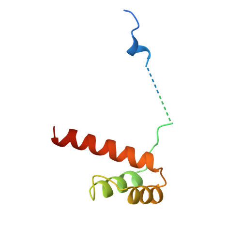
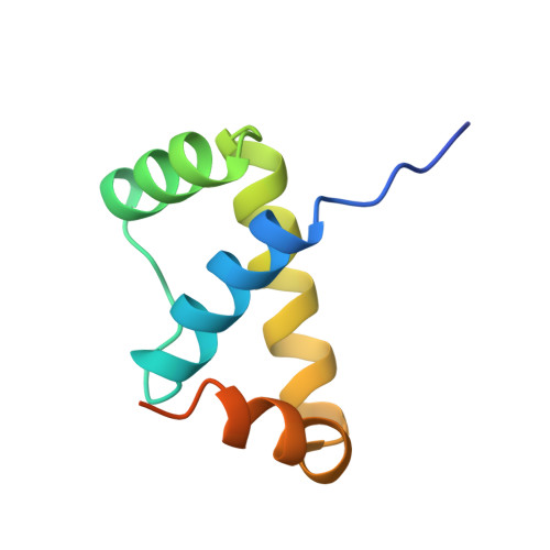
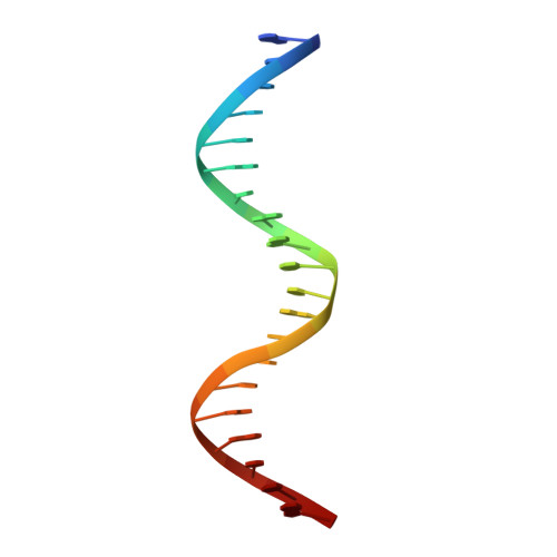
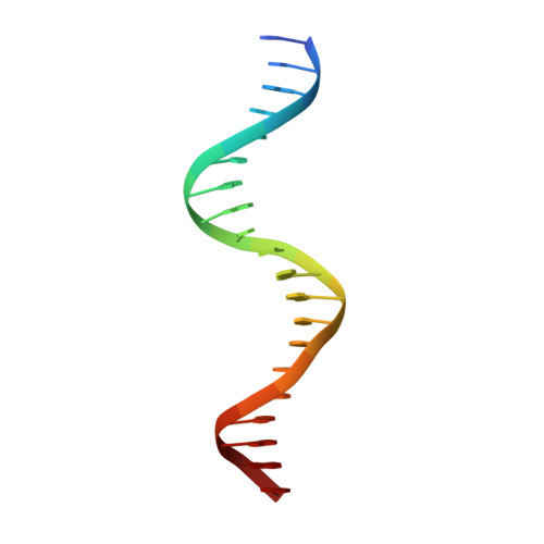
    Structure of a HoxB1-Pbx1 heterodimer bound to DNA: role of the hexapeptide and a fourth homeodomain helix in complex formation.

    Piper, D.E.Batchelor, A.H.Chang, C.P.Cleary, M.L.Wolberger, C.

    (1999) Cell 96: 587-597

    • DOI: https://doi.org/10.1016/s0092-8674(00)80662-5
    • Primary Citation of Related Structures:  
      1B72

    • PubMed Abstract: 

      Hox homeodomain proteins are developmental regulators that determine body plan in a variety of organisms. A majority of the vertebrate Hox proteins bind DNA as heterodimers with the Pbx1 homeodomain protein. We report here the 2.35 A structure of a ternary complex containing a human HoxB1-Pbx1 heterodimer bound to DNA. Heterodimer contacts are mediated by the hexapeptide of HoxB1, which binds in a pocket in the Pbx1 protein formed in part by a three-amino acid insertion in the Pbx1 homeodomain. The Pbx1 DNA-binding domain is larger than the canonical homeodomain, containing an additional alpha helix that appears to contribute to binding of the HoxB1 hexapeptide and to stable binding of Pbx1 to DNA. The structure suggests a model for modulation of Hox DNA binding activity by Pbx1 and related proteins.


    • Organizational Affiliation
      • Department of Biophysics and Biophysical Chemistry and Howard Hughes Medical Institute, Johns Hopkins School of Medicine, Baltimore, Maryland 21205, USA.

    Macromolecule Content 

    • Total Structure Weight: 33.78 kDa 
    • Atom Count: 2,010 
    • Modeled Residue Count: 181 
    • Deposited Residue Count: 224 
    • Unique protein chains: 2
    • Unique nucleic acid chains: 2

    Macromolecules

    Find similar proteins by: (by identity cutoff) | 3D Structure
    Entity ID: 3
    MoleculeChains Sequence LengthOrganismDetailsImage
    PROTEIN (HOMEOBOX PROTEIN HOX-B1)C [auth A] 97Homo sapiens Mutation(s): 0 
    Gene Names: HOXB-1
    UniProt & NIH Common Fund Data Resources
    Find proteins for P14653 (Homo sapiens)
    Explore P14653  
    Go to UniProtKB:  P14653
    PHAROS:  P14653
    GTEx:  ENSG00000120094  
    Entity Groups  
    Sequence Clusters30% Identity 50% Identity 70% Identity 90% Identity 95% Identity 100% Identity
    UniProt GroupP14653
    Sequence Annotations
    Expand
    • Reference Sequence
    Find similar proteins by: (by identity cutoff) | 3D Structure
    Entity ID: 4
    MoleculeChains Sequence LengthOrganismDetailsImage
    PROTEIN (PBX1)D [auth B] 87Homo sapiens Mutation(s): 0 
    Gene Names: PBX1
    UniProt & NIH Common Fund Data Resources
    Find proteins for P40424 (Homo sapiens)
    Explore P40424  
    Go to UniProtKB:  P40424
    PHAROS:  P40424
    GTEx:  ENSG00000185630  
    Entity Groups  
    Sequence Clusters30% Identity 50% Identity 70% Identity 90% Identity 95% Identity 100% Identity
    UniProt GroupP40424
    Sequence Annotations
    Expand
    • Reference Sequence

    Find similar nucleic acids by: Sequence | 3D Structure

    Entity ID: 1
    MoleculeChains LengthOrganismImage
    DNA (5'-D(*AP*CP*TP*CP*TP*AP*TP*GP*AP*TP*TP*GP*AP*TP*CP*GP*GP*CP*TP*G)-3')A [auth D] 20N/A
    Sequence Annotations
    Expand
    • Reference Sequence

    Find similar nucleic acids by: Sequence | 3D Structure

    Entity ID: 2
    MoleculeChains LengthOrganismImage
    DNA (5'-D(*TP*CP*AP*GP*CP*CP*GP*AP*TP*CP*AP*AP*TP*CP*AP*TP*AP*GP*AP*G)-3')B [auth E] 20N/A
    Sequence Annotations
    Expand
    • Reference Sequence
    Experimental Data & Validation

    Experimental Data

    • Method: X-RAY DIFFRACTION
    • Resolution: 2.35 Å
    • R-Value Free:  0.277 (Depositor) 
    • R-Value Work:  0.243 (Depositor), 0.240 (DCC) 
    • R-Value Observed: 0.243 (Depositor) 
    Space Group: P 21 21 21
    Unit Cell:
    Length ( Å )Angle ( ̊ )
    a = 58.67α = 90
    b = 64.58β = 90
    c = 81.91γ = 90
    Software Package:
    Software NamePurpose
    MLPHAREphasing
    CNSrefinement
    DENZOdata reduction
    SCALEPACKdata scaling

    Structure Validation

    View Full Validation Report


    Entry History 

    Deposition Data

    Revision History (Full details and data files)

    • Version 1.0: 1999年02月19日
      Type: Initial release
    • Version 1.1: 2008年04月26日
      Changes: Version format compliance
    • Version 1.2: 2011年07月13日
      Changes: Version format compliance
    • Version 1.3: 2023年12月27日
      Changes: Data collection, Database references

    AltStyle によって変換されたページ (->オリジナル) /