This site needs JavaScript to work properly. Please enable it to take advantage of the complete set of features!
Skip to main page content
U.S. flag

An official website of the United States government

Dot gov

The .gov means it’s official.
Federal government websites often end in .gov or .mil. Before sharing sensitive information, make sure you’re on a federal government site.

Https

The site is secure.
The https:// ensures that you are connecting to the official website and that any information you provide is encrypted and transmitted securely.

NIH NLM Logo
Log in
Access keys NCBI Homepage MyNCBI Homepage Main Content Main Navigation
. 1997 Jul 8;94(14):7577-82.
doi: 10.1073/pnas.94.14.7577.

Cloning, expression, and characterization of a cDNA encoding a novel human growth factor for primitive hematopoietic progenitor cells

Affiliations

Cloning, expression, and characterization of a cDNA encoding a novel human growth factor for primitive hematopoietic progenitor cells

A Hiraoka et al. Proc Natl Acad Sci U S A. .

Abstract

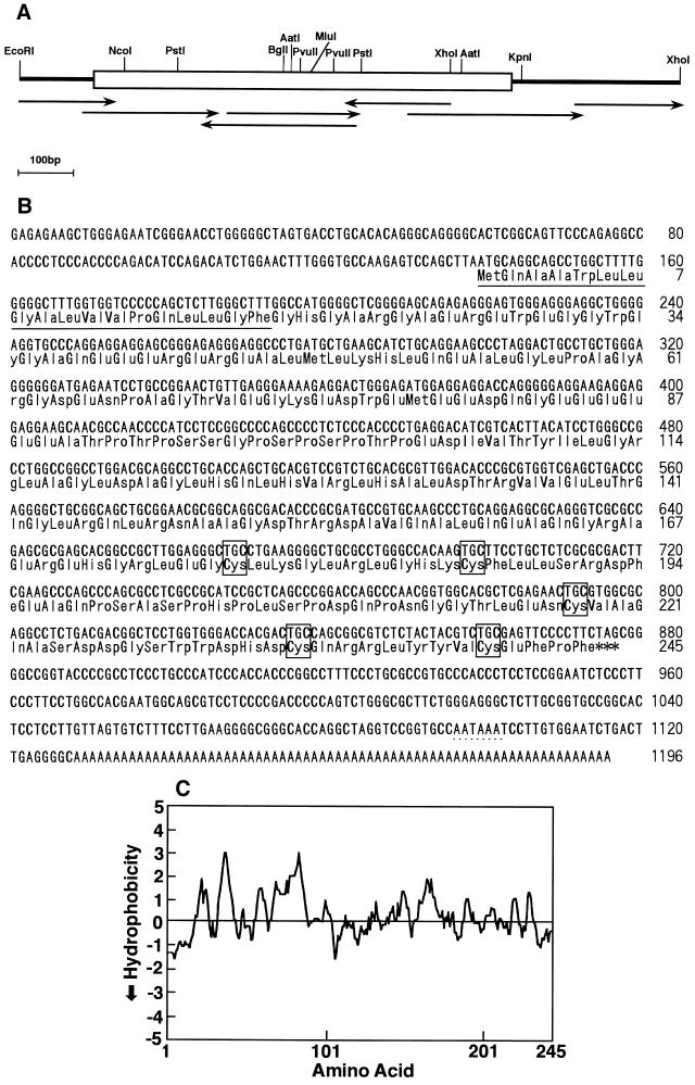
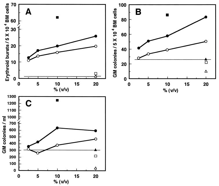
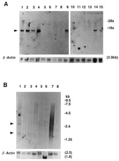
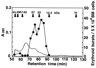
Multiple growth factors synergistically stimulate proliferation of primitive hematopoietic progenitor cells. A human myeloid cell line, KPB-M15, constitutively produces a novel hematopoietic cytokine, termed stem cell growth factor (SCGF), possessing species-specific proliferative activities. Here we report the molecular cloning, expression, and characterization of a cDNA encoding human SCGF using a newly developed lambdaSHDM vector that is more efficient for differential and expression cloning. cDNA for SCGF encodes a 29-kDa polypeptide without N-linked glycosylation. SCGF transiently produced by COS-1 cells supports growth of hematopoietic progenitor cells through a short-term liquid culture of bone marrow cells and exhibits promoting activities on erythroid and granulocyte/macrophage progenitor cells in primary semisolid culture with erythropoietin and granulocyte/macrophage colony-stimulating factor, respectively. Expression of SCGF mRNA is restricted to myeloid cells and fibroblasts, suggesting that SCGF is a growth factor functioning within the hematopoietic microenvironment. SCGF could disclose some human-specific mechanisms as yet unidentified from studies on the murine hematopoietic system.


Figures

Figure 1
Figure 1
Structure of λSHDM vector for differential and expression cloning. HTLV, human T-lymphotropic virus; SV40, simain virus 40.
Figure 2
Figure 2
(A) Restriction endonuclease map and sequencing strategy for pSHDM(116-10C) cDNA. Lines with arrowheads indicate direction and extent of sequencing. A predicted open reading frame is boxed. (B) Nucleotide sequence of SCGF cDNA and deduced amino acid sequence. pSHDM(116-10C) cDNA was sequenced using sets of oligonucleotide sequencing primers and dideoxy terminator cycle sequencing AmpliTaq on an Applied Biosystems model 373A sequencer. Numbering is from the 5′ end of the DNA sequence and from initiation methionine of the amino acid sequence. The putative signal peptide sequence is underlined. Cysteine residues are boxed. The stop codon is indicated by asterisks. Poly(A) signal is dot-underlined in the 3′-untranslated region. (C) Hydrophobicity of SCGF amino acid sequence. Each plot was calculated by the method of Hopp and Wood (22) using the dnasis Mac program (Hitachi, Tokyo).
Figure 3
Figure 3
Hematopoietic activities of SCGF on human BM cells. CM samples were reevaluated at various concentrations for BPA (A), GPA (B), and ΔGPA (C), as was CM from COS-1 cells transfected with pSHDM(116-10C) cDNA (•), mock-transfected COS-1 cells (▴), COS-1 cells transfected with pSHDM (soluble rhSCF) cDNA (▪), KPB-M15 cells (しろまる), K562 cells (▵), and MOLT-4 cells (しろいしかく). In C, the number of preexisting CFU-GM before initial liquid culture was 293 per ml. Data are given as means of three dishes.
Figure 4
Figure 4
Northern blot hybridization. (A) pSHDM(116-10C) cDNA fragment was hybridized with total RNA (20 μg) from human cell lines. Lanes: 1, HL-60 (myeloid); 2, THP-1 (monocyte); 3, U-937 (monocyte); 4 KPB-M15 (myeloid); 5, U266Bl (B cell); 6, IM-9 (B cell); 7, MOLT-4 (T cell); 8, K562 (erythroid); 9, HEL (erythroid); 10, HeLa S3 (cervical carcinoma epithelial-like); 11, A431 (epidermoid carcinoma epithelial-like); 12, Bowes (melanoma cell); 13, 293 (adenovirus type 5-transformed embryonic kidney cell); 14, NHF (fibroblast); and 15, CCD-8Lu (fibroblast). (B) pSHDM(116-10C) cDNA fragment was hybridized with poly(A)+ RNA (2 μg) from human tissues. Lanes: 1, heart; 2, brain; 3, placenta; 4, lung; 5, liver; 6, skeletal muscle; 7, kidney; and 8, pancreas. The RNA blots were rehybridized with β-actin cDNA.
Figure 5
Figure 5
Sephacryl S-200HR gel filtration of SCGF, partially purified from KPB-M15-CM through DEAE-Sephacel (pH 6) and Cu2+ chelating-Sepharose column chromatography. Each fraction was monitored by BPA (•). A solid line indicates A280 nm, and Mr markers are given as arrows.

References

    1. Metcalf D. Blood. 1993;82:3515–3523. - PubMed
    1. Zsebo K M, Wypych J, McNiece I K, Lu H S, Smith K A, Karkare S B, Sachdev R K, Yuschenkoff V N, Birkett N C, Williams L R, Satyagal V N, Tung W, Bosselman R A, Mendiaz E A, Langley K E. Cell. 1990;63:195–201. - PubMed
    1. Huang E, Nocka K, Beier D R, Chu T-Y, Buck J, Lahm H-W, Wellner D, Leder P, Besmer P. Cell. 1990;63:225–233. - PubMed
    1. Anderson D M, Lyman S D, Baird A, Wignall J M, Eisenman J, Rauch C, March C J, Boswell H S, Gimpel S D, Cosman D, Williams D E. Cell. 1990;63:235–243. - PubMed
    1. Lyman S D, James L, Bos T V, de Vries P, Brasel K, Gliniak B, Hollingsworth L T, Picha K S, McKenna H J, Splett R R, Fletcher F A, Maraskovsky E, Farrah T, Foxworthe D, Williams D E, Beckmann M P. Cell. 1993;75:1157–1167. - PubMed

MeSH terms

Substances

Associated data

LinkOut - more resources

Cite

AltStyle によって変換されたページ (->オリジナル) /