This site needs JavaScript to work properly. Please enable it to take advantage of the complete set of features!
Skip to main page content
U.S. flag

An official website of the United States government

Dot gov

The .gov means it’s official.
Federal government websites often end in .gov or .mil. Before sharing sensitive information, make sure you’re on a federal government site.

Https

The site is secure.
The https:// ensures that you are connecting to the official website and that any information you provide is encrypted and transmitted securely.

NIH NLM Logo
Log in
Access keys NCBI Homepage MyNCBI Homepage Main Content Main Navigation
Review
. 2022 Jun 15;86(2):e0002522.
doi: 10.1128/mmbr.00025-22. Epub 2022 Apr 12.

Transcending Dimensions in Apicomplexan Research: from Two-Dimensional to Three-Dimensional In Vitro Cultures

Affiliations
Review

Transcending Dimensions in Apicomplexan Research: from Two-Dimensional to Three-Dimensional In Vitro Cultures

Carlos J Ramírez-Flores et al. Microbiol Mol Biol Rev. .

Abstract

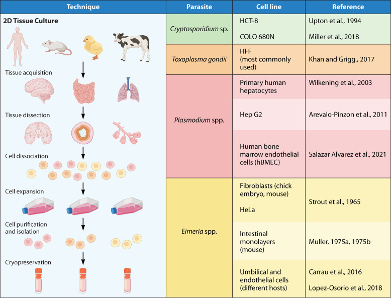
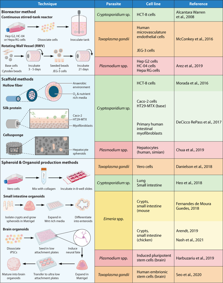
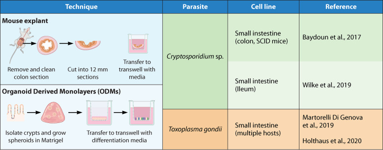
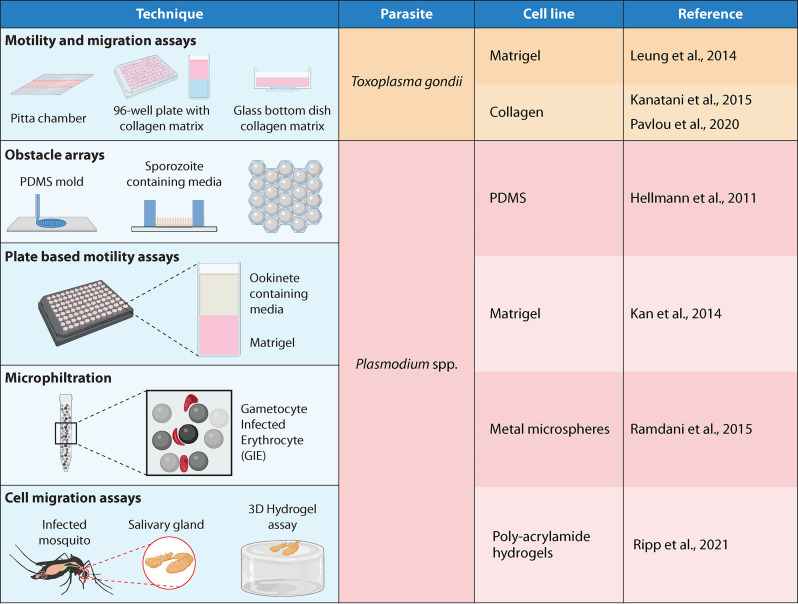
Parasites belonging to the Apicomplexa phylum are among the most successful pathogens known in nature. They can infect a wide range of hosts, often remain undetected by the immune system, and cause acute and chronic illness. In this phylum, we can find parasites of human and veterinary health relevance, such as Toxoplasma, Plasmodium, Cryptosporidium, and Eimeria. There are still many unknowns about the biology of these pathogens due to the ethical and practical issues of performing research in their natural hosts. Animal models are often difficult or nonexistent, and as a result, there are apicomplexan life cycle stages that have not been studied. One recent alternative has been the use of three-dimensional (3D) systems such as organoids, 3D scaffolds with different matrices, microfluidic devices, organs-on-a-chip, and other tissue culture models. These 3D systems have facilitated and expanded the research of apicomplexans, allowing us to explore life stages that were previously out of reach and experimental procedures that were practically impossible to perform in animal models. Human- and animal-derived 3D systems can be obtained from different organs, allowing us to model host-pathogen interactions for diagnostic methods and vaccine development, drug testing, exploratory biology, and other applications. In this review, we summarize the most recent advances in the use of 3D systems applied to apicomplexans. We show the wide array of strategies that have been successfully used so far and apply them to explore other organisms that have been less studied.

Keywords: 2D culture; 3D cultures; apicomplexan parasites; host-parasite interaction; organ-on-a-chip; organoids.


Conflict of interest statement

The authors declare no conflict of interest.

Figures

FIG 1
FIG 1
2D tissue culture systems: list of the most commonly used cell lines for in vitro culture of apicomplexan parasites. The sources shown are references (Upton et al., 1994), (Miller et al., 2018), (Khan and Grigg, 2017), (Arévalo-Pinzón et al., 2011), (Salazar Alvarez et al., 2021), (Wilkening et al., 2003), (Strout et al., 1965), (Müller, 1975a), (Müller, 1975b), (López-Osorio et al., 2018), and (Carrau et al., 2016).
FIG 2
FIG 2
3D tissue culture systems: list of cell lines and tissues most commonly used for the generation of in vitro 3D structures in apicomplexan research. The sources shown are references (Chua et al., 2019), (DeCicco RePass et al., 2017), (Alcantara Warren et al., 2008), (Morada et al., 2016), (Heo et al., 2018), (McConkey et al., 2016), (Danielson et al., 2018), (Seo et al., 2020), (Arez et al., 2019), (Harbuzariu et al., 2019), (Fernandes de Moura Guedes, 2018), (Arendt, 2019), and (Nash et al., 2021).
FIG 3
FIG 3
2D-3D hybrid models: these are techniques that are neither 3D culture nor 2D culture but a combination of the two. Here, we find the use of mouse explants and organ-derived monolayers (ODMs). The sources shown are references (Baydoun et al., 2017), (Wilke et al., 2019), (Martorelli Di Genova et al., 2019), and (Holthaus et al., 2020).
FIG 4
FIG 4
Other 3D systems: here, we find gel-based and mechanical systems devoid of any cell line, except for the organism of study. These include hydrogels, 3D printed arrays, and filtration systems. The sources shown are references (Leung et al., 2014), (Pavlou et al., 2020), (Kanatani et al., 2015), (Hellmann et al., 2011), (Ripp et al., 2021), (Ramdani et al., 2015), and (Kan et al., 2014).

References

    1. Visvesvara GS, Garcia LS. 2002. Culture of protozoan parasites. Clin Microbiol Rev 15:327–328. 10.1128/CMR.15.3.327-328.2002. - DOI - PMC - PubMed
    1. Justice MJ, Dhillon P. 2016. Using the mouse to model human disease: increasing validity and reproducibility. Dis Model Mech 9:101–103. 10.1242/dmm.024547. - DOI - PMC - PubMed
    1. Murillo-Cuesta S, Artuch R, Asensio F, de la Villa P, Dierssen M, Enríquez JA, Fillat C, Fourcade S, Ibáñez B, Montoliu L, Oliver E, Pujol A, Salido E, Vallejo M, Varela-Nieto I. 2020. The value of mouse models of rare diseases: a Spanish experience. Front Genet 11:583932. 10.3389/fgene.2020.583932. - DOI - PMC - PubMed
    1. Bayir E, Sendemir A, Missirlis YF. 2019. Mechanobiology of cells and cell systems, such as organoids. Biophys Rev 11:721–728. 10.1007/s12551-019-00590-7. - DOI - PMC - PubMed
    1. Co JY, Margalef-Català M, Li X, Mah AT, Kuo CJ, Monack DM, Amieva MR. 2019. Controlling epithelial polarity: a human enteroid model for host-pathogen interactions. Cell Rep 26:2509–2520.e4. 10.1016/j.celrep.2019年01月10日8. - DOI - PMC - PubMed

Publication types

Cite

AltStyle によって変換されたページ (->オリジナル) /