This site needs JavaScript to work properly. Please enable it to take advantage of the complete set of features!
Skip to main page content
U.S. flag

An official website of the United States government

Dot gov

The .gov means it’s official.
Federal government websites often end in .gov or .mil. Before sharing sensitive information, make sure you’re on a federal government site.

Https

The site is secure.
The https:// ensures that you are connecting to the official website and that any information you provide is encrypted and transmitted securely.

NIH NLM Logo
Log in
Access keys NCBI Homepage MyNCBI Homepage Main Content Main Navigation
. 2021 Jan 14;11(1):1407.
doi: 10.1038/s41598-021-80959-5.

The role of Toll-like receptor 9 in a murine model of Cryptococcus gattii infection

Affiliations

The role of Toll-like receptor 9 in a murine model of Cryptococcus gattii infection

Elias Barbosa da Silva-Junior et al. Sci Rep. .

Abstract

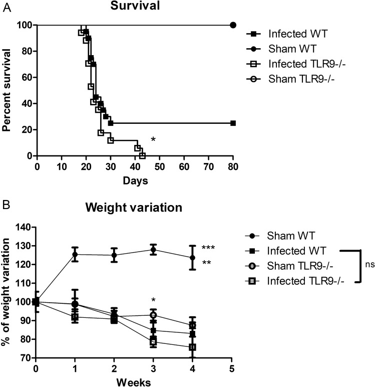
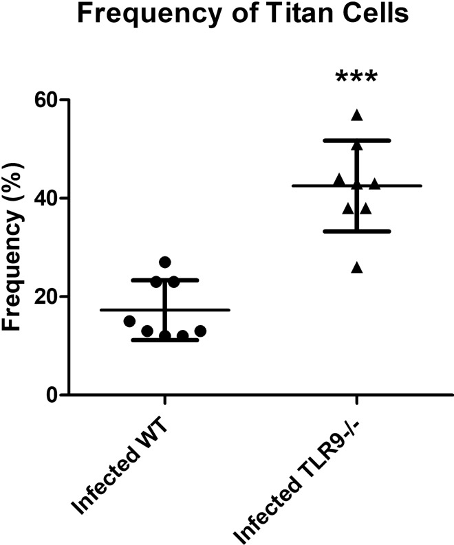
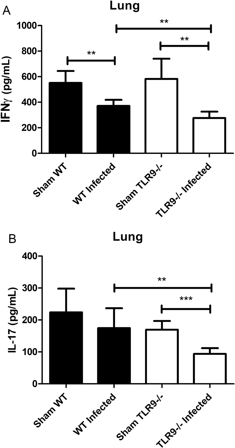
Toll-like receptor 9 (TLR9) is crucial to the host immune response against fungi, such as Candida albicans, Aspergillus fumigatus and Cryptococcus neoformans, but its importance in Cryptococcus gattii infection is unknown. Our study aimed to understand the role of TLR9 during the course of experimental C. gattii infection in vivo, considering that the cryptococcal DNA interaction with the receptor could contribute to host immunity even in an extremely susceptible model. We inoculated C57BL/6 (WT) and TLR9 knock-out (TLR9-/-) mice intratracheally with 104 C. gattii yeast cells. TLR9-/- mice had a higher mortality rate compared to WT mice and more yeast cells that had abnormal size, known as titan cells, in the lungs. TLR9-/- mice also had a greater number of CFUs in the spleen and brain than WT mice, in addition to having lower levels of IFN-γ and IL-17 in the lung. With these markers of aggressive cryptococcosis, we can state that TLR9-/- mice are more susceptible to C. gattii, probably due to a mechanism associated with the decrease of a Th1 and Th17-type immune response that promotes the formation of titan cells in the lungs. Therefore, our results indicate the participation of TLR9 in murine resistance to C. gattii infection.


Conflict of interest statement

The authors declare no competing interests.

Figures

Figure 1
Figure 1
Analysis of susceptibility of TLR9−/− and WT mice to C. gattii infection. (A) Survival curves of infected C57BL/6 WT (n = 20) and TLR9−/− (n = 17) mice. Mice were infected with 104 C. gattii R265 belonging to serotype IIa. Sham WT (n = 6) and TLR9−/− (n = 6) mice given with vehicle (PBS) only were used as controls. Log-rank test (Mantel-Cox test): *P ≤ 0.05. (B) The percentage change in weight of infected WT (n = 11) and TLR9−/− (n = 10) mice, as well as the Sham groups (Sham WT and Sham TLR9−/−) (n = 5 per group), was determined through weekly mass measurements over 28 days (0, 7, 14, 21 and 28 days after inoculation). Black squares represent infected WT mice, black balls represent uninfected WT mice, white squares represent infected TLR9−/− mice, and white balls represent uninfected TLR9−/− animals. One-Way ANOVA (**P ≤ 0.01) and Two-Way ANOVA with Bonferroni posttest (*P ≤ 0.05; ***P ≤ 0.001); ns = not statistically significant. Graphs show the union of the results from 4 similar and independent experiments.
Figure 2
Figure 2
Comparison of CFU counts in different target organs of C. gattii infection in WT and TLR9−/− mice. Analysis of fungal load after 21 days of C. gattii infection through maceration of tissues taken from WT (n = 6) and TLR9−/− mice (n = 7). (AC) The CFUs in the lungs, brain and spleen, respectively. Black bars represent infected C57BL/6 WT mice and white bars represent infected C57BL/6 TLR9−/− mice. Organ homogenates were obtained in 5 mL PBS, the lungs and brain homogenates were then diluted ×ばつ10,000 and ×ばつ100, respectively, and 50 μL was spread on Petridishes containing Sabouraud's agar. Colonies were counted 72 h after incubation at 37 °C. CFU colony forming units. The graphs show a representative result of six similar and independent experiments. Student T-test and Mann–Whitney U test: *P ≤ 0.05; **P ≤ 0.01; ns = not statistically significant.
Figure 3
Figure 3
Histological sections of lungs of C. gattii-infected WT and TLR9−/− animals. Micrographs of histological sections of lungs obtained from WT and TLR9−/− animals infected with C. gattii after 21 days (CF) or given sterile PBS (Sham) (A, B). In the left column, the histological sections of the lungs of WT mice (A, C, E) (n = 3). In the right column, histological sections of the TLR9−/− mice (B, D, F) (n = 4). Magnification, ×ばつ40 (AD); Magnification ×ばつ100 (E, F). The arrows show normal C. gattii yeasts and the arrowheads show C. gattii titan cells in the lungs of both WT and TLR9−/− mice. The histological sections were stained with hematoxylin, eosin and Alcian Blue and photographed under a light microscope.
Figure 4
Figure 4
Percentage of titan cells in the lung of C. gattii-infected mice. Titan cell (> 10 μm cell size) counts per 100 yeast cells observed in defined quadrants in the histological sections of the lungs of WT (n = 3) and TLR9−/− (n = 4) mice after 21 days of C. gattii infection. Each lung (right and left) was divided into two quadrants (4 quadrants for each animal). Titan cells were counted in each quadrant and each result was considered as an absolute value for the respective quadrant. The graphs show a representative result. Student T-test: ***P ≤ 0.0001.
Figure 5
Figure 5
Levels of IFN-γ and IL-17 in total lung homogenates of C. gattii-infected mice. Measurement of IFN-γ and IL-17 levels by ELISA of total lung homogenates of WT and TLR9−/− mice, infected with C. gattii or given PBS, after 21 days. (A) Dosage of IFN-γ and (B) IL-17. The graphs show a representative result of two similar and independent experiments. Sham WT (n = 4), Sham TLR9−/− (n = 4), infected WT (n = 6), infected TLR9−/− (n = 7). **P < 0.005; ***P < 0.001; ns = not statistically significant.

References

    1. Rozenbaum R, Goncalves AJ. Clinical epidemiological study of 171 cases of cryptococcosis. Clin. Infect. Dis. 1994;18:369–380. doi: 10.1093/clinids/18.3.369. - DOI - PubMed
    1. Speed B, Dunt D. Clinical and host differences between infections with the two varieties of Cryptococcus neoformans. Clin. Infect. Dis. 1995;21:28–34. doi: 10.1093/clinids/21.1.28. - DOI - PubMed
    1. Zaragoza O. Basic principles of the virulence of Cryptococcus. Virulence. 2019;10:490–501. doi: 10.1080/21505594.2019.1614383. - DOI - PMC - PubMed
    1. Villena SN, et al. Capsular polysaccharides galactoxylomannan and glucuronoxylomannan from Cryptococcus neoformans induce macrophage apoptosis mediated by Fas ligand. Cell. Microbiol. 2008;10:1274–1285. doi: 10.1111/j.1462-5822.2008.01125.x. - DOI - PubMed
    1. Rocha JD, et al. Capsular polysaccharides from Cryptococcus neoformans modulate production of neutrophil extracellular traps (NETs) by human neutrophils. Sci. Rep. 2015;5:8008. doi: 10.1038/srep08008. - DOI - PMC - PubMed

Publication types

LinkOut - more resources

Cite

AltStyle によって変換されたページ (->オリジナル) /