This site needs JavaScript to work properly. Please enable it to take advantage of the complete set of features!
Skip to main page content
U.S. flag

An official website of the United States government

Dot gov

The .gov means it’s official.
Federal government websites often end in .gov or .mil. Before sharing sensitive information, make sure you’re on a federal government site.

Https

The site is secure.
The https:// ensures that you are connecting to the official website and that any information you provide is encrypted and transmitted securely.

NIH NLM Logo
Log in
Access keys NCBI Homepage MyNCBI Homepage Main Content Main Navigation
. 2020 Nov 24;11(6):e01821-20.
doi: 10.1128/mBio.01821-20.

Phagosomal F-Actin Retention by Cryptococcus gattii Induces Dendritic Cell Immunoparalysis

Affiliations

Phagosomal F-Actin Retention by Cryptococcus gattii Induces Dendritic Cell Immunoparalysis

Khusraw Jamil et al. mBio. .

Abstract

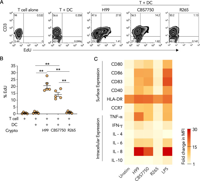
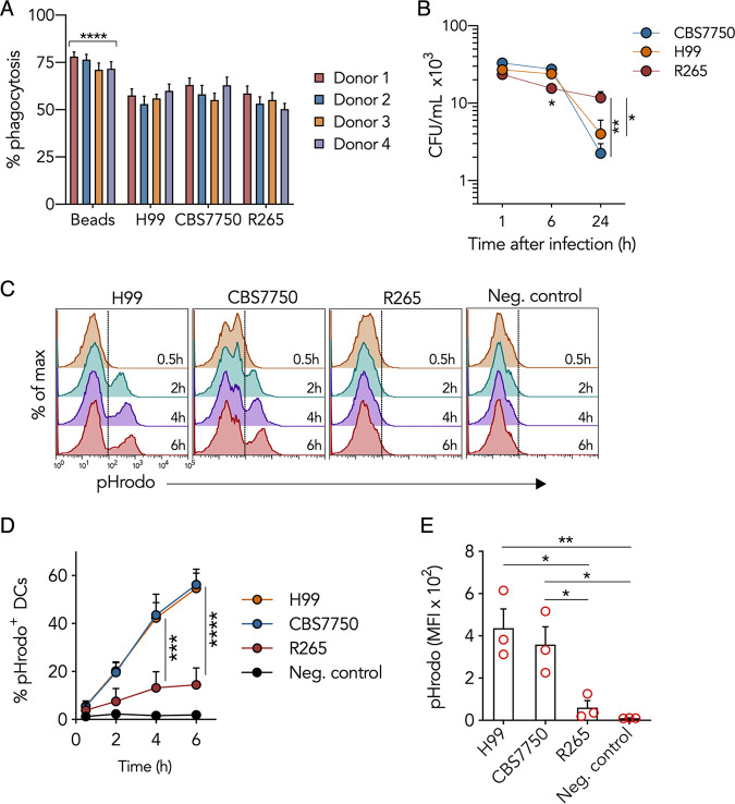
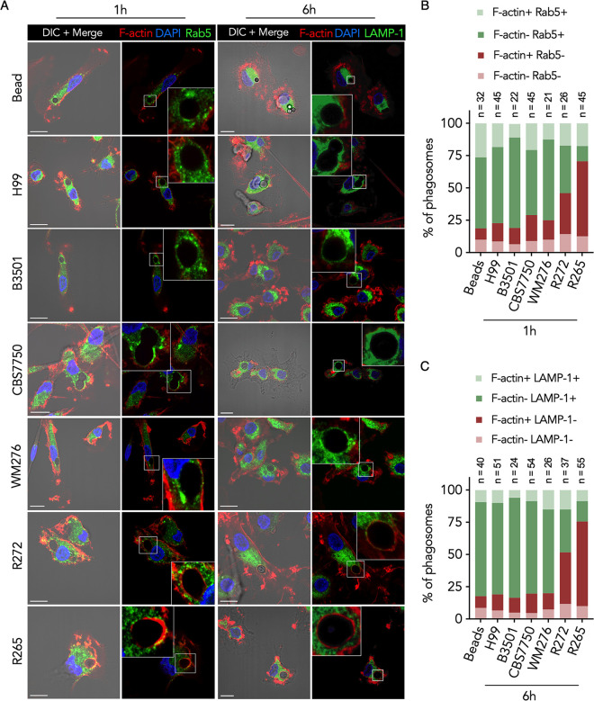
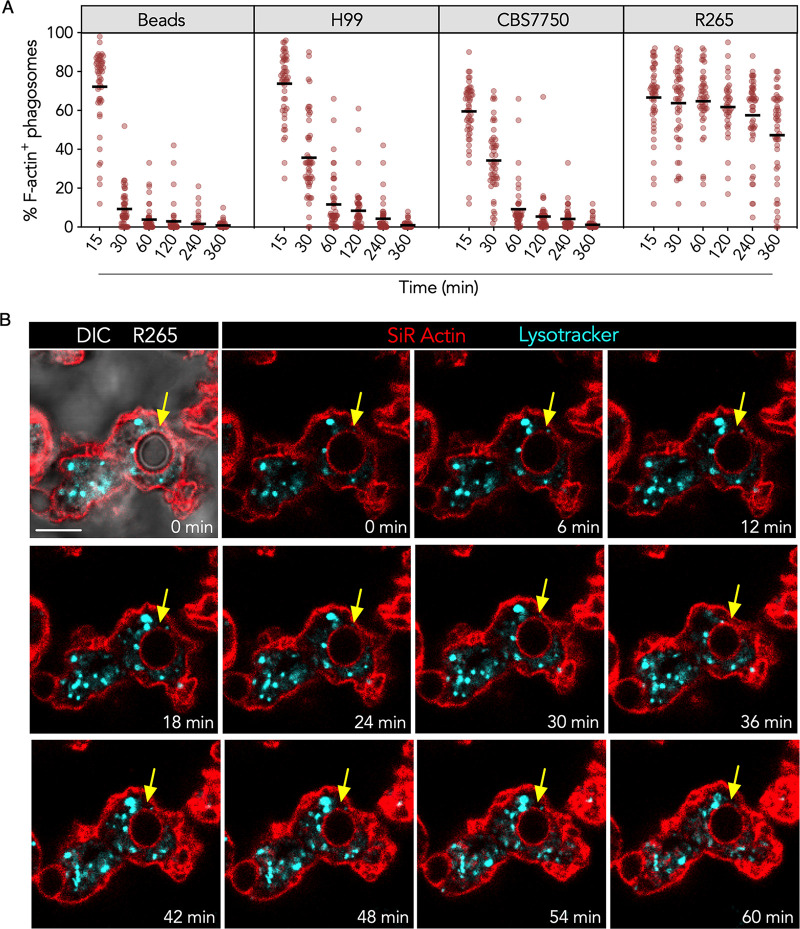
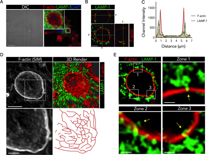
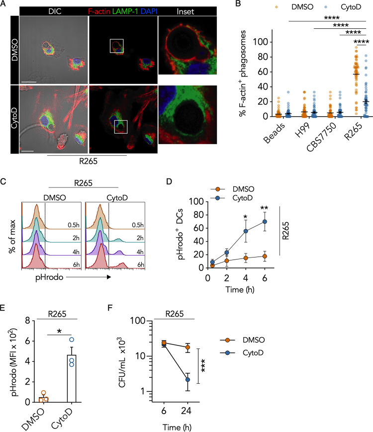
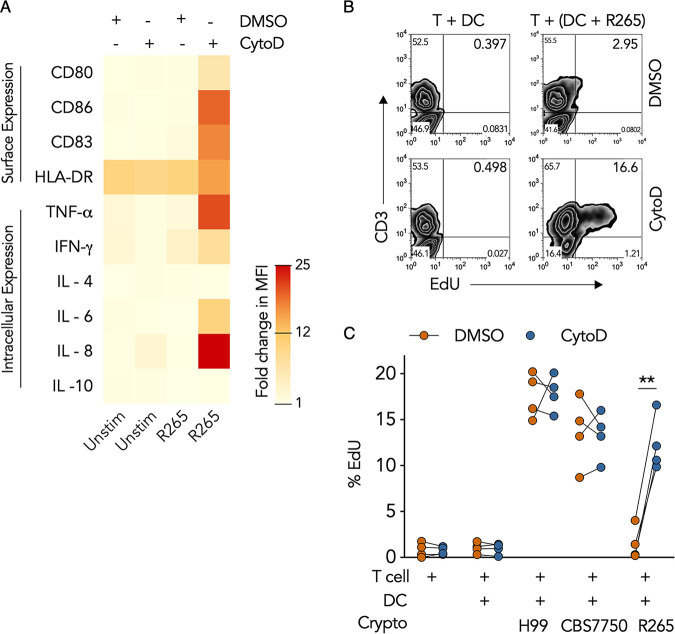
Cryptococcus gattii is a major cause of life-threatening mycosis in immunocompetent individuals and responsible for the ongoing epidemic outbreak of cryptococcosis in the Pacific Northwest of North America. This deadly fungus is known to evade important host immune responses, including dendritic cell (DC) maturation and concomitant T cell immunity, via immune evasion mechanisms that remain unclear. Here, we demonstrate that primary human DCs phagocytose C. gattii but the maturation of phagosomes to phagolysosomes was blocked as a result of sustained filamentous actin (F-actin) that entrapped and concealed the phagosomes from recognition. Superresolution structured illumination microscopy (SR-SIM) revealed that the persistent phagosomal F-actin formed a cage-like structure that sterically hindered and functionally blocked the fusion of lysosomes. Blocking lysosome fusion was sufficient to inhibit phagosomal acidification and subsequent intracellular fungal killing by DCs. Retention of phagosomal F-actin by C. gattii also caused DC immunoparalysis. Disrupting the retained F-actin cage with cytochalasin D not only restored DC phagosomal maturation but also promoted DC costimulatory maturation and robust T cell activation and proliferation. Collectively, these results reveal a unique mechanism of DC immune evasion that enhances intracellular fungal pathogenicity and may explain suppressed cell-mediated immunity.IMPORTANCECryptococcus yeast species typically display characteristics of opportunistic pathogens, with the exception of C. gattii, which can cause life-threatening respiratory and disseminated brain infections in otherwise healthy people. The pathogenesis of C. gattii is not well understood, but an important characteristic is that C. gattii is capable of evading host cell-mediated immune defenses initiated by DCs. Here, we report that when virulent C. gattii becomes ingested by a DC, the intracellular compartment containing the fungi is covered by a persistent protein cage structure consisting of F-actin. This F-actin cage acts as a barrier to prevent interaction with other intracellular compartments, and as a result, the DC fails to kill the fungi and activate important cell-mediated immune responses. We propose that this unique immune evasion mechanism permits C. gattii to remain unchallenged within host cells, leading to persistent infection.

Keywords: Cryptococcus gattii; dendritic cells; immune evasion; immunoparalysis; phagosomal F-actin.


Figures

FIG 1
FIG 1
Hypervirulent Cryptococcus gattii suppresses DC immunity. (A) Representative flow cytometry plot of EdU incorporation (measure of cell proliferation) in peripheral blood T cells cultured for 6 days either alone, with resting DCs, or with DCs infected with R265, H99, or CBS7750 (6 h). (B) Quantification of data in panel A as percent EdU staining in CD3+ T cells. Data are presented as means ± standard errors of the means (SEM) from 5 donors, shown individually as dots. **, P < 0.001 by one-way ANOVA (adjusted P values). (C) Heat map of the fold changes in the mean fluorescence intensity (MFI) of surface and intracellular markers in resting (Unstim) DCs, Cryptococcus-infected DCs (6 h), and LPS-stimulated DCs (6 h). Data are presented as the average fold changes in MFI, standardized to isotype-matched controls, from 5 different donors. The color spectrum represents the minimum and maximum fold changes in the MFI.
FIG 2
FIG 2
Hypervirulent Cryptococcus gattii attenuates DC phagosomal maturation. (A) Phagocytic frequency 1 h after infection (MOI of 10 for 2 h), measured by confocal microscopy in DCs infected with Cryptococcus or fed polystyrene beads. The frequency was calculated as the number of DCs with internalized cryptococci or beads (confirmed by phalloidin labeling of cortical actin) relative to the total number of DCs in a field of view (FOV). Data are presented as means ± SEM from 25 to 30 FOVs per donor. ****, P < 0.0001 (by one-way ANOVA [adjusted P values] of beads compared to Cryptococcus). (B) Intracellular Cryptococcus survival within DCs at 1 h, 6 h, or 24 h postinfection (MOI of 10 for 2 h). Data are presented as means ± SEM on a log10 scale and are averages from 4 donors. *, P < 0.03; **, P = 0.0062 (by one-way ANOVA [adjusted P values] at each time point). (C) Representative flow cytometric histograms (gated on total DCs) of pHrodo fluorescence at different times after the 2-h infection period. DCs were cultured with pHrodo-labeled cryptococci or given unlabeled beads (negative [Neg.] control) for 2 h. (D) Percent change in pHrodo+ DCs, similar to panel C. Data are presented as means ± SEM from 3 donors. ***, P < 0.001; ****, P < 0.0001 (by two-way ANOVA [adjusted P values]). (E) MFI of pHrodo 6 h after infection in DCs cultured with cryptococci or fed unlabeled beads (negative control). *, P < 0.05; **, P < 0.01 (by one-way ANOVA [adjusted P values]). Data are presented as means ± SEM from 3 donors (represented as dots).
FIG 3
FIG 3
DC phagosomes containing PNW outbreak strains of Cryptococcus gattii are enriched with an F-actin coat. (A) Confocal micrographs of DCs infected with Cryptococcus or fed polystyrene beads and immunolabeled for Rab5 (green) 1 h or LAMP-1 (green) 6 h after the 2-h infection period. In both cases, coverslips were stained with phalloidin (DyLight 554) to label F-actin (red) and DAPI to stain the nucleus (blue). Images are shown as maximum-intensity projections (MIPs) in the merge panels and as a single confocal stack in the digitally magnified region represented by a white box. DIC, differential interference contrast. Bars = 10 μm. (B) Quantification of the data in panel A at 1 h postinfection. Data are presented as stacked bar graphs of the average proportions of phagosomes per FOV that are enriched for F-actin, Rab5, both, or neither. (C) Quantification of the data in panel A at 6 h postinfection. Data are presented as stacked bar graphs of the average proportions of phagosomes per FOV that are enriched for F-actin, LAMP-1, both, or neither. The number of FOVs (n) is shown under each condition. Data are representative of results from 3 donors.
FIG 4
FIG 4
DC phagosomal F-actin is retained and does not fluctuate over time in response to hypervirulent Cryptococcus gattii. (A) Frequency of phagosomal F-actin enrichment at various times. DCs were infected with Cryptococcus or given polystyrene beads for 2 h and fixed at increasing times after infection (minutes). Fixed samples were imaged by confocal microscopy, and F-actin was labeled with DyLight 554 phalloidin. Each data point is the proportion of phagosomes per FOV (50 to 65 FOVs pooled from 3 donors) that are enriched for F-actin. (B) Time-lapse confocal micrographs of DCs infected with C. gattii R265 corresponding to Movie S1 in the supplemental material. DCs were labeled with SiR-actin (red) to stain F-actin and with LysoTracker (cyan) to stain the lysosomes prior to coculture with the cryptococci for 30 min. Images are presented as single confocal stacks at 6-min intervals over a course of 1 h. The yellow arrows point to the position of the phagosome containing R265, as determined by DIC. Data are representative of results from 3 separate experiments.
FIG 5
FIG 5
Hypervirulent Cryptococcus gattii forms a porous cage-like F-actin structure on DC phagosomes that limits lysosome fusion. (A) Confocal micrograph (MIP) of R265-infected DCs and immunolabeled LAMP-1 (green), phalloidin-labeled F-actin (red), and the DAPI-stained nucleus (blue) 4 h after the 2-h infection period. Bar = 15 μm. (B) SR-SIM image (single z-stack) of the phagosomal region (white box in panel A) shown as a cross section and orthogonal sections. Bar = 2 μm. (C) Positional pixel intensity graph of LAMP-1 (green) and F-actin (red) on a single line depicted by "y" in panel B from left (0 μm) to right (7 μm). (D) SR-SIM image (MIP) of F-actin (top left) and 3D reconstruction of F-actin and LAMP-1 (top right) of the phagosomal region (white box in panel A). A digital magnification (bottom left) and a pixel map (bottom right) of the F-actin SR-SIM image (dashed square, top left) are shown. Bars = 0.5 μm. (E) SR-SIM image (single stack) of the phagosome (white box in panel A), with magnified regions indicated by the corresponding numbers. Bars = 0.2 μm. Data are representative of results from 3 donors.
FIG 6
FIG 6
The hypervirulent Cryptococcus gattii phagosomal F-actin cage subverts DC phagosomal processing and maturation. (A) Confocal micrographs (MIPs) and digital magnifications (white boxes) of R265-infected DCs that were pulsed with cytochalasin D (CytoD) or DMSO (vehicle control) for 2 h and cultured for an additional 4 h in medium alone. Cells were immunostained for LAMP-1 (green), phalloidin to label F-actin (red), and DAPI to stain the nucleus (blue). Bars = 10 μm. (B) Quantification of the data in panel A as the proportion of F-actin-enriched phagosomes containing cryptococci or beads per FOV in response to cytochalasin D treatment. Data are presented as a scatterplot of 50 to 55 FOVs from 3 donors (the mean is represented by a black line). ****, P < 0.0001 (by one-way ANOVA [adjusted P values]). (C) Representative flow cytometric histograms of DCs cultured with pHrodo-labeled R265 and treated with cytochalasin D or DMSO at 0.5 to 6 h posttreatment. (D) Quantification of the data in panel C. Data are presented as means ± SEM of the percentage of pHrodo+ DCs (averages from 3 donors). *, P = 0.033; **, P = 0.00053 (compared to the control [DMSO] by two-way ANOVA [adjusted P values]). (E) MFI of DCs cultured with pHrodo-labeled R265 6 h after cytochalasin D or DMSO treatment. Data are presented as means ± SEM of results from 3 donors (shown as dots). *, P = 0.023 by unpaired Welch’s t test (two-tailed P value). (F) Intracellular survival of R265 within DCs 1 h or 24 h following cytochalasin D or DMSO treatment. Data are presented as means ± SEM from 3 donors in a before-after graph. ***, P < 0.001 (by one-way ANOVA [adjusted P values]).
FIG 7
FIG 7
Immunoparalysis of DCs by hypervirulent Cryptococcus gattii is caused by the retention of the phagosomal F-actin cage. (A) Heat map of the fold changes in the MFIs of surface and intracellular markers in resting DCs (Unstim) or DCs infected with R265 that were pulsed with cytochalasin D or DMSO for 2 h and cultured for an additional 4 h in medium alone. Data are presented as the average fold changes in MFIs, standardized to isotype-matched controls, from 4 different donors. The color spectrum represents the minimum and maximum fold changes in MFIs. (B) Representative flow cytometry plot of EdU incorporation in peripheral blood T cells cultured for 6 days with uninfected DCs or DCs infected with R265 and pulse-treated with cytochalasin D or DMSO (similar to panel A) prior to coculture with T cells. (C) Quantification of data in panel B as percent EdU staining in CD3+ T cells in response to DCs treated with cytochalasin D or DMSO. Data are presented in a before-after graph where each connected pair of dots represents data for each donor (total of 4 donors). **, P = 0.0014 (by one-way ANOVA [adjusted P values]).

References

    1. Kidd SE, Hagen F, Tscharke RL, Huynh M, Bartlett KH, Fyfe M, MacDougall L, Boekhout T, Kwon-Chung KJ, Meyer W. 2004. A rare genotype of Cryptococcus gattii caused the cryptococcosis outbreak on Vancouver Island (British Columbia, Canada). Proc Natl Acad Sci U S A 101:17258–17263. doi:10.1073/pnas.0402981101. - DOI - PMC - PubMed
    1. Harris JR, Lockhart SR, Sondermeyer G, Vugia DJ, Crist MB, D’Angelo MT, Sellers B, Franco-Paredes C, Makvandi M, Smelser C, Greene J, Stanek D, Signs K, Nett RJ, Chiller T, Park BJ. 2013. Cryptococcus gattii infections in multiple states outside the US Pacific Northwest. Emerg Infect Dis 19:1620–1626. doi:10.3201/eid1910.130441. - DOI - PMC - PubMed
    1. Harris JR, Lockhart SR, Debess E, Marsden-Haug N, Goldoft M, Wohrle R, Lee S, Smelser C, Park B, Chiller T. 2011. Cryptococcus gattii in the United States: clinical aspects of infection with an emerging pathogen. Clin Infect Dis 53:1188–1195. doi:10.1093/cid/cir723. - DOI - PubMed
    1. Leongson K, Cousineau-Cote V, Goupil M, Aumont F, Senechal S, Gaboury L, Jolicoeur P, Kronstad JW, de Repentigny L. 2013. Altered immune response differentially enhances susceptibility to Cryptococcus neoformans and Cryptococcus gattii infection in mice expressing the HIV-1 transgene. Infect Immun 81:1100–1113. doi:10.1128/IAI.01339-12. - DOI - PMC - PubMed
    1. Mody CH, Lipscomb MF, Street NE, Toews GB. 1990. Depletion of CD4+ (L3T4+) lymphocytes in vivo impairs murine host defense to Cryptococcus neoformans. J Immunol 144:1472–1477. - PubMed

Publication types

Cite

AltStyle によって変換されたページ (->オリジナル) /