Differential susceptibility of Onchocerca volvulus microfilaria to ivermectin in two areas of contrasting history of mass drug administration in Cameroon: relevance of microscopy and molecular techniques for the monitoring of skin microfilarial repopulation within six months of direct observed treatment
- PMID: 33008333
- PMCID: PMC7530974
- DOI: 10.1186/s12879-020-05444-2
Differential susceptibility of Onchocerca volvulus microfilaria to ivermectin in two areas of contrasting history of mass drug administration in Cameroon: relevance of microscopy and molecular techniques for the monitoring of skin microfilarial repopulation within six months of direct observed treatment
Erratum in
-
Correction to: Differential susceptibility of Onchocerca volvulus microfilaria to ivermectin in two areas of contrasting history of mass drug administration in Cameroon: relevance of microscopy and molecular techniques for the monitoring of skin microfilarial repopulation within six months of direct observed treatment.Abong RA, Amambo GN, Chounna Ndongmo PW, Njouendou AJ, Ritter M, Beng AA, Esum ME, Deribe K, Fru-Cho J, Fombad FF, Nji TM, Enyong PI, Poole CB, Pfarr K, Hoerauf A, Carlow CKS, Wanji S. Abong RA, et al. BMC Infect Dis. 2021 Jan 18;21(1):81. doi: 10.1186/s12879-021-05797-2. BMC Infect Dis. 2021. PMID: 33461481 Free PMC article. No abstract available.
Abstract
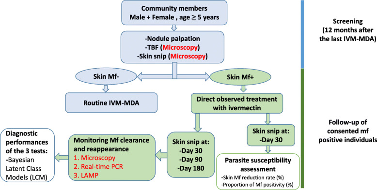
Background: Ivermectin is an excellent microfilaricide against Onchocerca volvulus. However, in some regions, long term use of ivermectin has resulted in sub-optimal responses to the treatment. More data to properly document the phenomenon in various contexts of ivermectin mass drug administration (IVM-MDA) is needed. Also, there is a need to accurately monitor a possible repopulation of skin by microfilariae following treatment. Skin snip microscopy is known to have a low sensitivity in individuals with light infections, which can be the case following treatment. This study was designed with two complementary objectives: (i) to assess the susceptibility of O. volvulus microfilariae to ivermectin in two areas undergoing IVM-MDA for different lengths of time, and (ii) to document the repopulation of skin by the O. volvulus microfilariae following treatment, using 3 independent diagnostic techniques.
Method: Identified microfilaridermic individuals were treated with ivermectin and re-examined after 1, 3, and 6 months using microscopy, actin real-time PCR (actin-qPCR) and O-150 LAMP assays. Susceptibility to ivermectin and trends in detecting reappearance of skin microfilariae were determined using three techniques. Microscopy was used as an imperfect gold standard to determine the performance of actin-qPCR and LAMP.
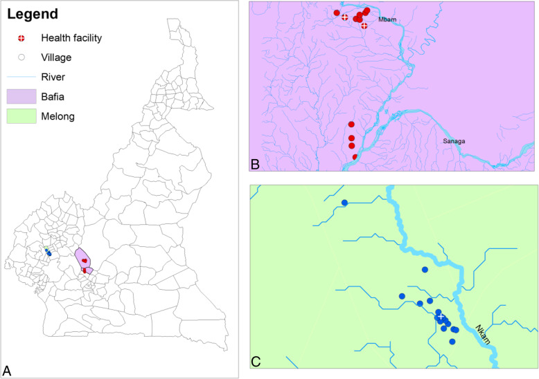
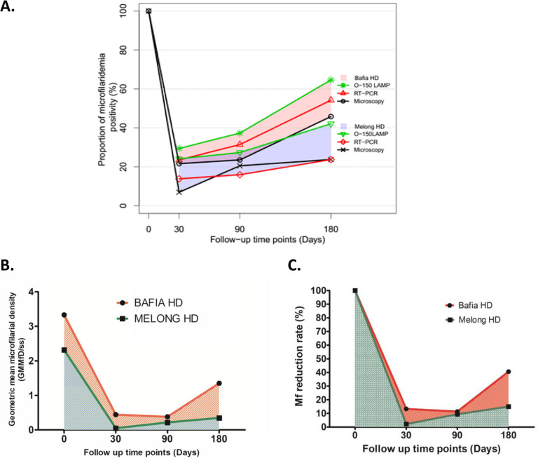
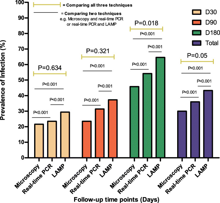
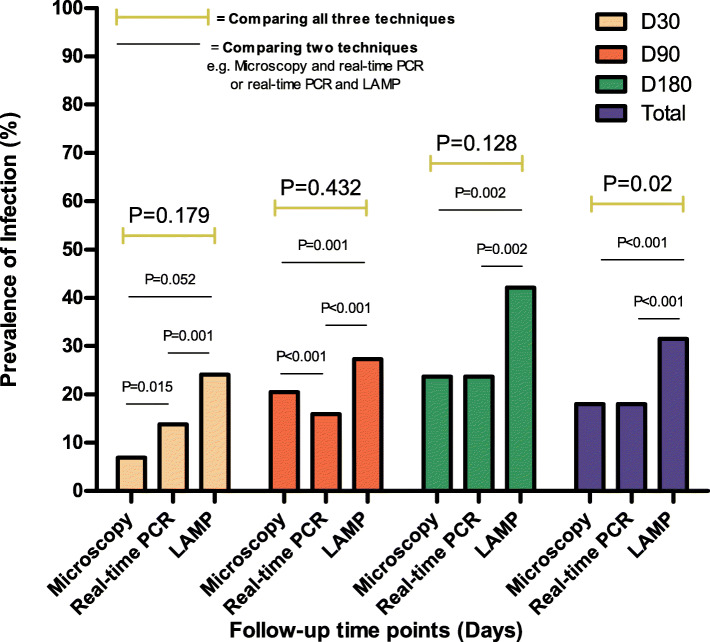
Results: In Bafia with over 20 years of IVM-MDA, 11/51 (21.6%) direct observe treated microfilaridemic participants were still positive for skin microfilariae after 1 month. In Melong, with 10 years of IVM-MDA, 2/29 (6.9%) treated participants were still positive. The microfilarial density reduction per skin biopsy within one month following treatment was significantly lower in participants from Bafia. In both study sites, the molecular techniques detected higher proportions of infected individuals than microscopy at all monitoring time points. LAMP demonstrated the highest levels of sensitivity and real-time PCR was found to have the highest specificity.
Conclusion: Patterns in skin mirofilariae clearance and repopulation were established. O. volvulus worms from Bafia with higher number of annual MDA displayed a lower clearance and higher repopulation rate after treatment with ivermectin. Molecular assays displayed higher sensitivity in monitoring O. volvulus microfilaridemia within six months following treatment.
Keywords: LAMP; Microfilaricides; Microfilaridemia; Microscopy; Monitoring; O. volvulus; Real-time PCR; Susceptibility.
Conflict of interest statement
The authors declare that they have no competing interests.
Figures
References
-
- WHO: Uniting to Combat Neglected Tropical Diseases. River blindness (Onchocerciasis). 2020 WHO roadmap target: Elimination. https://unitingtocombatntds.org/ntds/onchocerciasis/. (Accessed 20 Apr 2020). In.; 2020.
-
- Adewole SOaA, S.K. Clinical manifestation of Onchocerciasis in Ise - Orun local government, Ekiti state. Nigeria Pakistan J Nutri. 2009;8:122–124. doi: 10.3923/pjn.2009.122.124. - DOI
-
- Edungbola LD, Watts SJ, Kayode OO. Endemicity and striking manifestations of onchocerciasis in Shao, Kwara state, Nigeria. Afr J Med Med Sci. 1987;16(3):147–156. - PubMed
-
- Katawa G, Layland LE, Debrah AY, von Horn C, Batsa L, Kwarteng A, Arriens S, D WT. Specht S, Hoerauf A, et al. Hyperreactive onchocerciasis is characterized by a combination of Th17-Th2 immune responses and reduced regulatory T cells. PLoS Negl Trop Dis. 2015;9(1):e3414. doi: 10.1371/journal.pntd.0003414. - DOI - PMC - PubMed
MeSH terms
Substances
Grants and funding
LinkOut - more resources
Full Text Sources