T Lymphocyte-Mediated Liver Immunopathology of Schistosomiasis
- PMID: 32132991
- PMCID: PMC7040032
- DOI: 10.3389/fimmu.2020.00061
T Lymphocyte-Mediated Liver Immunopathology of Schistosomiasis
Abstract
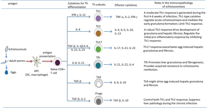
The parasitic worms, Schistosoma mansoni and Schistosoma japonicum, reside in the mesenteric veins, where they release eggs that induce a dramatic granulomatous response in the liver and intestines. Subsequently, infection may further develop into significant fibrosis and portal hypertension. Over the past several years, uncovering the mechanism of immunopathology in schistosomiasis has become a major research objective. It is known that T lymphocytes, especially CD4+ T cells, are essential for immune responses against Schistosoma species. However, obtaining a clear understanding of how T lymphocytes regulate the pathological process is proving to be a daunting challenge. To date, CD4+ T cell subsets have been classified into several distinct T helper (Th) phenotypes including Th1, Th2, Th17, T follicular helper cells (Tfh), Th9, and regulatory T cells (Tregs). In the case of schistosomiasis, the granulomatous inflammation and the chronic liver pathology are critically regulated by the Th1/Th2 responses. Animal studies suggest that there is a moderate Th1 response to parasite antigens during the acute stage, but then, egg-derived antigens induce a sustained and dominant Th2 response that mediates granuloma formation and liver fibrosis. In addition, the newly discovered Th17 cells also play a critical role in the hepatic immunopathology of schistosomiasis. Within the liver, Tregs are recruited to hepatic granulomas and exert an immunosuppressive role to limit the granulomatous inflammation and fibrosis. Moreover, recent studies have shown that Tfh and Th9 cells might also promote liver granulomas and fibrogenesis in the murine schistosomiasis. Thus, during infection, T-cell subsets undergo complicated cross-talk with antigen presenting cells that then defines their various roles in the local microenvironment for regulating the pathological progression of schistosomiasis. This current review summarizes a vast body of literature to elucidate the contribution of T lymphocytes and their associated cytokines in the immunopathology of schistosomiasis.
Keywords: T lymphocyte; immunopathology; liver fibrosis; schistosomiasis; soluble egg antigen.
Copyright © 2020 Zheng, Zhang, Chen, Nie, Miller, Gong and Liu.
Figures
References
-
- Vos T, Flaxman AD, Naghavi M, Lozano R, Michaud C, Ezzati M, et al. . Years lived with disability (YLDs) for 1160 sequelae of 289 diseases and injuries 1990–2010: a systematic analysis for the Global Burden of Disease Study 2010. Lancet. (2012) 380:2163–96. 10.1016/S0140-6736(12)61729-2 - DOI - PMC - PubMed
Publication types
MeSH terms
Substances
LinkOut - more resources
Full Text Sources
Research Materials
Miscellaneous