This site needs JavaScript to work properly. Please enable it to take advantage of the complete set of features!
Skip to main page content
U.S. flag

An official website of the United States government

Dot gov

The .gov means it’s official.
Federal government websites often end in .gov or .mil. Before sharing sensitive information, make sure you’re on a federal government site.

Https

The site is secure.
The https:// ensures that you are connecting to the official website and that any information you provide is encrypted and transmitted securely.

NIH NLM Logo
Log in
Access keys NCBI Homepage MyNCBI Homepage Main Content Main Navigation
. 2019 Aug 27;10(4):e01867-19.
doi: 10.1128/mBio.01867-19.

A Novel Role of Fungal Type I Myosin in Regulating Membrane Properties and Its Association with d-Amino Acid Utilization in Cryptococcus gattii

Affiliations

A Novel Role of Fungal Type I Myosin in Regulating Membrane Properties and Its Association with d-Amino Acid Utilization in Cryptococcus gattii

Ami Khanal Lamichhane et al. mBio. .

Abstract

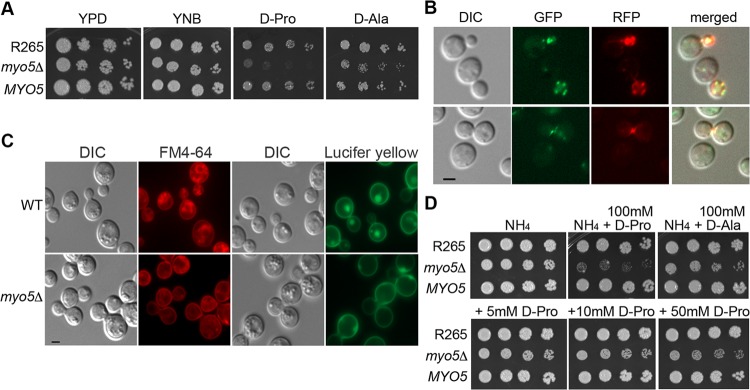
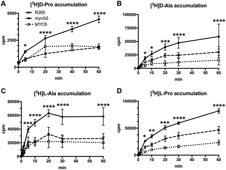
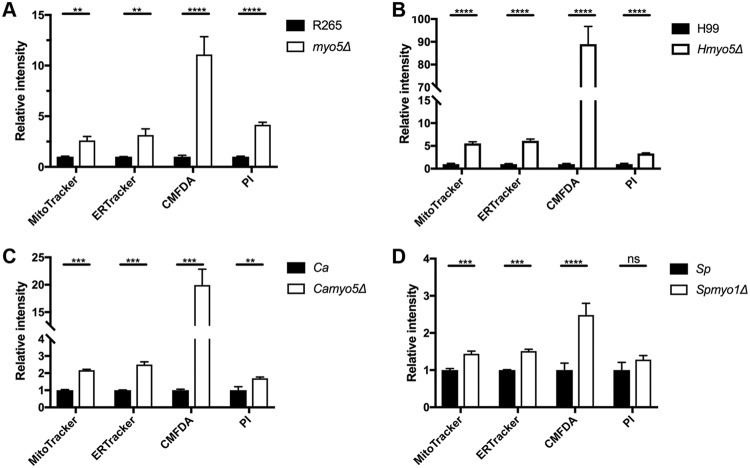
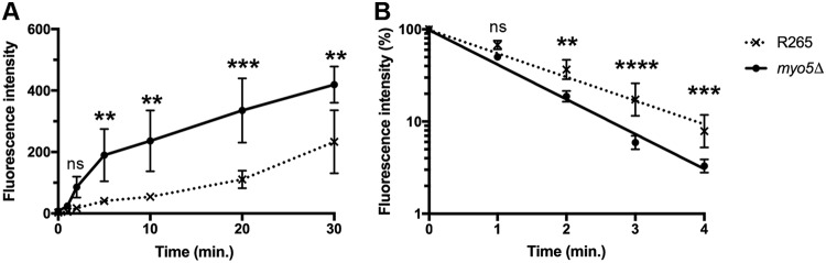
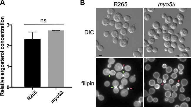
We found a novel role of Myo5, a type I myosin (myosin-I), and its fortuitous association with d-amino acid utilization in Cryptococcus gattii Myo5 colocalized with actin cortical patches and was required for endocytosis. Interestingly, the myo5Δ mutant accumulated high levels of d-proline and d-alanine which caused toxicity in C. gattii cells. The myo5Δ mutant also accumulated a large set of substrates, such as membrane-permeant as well as non-membrane-permeant dyes, l-proline, l-alanine, and flucytosine intracellularly. Furthermore, the efflux rate of fluorescein was significantly increased in the myo5Δ mutant. Importantly, the endocytic defect of the myo5Δ mutant did not affect the localization of the proline permease and flucytosine transporter. These data indicate that the substrate accumulation phenotype is not solely due to a defect in endocytosis, but the membrane properties may have been altered in the myo5Δ mutant. Consistent with this, the sterol staining pattern of the myo5Δ mutant was different from that of the wild type, and the mutant was hypersensitive to amphotericin B. It appears that the changes in sterol distribution may have caused altered membrane permeability in the myo5Δ mutant, allowing increased accumulation of substrate. Moreover, myosin-I mutants generated in several other yeast species displayed a similar substrate accumulation phenotype. Thus, fungal type I myosin appears to play an important role in regulating membrane permeability. Although the substrate accumulation phenotype was detected in strains with mutations in the genes involved in actin nucleation, the phenotype was not shared in all endocytic mutants, indicating a complicated relationship between substrate accumulation and endocytosis.IMPORTANCECryptococcus gattii, one of the etiological agents of cryptococcosis, can be distinguished from its sister species Cryptococcus neoformans by growth on d-amino acids. C. gattiiMYO5 affected the growth of C. gattii on d-amino acids. The myo5Δ cells accumulated high levels of various substrates from outside the cells, and excessively accumulated d-amino acids appeared to have caused toxicity in the myo5Δ cells. We provide evidence on the alteration of membrane properties in the myo5Δ mutants. Additionally, alteration in the myo5Δ membrane permeability causing higher substrate accumulation is associated with the changes in the sterol distribution. Furthermore, myosin-I in three other yeasts also manifested a similar role in substrate accumulation. Thus, while fungal myosin-I may function as a classical myosin-I, it has hitherto unknown additional roles in regulating membrane permeability. Since deletion of fungal myosin-I causes significantly elevated susceptibility to multiple antifungal drugs, it could serve as an effective target for augmentation of fungal therapy.

Keywords: Cryptococcus; actin patches; endocytosis; membrane permeability; myosin-I.


Figures

FIG 1
FIG 1
Identification of MYO5 function. (A) MYO5 is important for d-proline utilization. Cells of the wild type, the myo5Δ mutant, and the myo5Δ complemented strain (MYO5) were serially diluted and spotted on the indicated media. Plates were incubated at 30°C for 3 days and photographed. (B) Myo5 is colocalized with actin cortical patches. Myo5 was tagged with green fluorescent protein (GFP) (green color). Actin was visualized with Lifeact-RFP (red color). Fluorescent and differential interference contrast (DIC) images were taken from log-phase cells. (C) MYO5 is involved in endocytosis. Cells were stained with FM4-64 to determine membrane internalization (DIC and FM4-64 columns) or stained with Lucifer yellow to determine fluid phase endocytosis (DIC and Lucifer yellow columns). For FM4-64 staining, cells were stained with 16 μM FM4-64 on ice for 15 min, washed, and incubated in YEPD for 80 min before imaging. Bar = 2 μm. WT, wild type. (D) d-Amino acids are toxic to the myo5Δ mutant. Cells were serially diluted and spotted on media containing 10 mM ammonium sulfate (NH4) with or without supplementation with 100 mM d-proline or d-alanine (top row). Cells were also spotted on NH4 media supplemented with 5, 10, or 50 mM d-proline (bottom row). Plates were incubated at 30°C for 3 days and photographed.
FIG 2
FIG 2
The myo5Δ mutant accumulates high levels of amino acids. Log-phase cells of the wild type, myo5Δ mutant, and the myo5Δ complemented strain (MYO5) were incubated with the indicated 3H-labeled amino acids, and the time course of intracellular accumulation of each amino acid was determined. The experiments were repeated three times, and the error bars represent the standard deviations. Two-way ANOVA with Dunnett’s multiple-comparison test was used to compare the differences at the indicated time points between the corresponding R265 strain and myo5Δ mutant (*, P < 0.0332; **, P < 0.0021; ***, P < 0.0002; ****, P < 0.0001).
FIG 3
FIG 3
The myo5Δ mutant accumulates high levels of membrane-permeant and -nonpermeant dyes as determined by microscopy. (A to D) Cells were incubated with the indicated fluorescent dyes and observed with a microscope. (A) ER-Tracker Red. (B) CellTracker Green CMFDA. (C) MitoTracker Red CMXRos. (D) Propidium iodide (PI). Fluorescent and DIC images were photographed. The staining patterns of each dye were similar between the wild type (WT) and the myo5Δ mutant, but the fluorescent intensity was greater in the myo5Δ mutant than in the wild type. Bars = 5 μm.
FIG 4
FIG 4
Myosin-I mutants of different fungal species accumulate high levels of membrane-permeant or -nonpermeant dyes. Cells of indicated strains were incubated with each membrane-permeant or -nonpermeant dye, and accumulation of each dye was assessed by flow cytometry. The experiments were repeated three times, and the error bars represent the standard deviations. Ca, C. albicans; Sp, S. pombe. Values that are significantly different are indicated by bars and asterisks as follows: **, P ≤ 0.01; ***, P ≤ 0.001; ****, P ≤ 0.0001. Values that are not significantly different (P > 0.05) are indicated (ns).
FIG 5
FIG 5
Myosin-I mutants of different fungal species accumulate higher levels of flucytosine. Cells of the indicated strains were incubated with [3H]labeled flucytosine, and the time course of intracellular accumulation of 3H was determined. The experiments were repeated three times, and the error bars represent the standard deviations. Ca, C. albicans; Sp, S. pombe. Two-way ANOVA with Dunnett’s multiple-comparison test was used to compare the differences at the indicated time point between the corresponding R265 strain and the myo5Δ mutant (ns, not significant; *, P < 0.0332; **, P < 0.0021; ***, P < 0.0002; ****, P < 0.0001).
FIG 6
FIG 6
(A) FCY2 is required for resistance to flucytosine. Cells of the indicated strains were spotted on YNB medium with or without 0.5 μg/ml of flucytosine. (B) AAP9 is required for the growth in media containing d-/l-proline as the sole nitrogen source. Cells of the indicated strains were spotted on media containing 10 mM ammonium sulfate, l-proline, or d-proline. Plates were incubated at 30°C for 3 days and photographed. (C and D) Deletion of MYO5 does not affect the distribution of transporters for flucytosine or proline. The Fcy2 and Aap9 proteins were separately tagged with green fluorescent protein in the myo5Δ mutant and the wild type, respectively. Strains were grown in YPD (C) or YNB supplemented with 10 mM Asn as the sole nitrogen source (D). Fluorescent and DIC images were taken from log-phase cells. Bar = 2 μm.
FIG 7
FIG 7
Fluorescein accumulation and efflux rate are higher in the myo5Δ mutant. (A) Time course of intracellular fluorescein accumulation. Cells were incubated with 10 μM FDA, and cellular fluorescent intensity was determined at the indicated time by flow cytometry. (B) Kinetics of fluorescein efflux. Cells were loaded with similar amounts of fluorescein, and the efflux of fluorescein was determined by measuring the intracellular fluorescent intensity by flow cytometry. The experiments were repeated three times, and the error bars represent standard deviations. Two-way ANOVA with Dunnett’s multiple-comparison test was used to compare the differences at the indicated time point between the corresponding R265 strain and myo5Δ mutant (ns, not significant; **, P < 0.0021; ***, P < 0.0002; ****, P < 0.0001).
FIG 8
FIG 8
Ergosterol distribution appears to be altered in the myo5Δ mutant. (A) Ergosterol content is similar between the wild type and myo5Δ mutant. Ergosterol was extracted from cells and quantitated by LCMS. The experiments were repeated three times, and the error bars represent the standard deviations. (B) Filipin staining patterns are different between the wild type and the myo5Δ mutant. Log-phase grown cells were stained with 5 μg/ml filipin. The fluorescent and DIC images were photographed. Red arrowhead, emerging bud; green arrowhead, isthmus. Bar = 2 μm.
FIG 9
FIG 9
Several genes involved in endocytosis play a role in membrane permeability. (A) ARP2, ARP3, and VRP1 are important for d-proline utilization. Cells of the indicated mutants and the corresponding complemented strains (ARP2, ARP3, and VRP1) were serially diluted and spotted on the indicated media. Plates were incubated at 30°C for 3 days and photographed. (B and C) Deletion of ARP2, ARP3, and VRP1 causes accumulation of different fluorescent dyes (B) or [3H]flucytosine (C). (D and E) Not every endocytic mutant accumulates different fluorescent dyes (D) or [3H]flucytosine (E). (ns, not significant [P > 0.05]; *, P ≤ 0.05; **, P ≤ 0.01; ***, P ≤ 0.001; ****, P ≤ 0.0001).

References

    1. Sellers JR. 2000. Myosins: a diverse superfamily. Biochim Biophys Acta 1496:3–22. doi:10.1016/s0167-4889(00)00005-7. - DOI - PubMed
    1. Coluccio LM. 2008. Myosin I, p 95–124. In Coluccio LM. (ed), Myosins: a superfamily of molecular motors. Springer, Dordrecht, The Netherlands. doi:10.1007/978-1-4020-6519-4. - DOI
    1. Masters TA, Kendrick-Jones J, Buss F. 2017. Myosins: domain organisation, motor properties, physiological roles and cellular functions. Handb Exp Pharmacol 235:77–122. doi:10.1007/164_2016_29. - DOI - PubMed
    1. McIntosh BB, Ostap EM. 2016. Myosin-I molecular motors at a glance. J Cell Sci 129:2689–2695. doi:10.1242/jcs.186403. - DOI - PMC - PubMed
    1. Kim SV, Flavell RA. 2008. Myosin I: from yeast to human. Cell Mol Life Sci 65:2128–2137. doi:10.1007/s00018-008-7435-5. - DOI - PMC - PubMed

Publication types

LinkOut - more resources

Cite

AltStyle によって変換されたページ (->オリジナル) /