A Review of Zoonotic Pathogens of Dromedary Camels
- PMID: 31140075
- PMCID: PMC7087575
- DOI: 10.1007/s10393-019-01413-7
A Review of Zoonotic Pathogens of Dromedary Camels
Abstract
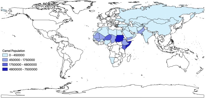
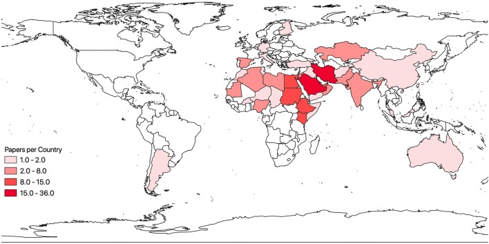
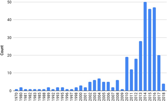
Dromedary, or one-humped, camels Camelus dromedarius are an almost exclusively domesticated species that are common in arid areas as both beasts of burden and production animals for meat and milk. Currently, there are approximately 30 million dromedary camels, with highest numbers in Africa and the Middle East. The hardiness of camels in arid regions has made humans more dependent on them, especially as a stable protein source. Camels also carry and may transmit disease-causing agents to humans and other animals. The ability for camels to act as a point source or vector for disease is a concern due to increasing human demands for meat, lack of biosafety and biosecurity protocols in many regions, and a growth in the interface with wildlife as camel herds become sympatric with non-domestic species. We conducted a literature review of camel-borne zoonotic diseases and found that the majority of publications (65%) focused on Middle East respiratory syndrome (MERS), brucellosis, Echinococcus granulosus, and Rift Valley fever. The high fatality from MERS outbreaks during 2012-2016 elicited an immediate response from the research community as demonstrated by a surge of MERS-related publications. However, we contend that other camel-borne diseases such as Yersinia pestis, Coxiella burnetii, and Crimean-Congo hemorrhagic fever are just as important to include in surveillance efforts. Camel populations, particularly in sub-Saharan Africa, are increasing exponentially in response to prolonged droughts, and thus, the risk of zoonoses increases as well. In this review, we provide an overview of the major zoonotic diseases present in dromedary camels, their risk to humans, and recommendations to minimize spillover events.
Keywords: Camel; Nomadic; One health; Pathogen; Spillover; Zoonoses.
Conflict of interest statement
The authors declare that they have no conflict of interest.
Figures
References
-
- Abushhewa MH, Abushhiwa MHS, Nolan MJ, Jex AR, Campbell BE, Jabbar A, Gasser RB. Genetic classification of Echinococcus granulosus cysts from humans, cattle and camels in Libya using mutation scanning-based analysis of mitochondrial loci. Mol Cell Probes. 2010;24:346–351. doi: 10.1016/j.mcp.201007005. - DOI - PubMed
Publication types
MeSH terms
Grants and funding
LinkOut - more resources
Full Text Sources
Medical