This site needs JavaScript to work properly. Please enable it to take advantage of the complete set of features!
Skip to main page content
U.S. flag

An official website of the United States government

Dot gov

The .gov means it’s official.
Federal government websites often end in .gov or .mil. Before sharing sensitive information, make sure you’re on a federal government site.

Https

The site is secure.
The https:// ensures that you are connecting to the official website and that any information you provide is encrypted and transmitted securely.

NIH NLM Logo
Log in
Access keys NCBI Homepage MyNCBI Homepage Main Content Main Navigation
. 2019 Mar 20;4(2):e00080-19.
doi: 10.1128/mSphere.00080-19.

A Novel Protocol for the Isolation of Fungal Extracellular Vesicles Reveals the Participation of a Putative Scramblase in Polysaccharide Export and Capsule Construction in Cryptococcus gattii

Affiliations

A Novel Protocol for the Isolation of Fungal Extracellular Vesicles Reveals the Participation of a Putative Scramblase in Polysaccharide Export and Capsule Construction in Cryptococcus gattii

Flavia C G Reis et al. mSphere. .

Abstract

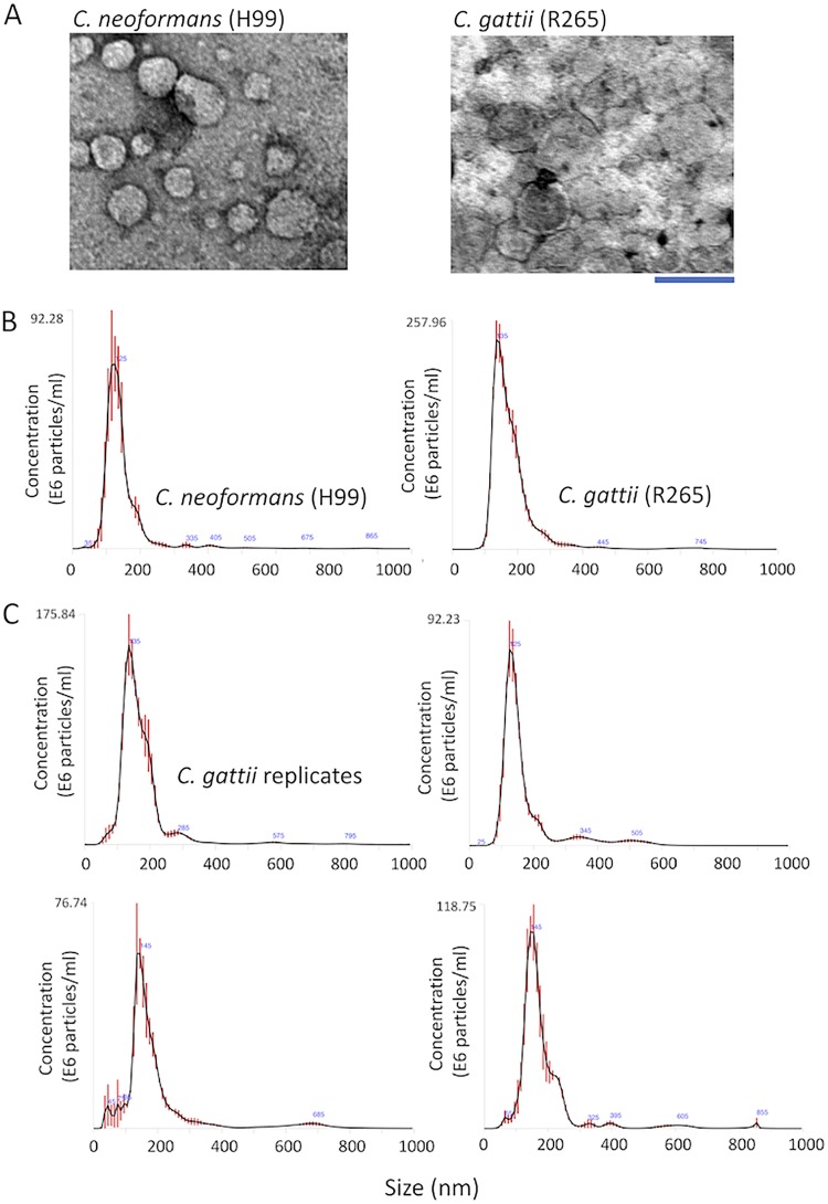
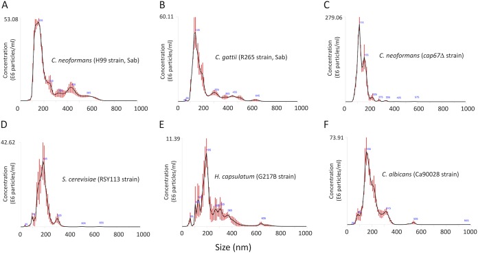
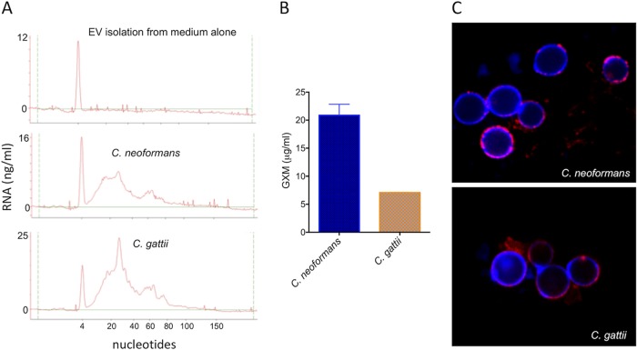
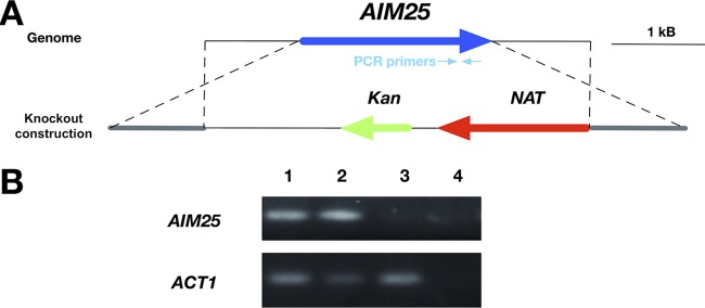
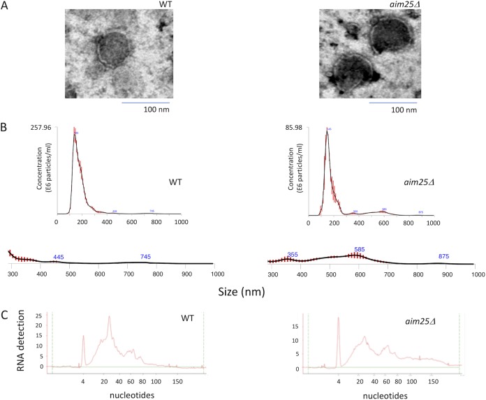
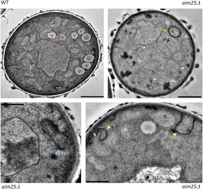
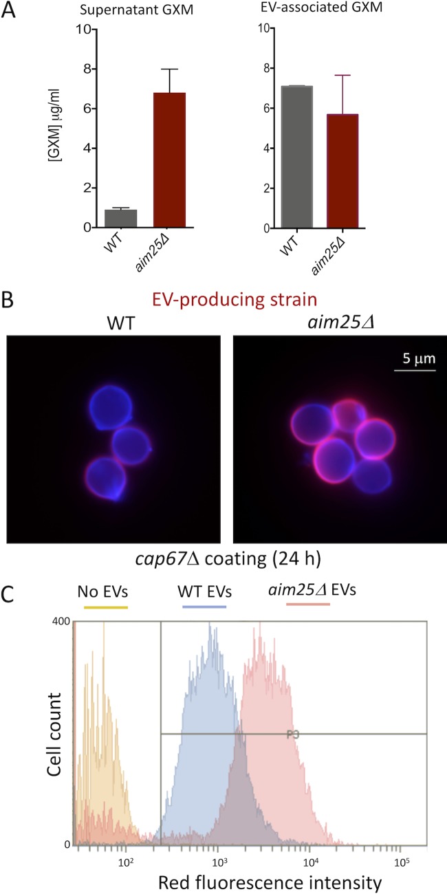
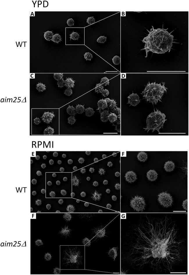
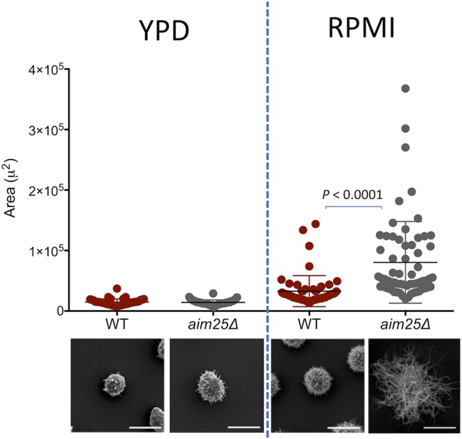
Regular protocols for the isolation of fungal extracellular vesicles (EVs) are time-consuming, hard to reproduce, and produce low yields. In an attempt to improve the protocols used for EV isolation, we explored a model of vesicle production after growth of Cryptococcus gattii and Cryptococcus neoformans on solid media. Nanoparticle tracking analysis in combination with transmission electron microscopy revealed that C. gattii and C. neoformans produced EVs in solid media. The properties of cryptococcal vesicles varied according to the culture medium used and the EV-producing species. EV detection was reproduced with an acapsular mutant of C. neoformans, as well as with isolates of Candida albicans, Histoplasma capsulatum, and Saccharomyces cerevisiae Cryptococcal EVs produced in solid media were biologically active and contained regular vesicular components, including the major polysaccharide glucuronoxylomannan (GXM) and RNA. Since the protocol had higher yields and was much faster than the regular methods used for the isolation of fungal EVs, we asked if it would be applicable to address fundamental questions related to cryptococcal secretion. On the basis that polysaccharide export in Cryptococcus requires highly organized membrane traffic culminating with EV release, we analyzed the participation of a putative scramblase (Aim25; CNBG_3981) in EV-mediated GXM export and capsule formation in C. gattii EVs from a C. gattiiaim25Δ strain differed from those obtained from wild-type (WT) cells in physical-chemical properties and cargo. In a model of surface coating of an acapsular cryptococcal strain with vesicular GXM, EVs obtained from the aim25Δ mutant were more efficiently used as a source of capsular polysaccharides. Lack of the Aim25 scramblase resulted in disorganized membranes and increased capsular dimensions. These results associate the description of a novel protocol for the isolation of fungal EVs with the identification of a previously unknown regulator of polysaccharide release.IMPORTANCE Extracellular vesicles (EVs) are fundamental components of the physiology of cells from all kingdoms. In pathogenic fungi, they participate in important mechanisms of transfer of antifungal resistance and virulence, as well as in immune stimulation and prion transmission. However, studies on the functions of fungal EVs are still limited by the lack of efficient methods for isolation of these compartments. In this study, we developed an alternative protocol for isolation of fungal EVs and demonstrated an application of this new methodology in the study of the physiology of the fungal pathogen Cryptococcus gattii Our results describe a fast and reliable method for the study of fungal EVs and reveal the participation of scramblase, a phospholipid-translocating enzyme, in secretory processes of C. gattii.

Keywords: Cryptococcus; extracellular vesicles; fungi; scramblase; secretion.


Figures

FIG 1
FIG 1
Isolation of fungal EVs from solid cultures of C. neoformans and C. gattii (strains H99 and R265, respectively). (A) Transmission electron microscopy of vesicular fractions obtained after growth of both pathogens on solid YPD. Scale bar, 200 nm. (B) Nanoparticle tracking analysis of the EV preparations illustrated in panel A, showing a concentration of vesicles in the range of 100 to 200 nm. (C) NTA profiles of four samples of C. gattii EVs obtained independently.
FIG 2
FIG 2
NTA profiles of EVs obtained from different fungal cultures in solid media. (A and B) Analysis of C. neoformans (A) and C. gattii (B) EVs obtained from Sabouraud cultures. (C) NTA of EVs obtained from an acapsular mutant of C. neoformans, suggesting that vesicle release in solid medium does not demand capsular structures. EVs were also detected by NTA after growth of S. cerevisiae (D), H. capsulatum (E), and C. albicans (F), indicating that the protocol is applicable to the study of different fungal pathogens.
FIG 3
FIG 3
Analysis of the cargo of fungal EVs obtained from solid cultures of C. neoformans and C. gattii (strains H99 and R265, respectively). (A) Analysis of nucleic acid content confirmed the presence of small RNAs in cryptococcal vesicles. In these panels, the y axis corresponds to RNA detection as a function of fluorescence intensity, while the x axis represents RNA size in nucleotides. The first, sharp peak at 4 nucleotides corresponds to the RNA size marker. No RNA was detected in control samples obtained from the culture medium. (B) Detection of GXM in vesicular samples obtained from C. neoformans and C. gattii by ELISA. The concentration of vesicular GXM was significantly higher in C. neoformans samples (P = 0.0099). (C) Functional analysis of vesicular GXM in samples obtained from solid medium. The cap67Δ mutant of C. neoformans efficiently incorporated GXM (red fluorescence) from EVs produced by both C. neoformans and C. gattii in solid medium into the cell wall (blue fluorescence). Results in all panels are representative of three independent experiments.
FIG 4
FIG 4
Construction of the aim25Δ mutant. (A) AIM25 knockout scheme. The genome locus containing the AIM25 gene and the knockout construct are shown in the upper and lower diagrams, respectively. NAT, cassette conferring nourseothricin resistance; Kan, cassette conferring kanamycin resistance for cloning purposes in Escherichia coli. Hybridization sites of the PCR primers are also shown. (B) Confirmation of AIM25 deletion by PCR. Genomic DNA (100 ng) from WT cells (lane 1), a transformant with ectopic integration of the knockout cassette (lane 2) and the aim25Δ mutant (lane 3) was submitted to PCR using primers to amplify a segment of AIM25 (upper panel) or the gene encoding action (ACT1 [lower panel, loading control]). Control reactions without template addition are also shown (lane 4).
FIG 5
FIG 5
Analysis of EVs obtained after growth of wild-type (WT) or mutant (aim25Δ) cells of C. gattii in solid medium. (A) Transmission electron microscopy of WT and mutant cells. (B) NTA of EVs produced by WT and mutant cells, suggesting an increased detection of larger EVs (300 to 900 nm) in mutant cultures. The 300- to 900-nm size range of EVs was amplified below each NTA histogram. (C) Analysis of small RNAs contained in EVs produced by WT and mutant cells. The results shown in panels B and C are representative of two and three independent experiments, respectively.
FIG 6
FIG 6
Transmission electron microscopy of wild-type (WT) and mutant (aim25Δ) cells of C. gattii. WT cells manifested the typical intracellular morphology of cryptococci, including well-defined vacuoles (V) and organized membranous compartments. In mutant cells, distorted membranes were abundantly detected. Phenotypic traits that were exclusive to mutant cells included a general lack of the typical cryptococcal vacuoles, highly-electron-dense membranous compartments (orange arrowhead), linearized membranes (white arrowheads), electron-dense, stacked membranes (blue arrowhead), and atypical invaginations of the plasma membrane (yellow arrowheads). Scale bars correspond to 200 nm.
FIG 7
FIG 7
Analysis of extracellular GXM in wild-type (WT) and mutant (aim25Δ) cells of C. gattii. (A) Determination of extracellular GXM in supernatant samples (A) demonstrated that mutant cells produced significantly increased polysaccharide concentrations (P = 0.0001). No significant changes in the GXM content (P = 0.41) were observed in EV samples. (B) Microscopic examination of the ability of cap67Δ cells to incorporate GXM obtained from C. gattii suggested that GXM incorporation by the acapsular strain was more efficient when aim25Δ vesicles were used. Blue fluorescence denotes cell wall staining with calcofluor white. Red fluorescence corresponds to GXM staining with MAb 18B7. (C) Flow cytometry analysis of acapsular cells under the conditions described in panel B, providing a quantitative confirmation of the visual observation resulting from microscopic analysis. Results are representative of two independent experiments.
FIG 8
FIG 8
Scanning electron microscopy of wild-type (WT) and mutant (aim25Δ) cells of C. gattii after growth in solid YPD (capsule repression) or incubation in RPMI (capsule induction). General views of WT (A and E) or aim25Δ (C and F) cells are shown for each condition in the left panels. Magnified views of WT (B and F) or aim25Δ (D and G) cells from the insets in the left panels are shown in the right panels. Scale bars correspond to 5 μm.
FIG 9
FIG 9
Analysis of cellular area as a consequence of capsular dimensions in wild-type (WT) and mutant (aim25Δ) cells of C. gattii after growth in solid YPD (capsule repression) or incubation in RPMI (capsule induction). The most representative phenotypes observed under each experimental condition are shown on the bottom as scanning electron microscopy images. Differences in capsular dimensions had no statistical significance, with the exception of the comparison between WT and mutant cells after incubation in RPMI. Scale bars correspond to 5 μm.

References

    1. Raposo G, Stoorvogel W. 2013. Extracellular vesicles: exosomes, microvesicles, and friends. J Cell Biol 200:373–383. doi:10.1083/jcb.201211138. - DOI - PMC - PubMed
    1. Rodrigues ML, Nimrichter L, Oliveira DL, Frases S, Miranda K, Zaragoza O, Alvarez M, Nakouzi A, Feldmesser M, Casadevall A. 2007. Vesicular polysaccharide export in Cryptococcus neoformans is a eukaryotic solution to the problem of fungal trans-cell wall transport. Eukaryot Cell 6:48–59. doi:10.1128/EC.00318-06. - DOI - PMC - PubMed
    1. Zarnowski R, Sanchez H, Covelli AS, Dominguez E, Jaromin A, Bernhardt J, Mitchell KF, Heiss C, Azadi P, Mitchell A, Andes DR. 2018. Candida albicans biofilm-induced vesicles confer drug resistance through matrix biogenesis. PLoS Biol 16:e2006872. doi:10.1371/journal.pbio.2006872. - DOI - PMC - PubMed
    1. Kabani M, Melki R. 2015. Sup35p in its soluble and prion states is packaged inside extracellular vesicles. mBio 6:e01017-15. doi:10.1128/mBio.01017-15. - DOI - PMC - PubMed
    1. Liu S, Hossinger A, Hofmann JP, Denner P, Vorberg IM. 2016. Horizontal transmission of cytosolic sup35 prions by extracellular vesicles. mBio 7:e00915-16. doi:10.1128/mBio.00915-16. - DOI - PMC - PubMed

Publication types

MeSH terms

LinkOut - more resources

Cite

AltStyle によって変換されたページ (->オリジナル) /