This site needs JavaScript to work properly. Please enable it to take advantage of the complete set of features!
Skip to main page content
U.S. flag

An official website of the United States government

Dot gov

The .gov means it’s official.
Federal government websites often end in .gov or .mil. Before sharing sensitive information, make sure you’re on a federal government site.

Https

The site is secure.
The https:// ensures that you are connecting to the official website and that any information you provide is encrypted and transmitted securely.

NIH NLM Logo
Log in
Access keys NCBI Homepage MyNCBI Homepage Main Content Main Navigation
Review
. 2018 Dec 14:9:763.
doi: 10.3389/fendo.2018.00763. eCollection 2018.

Role of Follicle-Stimulating Hormone in Spermatogenesis

Affiliations
Review

Role of Follicle-Stimulating Hormone in Spermatogenesis

Olayiwola O Oduwole et al. Front Endocrinol (Lausanne). .

Abstract

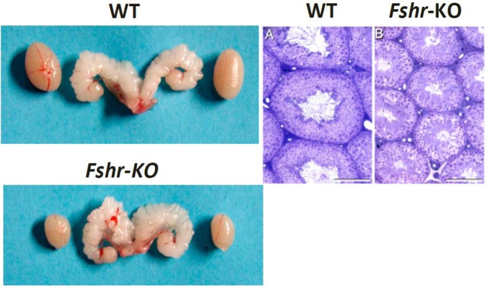
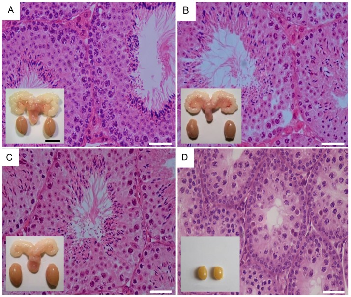
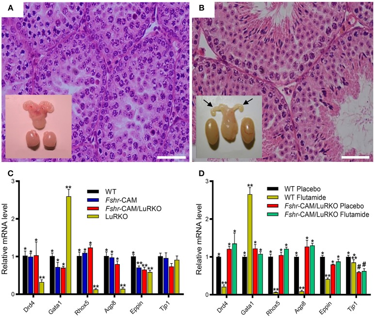
Spermatogenesis is a concerted sequence of events during maturation of spermatogonia into spermatozoa. The process involves differential gene-expression and cell-cell interplay regulated by the key endocrine stimuli, i.e., follicle-stimulating hormone (FSH) and luteinizing hormone (LH)-stimulated testosterone. FSH affects independently and in concert with testosterone, the proliferation, maturation and function of the supporting Sertoli cells that produce regulatory signals and nutrients for the maintenance of developing germ cells. Rodents are able to complete spermatogenesis without FSH stimulus, but its deficiency significantly decreases sperm quantity. Men carrying loss-of-function mutation in the gene encoding the ligand (FSHB) or its receptor (FSHR) present, respectively, with azoospermia or suppressed spermatogenesis. Recently, the importance of high intratesticular testosterone concentration for spermatogenesis has been questioned. It was established that it can be completed at minimal intratesticular concentration of the hormone. Furthermore, we recently demonstrated that very robust constitutive FSHR action can rescue spermatogenesis and fertility of mice even when the testosterone stimulus is completely blocked. The clinical relevance of these findings concerns a new strategy of high-dose FSH in treatment of spermatogenic failure.

Keywords: FSH; fertility; gonadotropins; sertoli cells; spermatogenesis; spermatogenic failure; testosterone.


Figures

Figure 1
Figure 1
Testes and seminal vesicles of adult wild type (WT) and FshrKO) mice (Left), and testicular histology of same genotypes (Right). No difference is observed in seminal vesicle sizes between the two genotypes, but the size of the FshrKO testes is about half that of WT. Also, while full spermatogenesis is visible in the histology of both testes, the tubular diameter is clearly narrower in the knockout testis. From (37) with permission.
Figure 2
Figure 2
Testicular histology and macroscopic views of testes and urogenital blocks of different mouse genotypes: (A) WT, (B) Fshr-CAM, (C) Fshr-CAM/LuRKO, and (D) LuRKO mice. (A–C) show normal spermatogenesis and testis and seminal vesicle (SV) sizes. In (D), spermatogenesis is arrested at the round spermatid (RS) stage, with small testes and rudimentary seminal vesicle (not visible). Scale bars: 50 μm; 10 mm (insets). From (31) with permission.
Figure 3
Figure 3
Effect of anti-androgen flutamide treatment on wild-type (WT) and genetically modified mice. (A,B) Testicular histology and macroscopic views of the testes and urogenital blocks of WT and Fshr-CAM/LuRKO mice. (A) The treatment arrested spermatogenesis at round spermatid stage in WT mice and reduced their testis and seminal vesicle sizes. (B) Identical treatment of Fshr-CAM/LuRKO mice had no apparent effect on their spermatogenesis and testis size but reduced seminal vesicle size (arrows in B). (C,D) Expression of selected target genes in untreated (A) and flutamide treated (B) mice. (A) Expression of androgen-regulated (Drd5, Rhox5, Eppin, and Tjp1), postmeiotic germ cell–specific (Aqp8), and germ cell–regulated (Gata1) genes in WT, Fshr-CAM, Fshr-CAM/LuRKO, and LuRKO testes. (B) Effect of flutamide treatment on expression of the same androgen-regulated genes in WT and Fshr-CAM/LuRKO mice. Data represent mean ± SEM. n = 3 samples/group. Bars with different symbols differ significantly from each other (P < 0.05; ANOVA/Newman-Keuls). The remarkable finding is that while flutamide treatment suppressed the expression of strictly androgen-dependent genes in WT mice, the same effect was not observed in the testis of Fshr-CAM/LuRKO mice. Scale bars: 50 μm. From (31) with permission.

References

    1. Mruk DD, Cheng CY. The mammalian blood-testis barrier: its biology and regulation. Endocr Rev. (2015) 36:564–91. 10.1210/er.2014-1101 - DOI - PMC - PubMed
    1. Steinberger E, Steinberger A. Spermatogenic function of the testis. In: Greep RO, Hamilton DW, editors. Handbook of Physiology. Washington DC: American Physiological Society; (1975). p. 1–10.
    1. Griswold MD. The central role of Sertoli cells in spermatogenesis. Semin Cell Dev Biol. (1998) 9:411–6. 10.1006/scdb.1998.0203 - DOI - PubMed
    1. Sharpe RM. Sperm counts and fertility in men: a rocky road ahead. science & Society Series on Sex and Science. EMBO Rep. (2012) 13:398–403. 10.1038/embor.2012.50 - DOI - PMC - PubMed
    1. Sharpe RM, McKinnell C, Kivlin C, Fisher JS. Proliferation and functional maturation of Sertoli cells, and their relevance to disorders of testis function in adulthood. Reproduction (2003) 125:769–84. 10.1530/rep.0.1250769 - DOI - PubMed
Cite

AltStyle によって変換されたページ (->オリジナル) /