This site needs JavaScript to work properly. Please enable it to take advantage of the complete set of features!
Skip to main page content
U.S. flag

An official website of the United States government

Dot gov

The .gov means it’s official.
Federal government websites often end in .gov or .mil. Before sharing sensitive information, make sure you’re on a federal government site.

Https

The site is secure.
The https:// ensures that you are connecting to the official website and that any information you provide is encrypted and transmitted securely.

NIH NLM Logo
Log in
Access keys NCBI Homepage MyNCBI Homepage Main Content Main Navigation
. 2018 Aug 2;12(8):e0006691.
doi: 10.1371/journal.pntd.0006691. eCollection 2018 Aug.

Infection of epididymal epithelial cells and leukocytes drives seminal shedding of Zika virus in a mouse model

Affiliations

Infection of epididymal epithelial cells and leukocytes drives seminal shedding of Zika virus in a mouse model

Erin M McDonald et al. PLoS Negl Trop Dis. .

Abstract

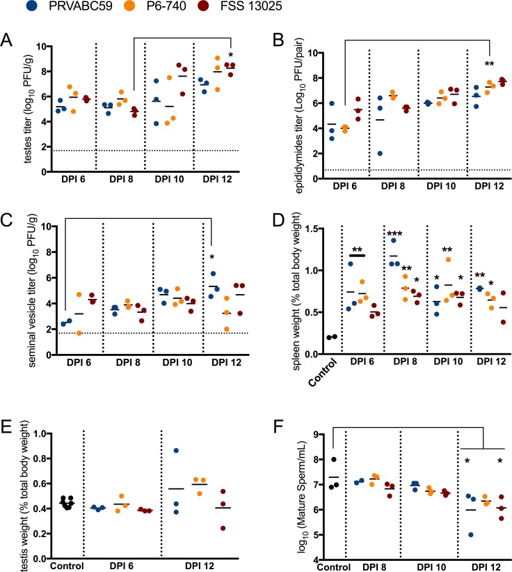
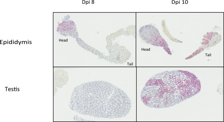
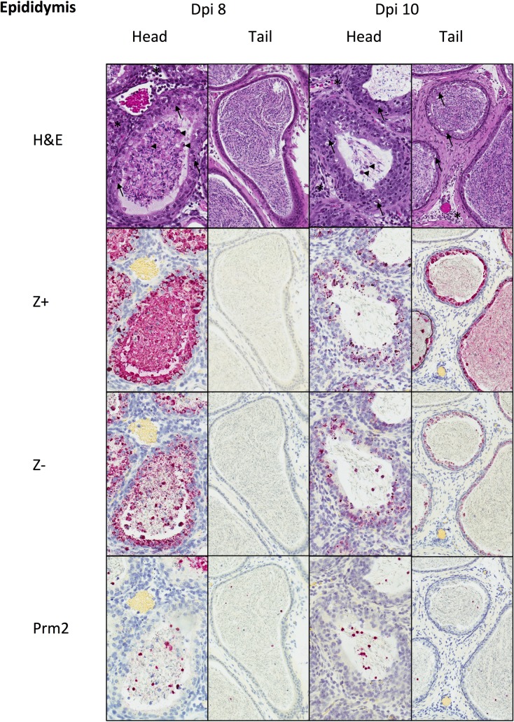
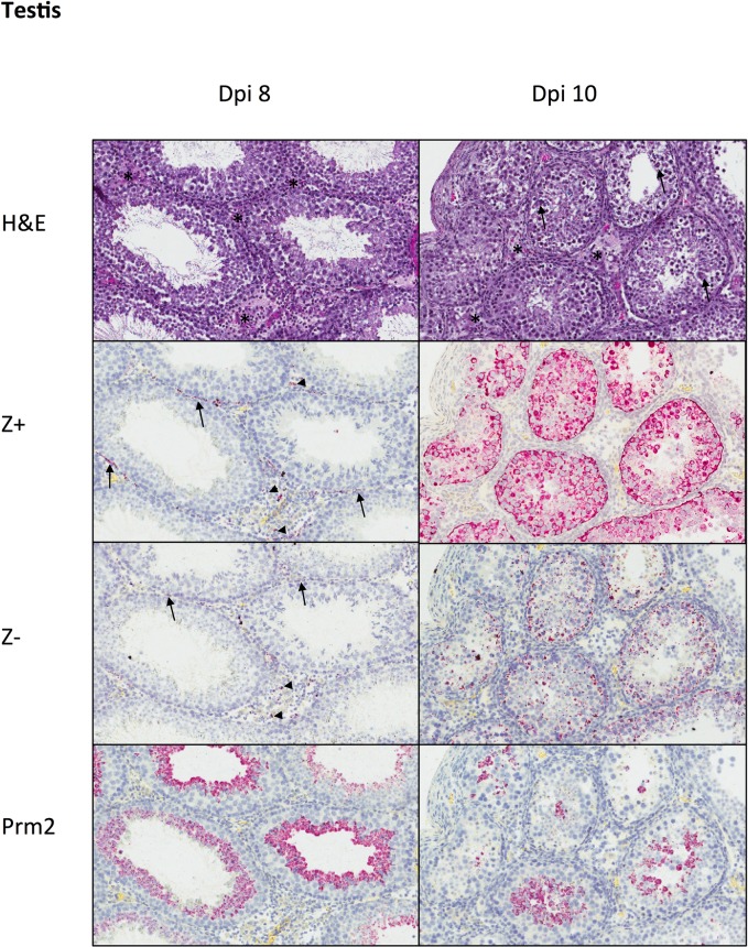
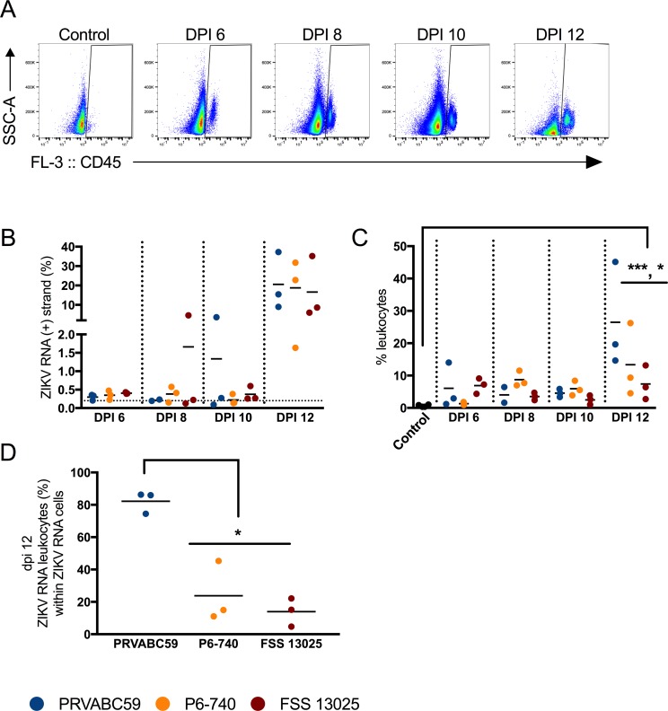
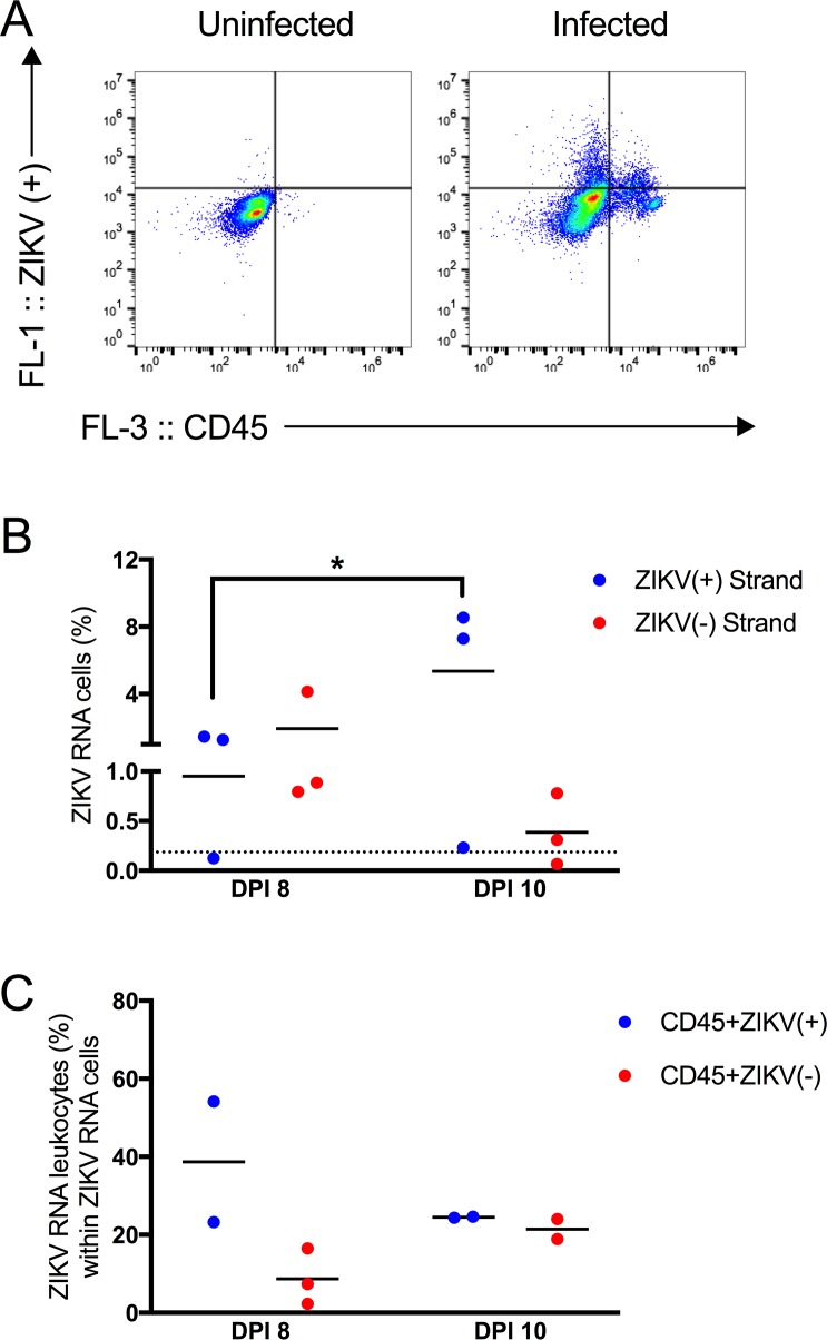
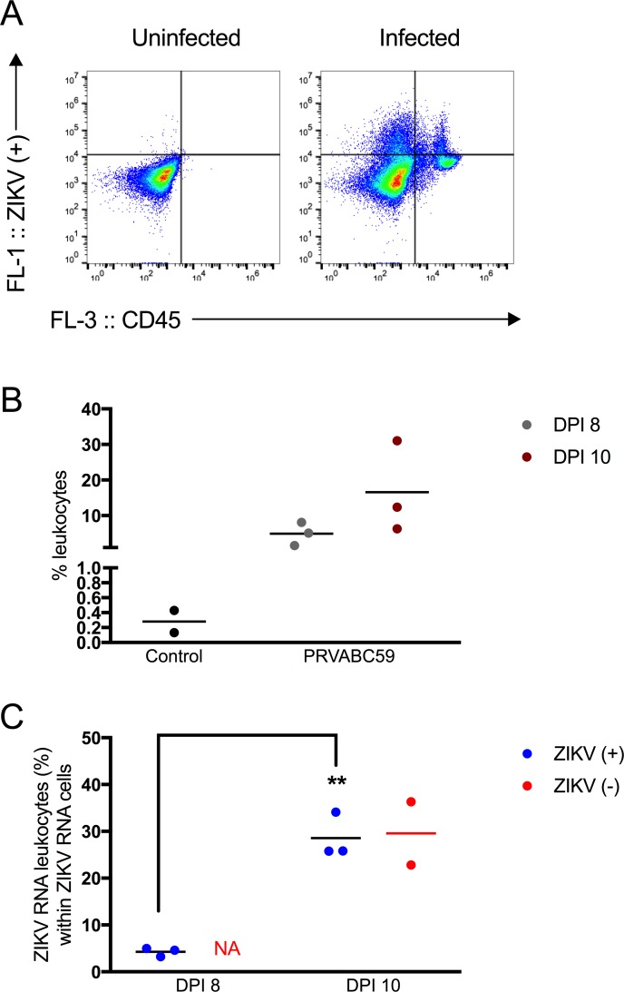
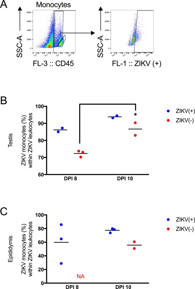
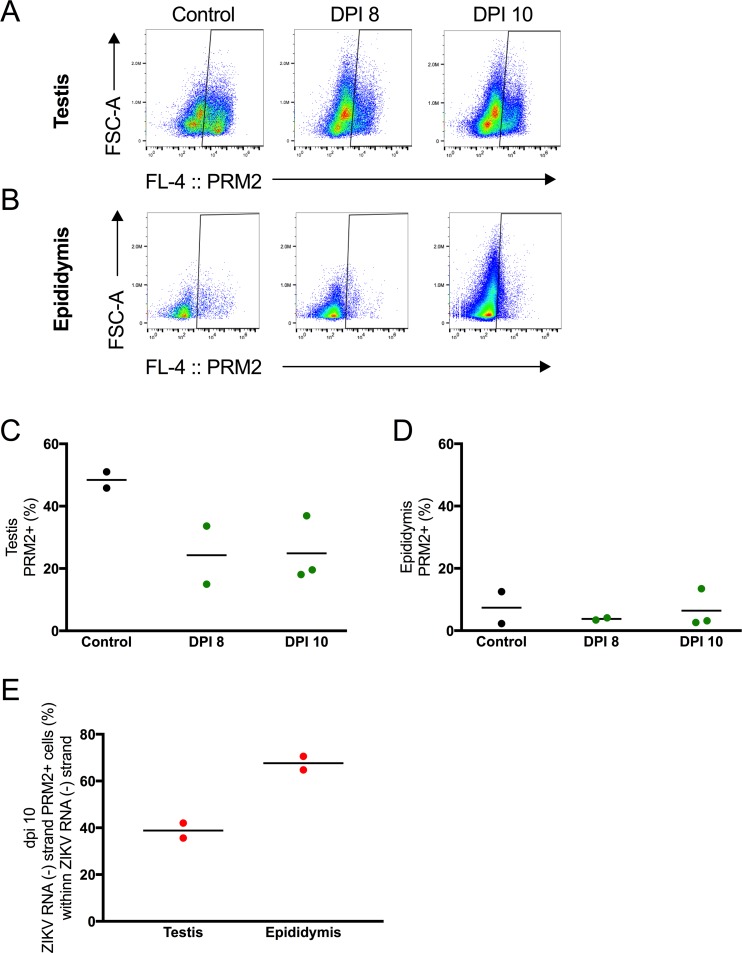
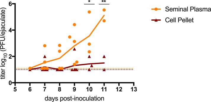
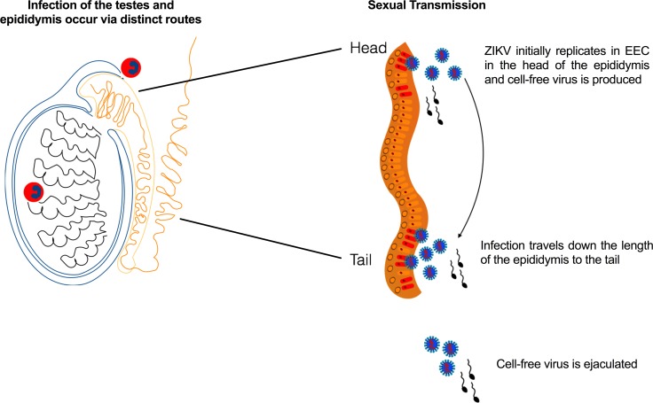
While primarily a mosquito-borne virus, Zika virus (ZIKV; genus Flavivirus in the Flaviviridae family) is capable of being sexually transmitted. Thirty to fifty percent of men with confirmed ZIKV infection shed ZIKV RNA in their semen, and prolonged viral RNA shedding in semen can occur for more than 6 months. The cellular reservoir of ZIKV in semen is unknown, although spermatozoa have been shown to contain ZIKV RNA and antigen. Yet, spermatozoa are not a requisite for sexual transmission, as at least one case of ZIKV sexual transmission involved a vasectomized man. To determine the cellular reservoirs of ZIKV in semen, an established animal model of sexual transmission was used. The majority of virus detected in the seminal fluid of infected mice during the peak timing of sexual transmission was from the supernatant fraction, suggesting cell-free ZIKV may be largely responsible for sexual transmission. However, some ZIKV RNA was cell-associated. In the testes and epididymides of infected mice, intracellular staining of ZIKV RNA was more pronounced in spermatogenic precursors (spermatocytes and spermatogonia) than in spermatids. Visualization of intracellular negative strand ZIKV RNA demonstrated ZIKV replication intermediates in leukocytes, immature spermatids and epididymal epithelial cells in the male urogenital tract. Epididymal epithelial cells were the principal source of negative-strand ZIKV RNA during the peak timing of sexual transmission potential, indicating these cells may be the predominant source of infectious cell-free ZIKV in seminal fluid. These data promote a more complete understanding of sexual transmission of ZIKV and will inform further model development for future studies on persistent ZIKV RNA shedding.


Conflict of interest statement

The authors have declared that no competing interests exist.

Figures

Fig 1
Fig 1. Splenomegaly and infection kinetics of the male reproductive tract during acute infection with Asian genotype ZIKV strains.
Mice were inoculated s.c. with 3 log10 PFU of ZIKV strains PRVABC59 (blue), P6-740 (yellow), and FSS13025 (maroon) (n = 12 per virus strain). At 6, 8, 10 and 12 days post-inoculation (dpi), three mice from each group were euthanized. (A) Viral titers in the testis. (B) Viral titers in the epididymis. The dashed line represents the limit of detection for the plaque assay. (C) Viral titers in the seminal vesicles. (D) Spleen weights were recorded and the ratio of (spleen to body weight) x 100 was measured. (E) Testicular weight as a percentage of total body weight. (F) Spermatozoa concentration decreased during acute infection. Epididymides were minced in warm PBS and a portion of the resulting cell suspension was stained with trypan blue and counted on a hemocytometer. Each symbol represents one mouse. P-values were determined by 2way ANOVA. *, p<0.05, ** p<0.01, ***p < 0.0001.
Fig 2
Fig 2. ZIKV genomic RNA localization in PRVABC59-inoculated mouse epididymis and testis at dpi 8 and 10.
At dpi 8, ZIKV RNA is localized to the head of the epididymis, and not visible in the testis at this magnification. At dpi 10, ISH signal is decreased in the head of the epididymis and strong in the body and tail of the epididymis, and within seminiferous tubules of the testis. ISH, Z (+) strand, hematoxylin counterstain of nuclei, Original magnification: 7X.
Fig 3
Fig 3. Histopathology and tissue ISH in PRVABC59-inoculated mouse epididymis at dpi 8 and 10.
At dpi 8, the head of the epididymis shows epithelial necrosis (arrows) and mild interstitial inflammation (*). Tubular lumens contain spermatozoa and degenerating round cells (arrowheads). The tail of the epididymis showed no epithelial changes and contained mature spermatozoa. RNA genomic RNA (Z+) and replicative intermediates (Z-) were present within epididymal epithelium and luminal immature spermatozoa and round cells in the head of the epididymis, but not in epithelium or mature spermatozoa in the tail of the epididymis. Many intraluminal round cells were Prm2+ spermatids. At dpi 10, epithelial necrosis and inflammation were similar in the head of the epididymis as seen at dpi 8, and these changes were also seen in the tail of the epididymis. ZIKV genomic RNA (Z+) was decreased in the epithelium and luminal cells of the head of the epididymis, and was also extensively present in the epithelium and spermatozoa of the tail of the epididymis. ZIKV replicative intermediates (Z-) were also present in the epididymal epithelium and in luminal round cells of both the head and the tail, but not in mature spermatozoa of the tail. Many intraluminal round cells were Prm2+ spermatids. H&E and ISH with hematoxylin counterstain of nuclei, Original magnifications: 400x (head), 200X (tail).
Fig 4
Fig 4. Histopathology and tissue ISH in PRVABC59-inoculated mouse testis at dpi 8 and 10.
At dpi 8, testis showed mild interstitial inflammation (*), without alterations in seminiferous epithelium. ZIKV genomic RNA (Z+) and replicative intermediates (Z-) were localized to scattered interstitial leukocytes (arrowheads) and peritubular myoid cells (arrows). Prm2+ staining showed round spermatids along the luminal surface of seminiferous tubules. At dpi 10, mild interstitial inflammation (*) was accompanied by patchy degeneration of seminiferous epithelium (arrows). Zika virus genomic RNA (Z+) and replicative intermediates (Z-) were present within all layers of seminiferous epithelium, and there was a reduction in the number of Prm2+ round spermatids compared to dpi 8. H&E and ISH with hematoxylin counterstain of nuclei, Original magnifications: 200X.
Fig 5
Fig 5. Testicular leukocytes contained genomic ZIKV RNA.
Mice were inoculated s.c. with 3 log10 PFU of ZIKV strains PRVABC59 (blue symbols), P6-740 (yellow), and FSS13025 (maroon) (n = 12 per virus strain). At six, eight, ten and twelve dpi, three mice from each group were euthanized. A single cell suspension of each testis was stained according to the PrimeFlow assay. (A) Representative dot plots showing testicular cells from uninfected control mice and ZIKV-infected mice stained with anti-CD45 antibody. (B) Percentage of ZIKV RNA (+) strand cells. Uninfected control mice were used to set gates for ZIKV RNA+ cells (gates were set such that 0.2% or less of the total population in the uninfected control was found in the ZIKV RNA+ gate.). (C) CD45+ leukocyte infiltration into the testes over time. (D) Percentage of ZIKV RNA (+) strand leukocytes within ZIKV RNA (+) strand cells. Each symbol represents one mouse. P-values were determined by 2way ANOVA. *, p<0.05, *** p<0.0001.
Fig 6
Fig 6. Testicular leukocytes contained replicating ZIKV RNA.
(A) Representative dot plots showing testicular cells stained with ZIKV RNA (+) strand probes (y-axis) and anti-CD45 antibody from an uninfected or infected mouse (x-axis). (B) Percentage of ZIKV RNA (+) or (-) strand cells. On dpi 8, none of the cells stained for ZIKV RNA (-) strand above background. On dpi 10, two of the three mice contained cells in the epididymal lumen that stained for ZIKV RNA (-) strand above background. (C) Percentage of ZIKV RNA leukocytes within ZIKV RNA cells. Each symbol represents one mouse. P-values were determined by 2way ANOVA. *, p<0.05.
Fig 7
Fig 7. Leukocytes in the epididymal lumen contained replicating ZIKV RNA.
(A) Representative dot plots showing cells from the epididymal lumen stained with ZIKV RNA (+) strand probes (y-axis) and anti-CD45 antibody (x-axis) from an uninfected or infected mouse. (B) Percentage of CD45+ leukocytes in control and infected animals. Leukocytes increase in the epididymal lumen upon ZIKV infection (black dots represent leukocytes from control animals; gray dots represent leukocyte from PRVABC59-inoculated mice on dpi 8 and maroon dots represent leukocytes from PRVABC59-inoculated mice on dpi 10). (C) Percentage of ZIKV RNA leukocytes within ZIKV RNA cells. On dpi 8, none of the cells stained for ZIKV RNA (-) strand above background. On dpi 10, two of the three mice contained cells in the epididymal lumen that stained for ZIKV RNA (-) strand above background. Each symbol represents one mouse. For the (+) strand, p-values were determined by unpaired t-test. *, p<0.05, ** p<0.01.
Fig 8
Fig 8. Monocytes were the major leukocyte population that contained ZIKV.
(A) Representative gating scheme. Monocytes are gated (high SSC in the CD45+ population) and then the ZIKV (+) RNA gate is applied to this population. (B) Percentage of ZIKV RNA monocytes out of ZIKV RNA CD45+ leukocytes in the testis. (C) Percentage of ZIKV RNA monocytes within ZIKV RNA leukocytes in the epididymis. Each symbol represents one mouse. P-values were determined by 2way ANOVA. *, p<0.05.
Fig 9
Fig 9. Immature spermatids contained genomic and replicative ZIKV RNA.
(A) Representative dot plots showing testicular cells from 8 and 10 dpi stained with probes to PRM2 mRNA. (B) Representative dot plots showing epididymal lumen cells from 8 and 10 dpi stained with probes to PRM2 mRNA. (C) Percentage of PRM2+ immature spermatids in the testis. (D) Percentage of PRM2+ immature spermatids in the epididymal lumen. (E) Percentage of ZIKV (-) strand RNA cells that co-stained with PRM2 in the testis and epididymal lumen. Each symbol represents one mouse.
Fig 10
Fig 10. Infectious ZIKV in seminal plasma versus cellular fractions from seminal fluids.
Mice were inoculated s.c. with 3 log10 PFU of ZIKV strain PRVABC59. Viral titers in seminal fluids collected from mice dpi 6 to 12. Individual ejaculates (n = 21) were partitioned into a cell pellet (maroon triangle) and a filtered supernatant (yellow circle). The colored lines represent the mean values across all mice and are used to make trends more visible. The limits of detection are represented by dashed lines (1.0 log10 for the cell pellet and 1.09 log10 for the filtered ejaculates). P-values were determined by 2way ANOVA. *, p<0.05; ** p<0.01.
Fig 11
Fig 11. Model of seminal shedding of ZIKV.
Tissue ISH suggests that the testes and epididymides are infected concurrently by trafficking leukocytes containing ZIKV. Once leukocytes transport ZIKV to the testis, peritubular myoid cells are the first cell type to become infected, and then the infection spreads to the seminiferous epithelium. In the epididymis, infected epididymal epithelial cells produce viral particles, which then spread intraluminally throughout the epididymis. Cell-free (and cell-associated virus) is ejaculated.

References

    1. Moreira J, Peixoto TM, Siqueira AM, Lamas CC. Sexually acquired Zika virus: a systematic review. Clinical microbiology and infection: the official publication of the European Society of Clinical Microbiology and Infectious Diseases. 2017;23(5):296–305. Epub 2017年01月08日. 10.1016/j.cmi.2016年12月02日7 . - DOI - PubMed
    1. Arsuaga M, Bujalance SG, Diaz-Menendez M, Vazquez A, Arribas JR. Probable sexual transmission of Zika virus from a vasectomised man. The Lancet Infectious diseases. 2016;16(10):1107 Epub 2016年09月28日. 10.1016/s1473-3099(16)30320-6 . - DOI - PubMed
    1. Mead PS, Duggal NK, Hook SA, Delorey M, Fischer M, Olzenak McGuire D, et al. Zika Virus Shedding in Semen of Symptomatic Infected Men. New England Journal of Medicine. 2018;378(15):1377–85. 10.1056/NEJMoa1711038 . - DOI - PubMed
    1. Duggal NK, Ritter JM, Pestorius SE, Zaki SR, Davis BS, Chang GJ, et al. Frequent Zika Virus Sexual Transmission and Prolonged Viral RNA Shedding in an Immunodeficient Mouse Model. Cell reports. 2017;18(7):1751–60. Epub 2017年02月16日. 10.1016/j.celrep.2017年01月05日6 . - DOI - PMC - PubMed
    1. McDonald EM, Duggal NK, Brault AC. Pathogenesis and sexual transmission of Spondweni and Zika viruses. PLoS neglected tropical diseases. 2017;11(10):e0005990 Epub 2017年10月07日. 10.1371/journal.pntd.0005990 . - DOI - PMC - PubMed

Publication types

Cite

AltStyle によって変換されたページ (->オリジナル) /