Molecular characterization of Vibrio cholerae responsible for cholera epidemics in Uganda by PCR, MLVA and WGS
- PMID: 29864113
- PMCID: PMC6002109
- DOI: 10.1371/journal.pntd.0006492
Molecular characterization of Vibrio cholerae responsible for cholera epidemics in Uganda by PCR, MLVA and WGS
Abstract
Background: For almost 50 years sub-Saharan Africa, including Uganda, has experienced several outbreaks due to Vibrio cholerae. Our aim was to determine the genetic relatedness and spread of strains responsible for cholera outbreaks in Uganda.
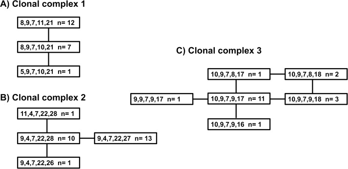
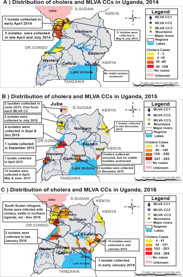
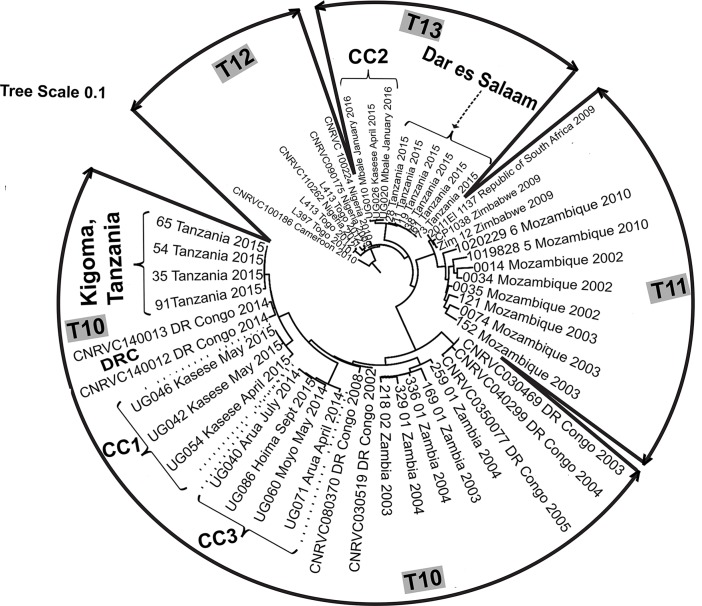
Methodology/principal findings: Sixty-three V. cholerae isolates collected from outbreaks in Uganda between 2014 and 2016 were tested using multiplex polymerase chain reaction (PCR), multi-locus variable number of tandem repeat analysis (MLVA) and whole genome sequencing (WGS). Three closely related MLVA clonal complexes (CC) were identified: CC1, 32% (20/63); CC2, 40% (25/63) and CC3, 28% (18/63). Each CC contained isolates from a different WGS clade. These clades were contained in the third wave of the 7th cholera pandemic strain, two clades were contained in the transmission event (T)10 lineage and other in T13. Analysing the dates and genetic relatedness revealed that V. cholerae genetic lineages spread between districts within Uganda and across national borders.
Conclusion: The V. cholerae strains showed local and regional transmission within Uganda and the East African region. To prevent, control and eliminate cholera, these countries should implement strong cross-border collaboration and regional coordination of preventive activities.
Conflict of interest statement
The authors have declared that no competing interests exist.
Figures
References
-
- Dick MH, Guillerm M, Moussy F, Chaignat C-L. Review of two decades of cholera diagnostics—how far have we really come? PLoS Negl. Trop. Dis. [Internet]. Public Library of Science; 2012. [cited 2017 Mar 2];6:e1845 Available from: http://www.ncbi.nlm.nih.gov/pubmed/23071851 doi: 10.1371/journal.pntd.0001845 - DOI - PMC - PubMed
-
- Jusatz HJ. 150 years pandemics of Asiatic cholera, 1831–1981. Zentralblatt fur Bakteriol. Mikrobiol. und Hyg. 1 Abt Orig. A Medizinische Mikrobiol. Infekt. und Parasitol. Int. J. Microbiol. Hyg. A Med. Microbiol. Infect. 1982;252:257–67. - PubMed
-
- Chatterji BP. On the discovery of the nature of cholera toxin in India. Vesalius acta Int. Hist. Med. 2010;16:16–8. - PubMed
-
- Ball L. Cholera and the Pump on Broad Street: Available from: http://www.ph.ucla.edu/epi/snow/Snow_Laura_Ball.pdf
Publication types
MeSH terms
Grants and funding
LinkOut - more resources
Full Text Sources
Other Literature Sources
Medical
Molecular Biology Databases
Research Materials