This site needs JavaScript to work properly. Please enable it to take advantage of the complete set of features!
Skip to main page content
U.S. flag

An official website of the United States government

Dot gov

The .gov means it’s official.
Federal government websites often end in .gov or .mil. Before sharing sensitive information, make sure you’re on a federal government site.

Https

The site is secure.
The https:// ensures that you are connecting to the official website and that any information you provide is encrypted and transmitted securely.

NIH NLM Logo
Log in
Access keys NCBI Homepage MyNCBI Homepage Main Content Main Navigation
. 2018 Apr 4;12(4):e0006382.
doi: 10.1371/journal.pntd.0006382. eCollection 2018 Apr.

Refractoriness of Sergentomyia schwetzi to Leishmania spp. is mediated by the peritrophic matrix

Affiliations

Refractoriness of Sergentomyia schwetzi to Leishmania spp. is mediated by the peritrophic matrix

Jovana Sadlova et al. PLoS Negl Trop Dis. .

Abstract

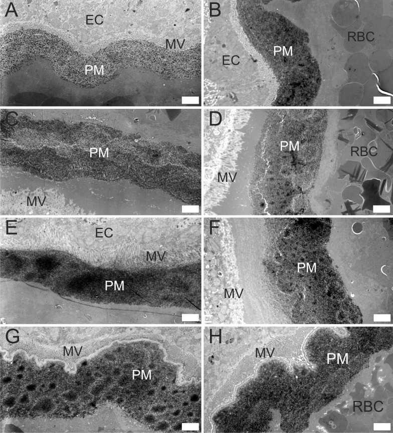
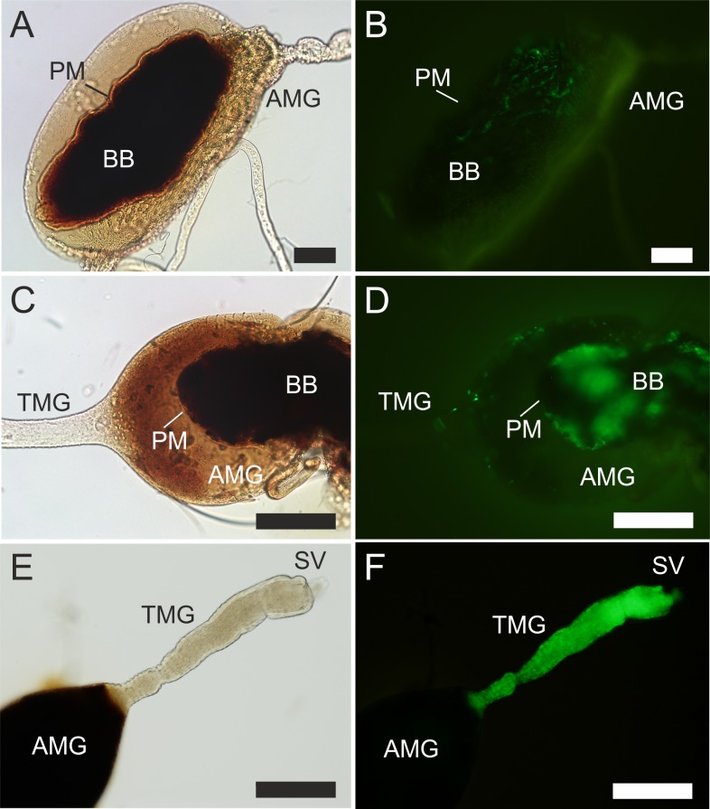
Background: The peritrophic matrix (PM) is an acellular chitin-containing envelope which in most blood sucking insects encloses the ingested blood meal and protects the midgut epithelium. Type I PM present in sand flies and other blood sucking batch feeders is secreted around the meal by the entire midgut in response to feeding. Here we tested the hypothesis that in Sergentomyia schwetzi the PM creates a physical barrier that prevents escape of Leishmania parasites from the endoperitrophic space.

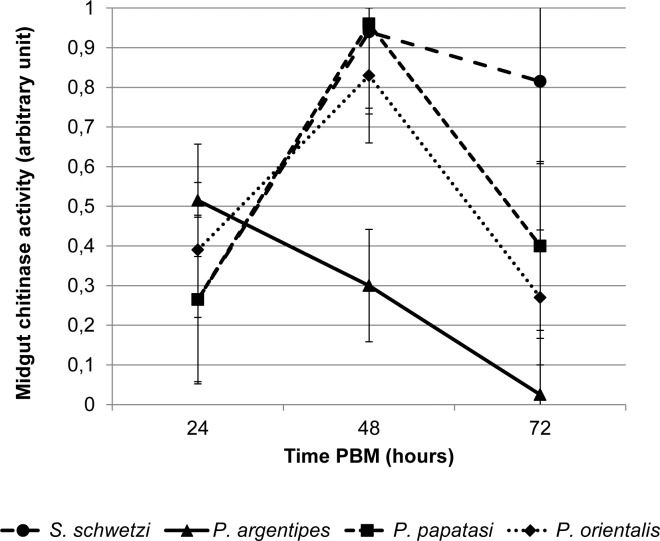
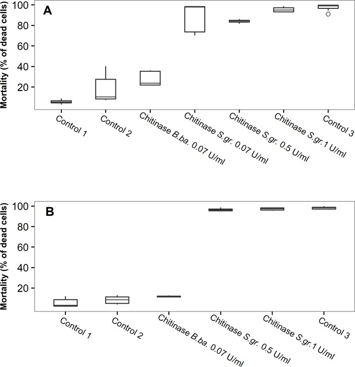
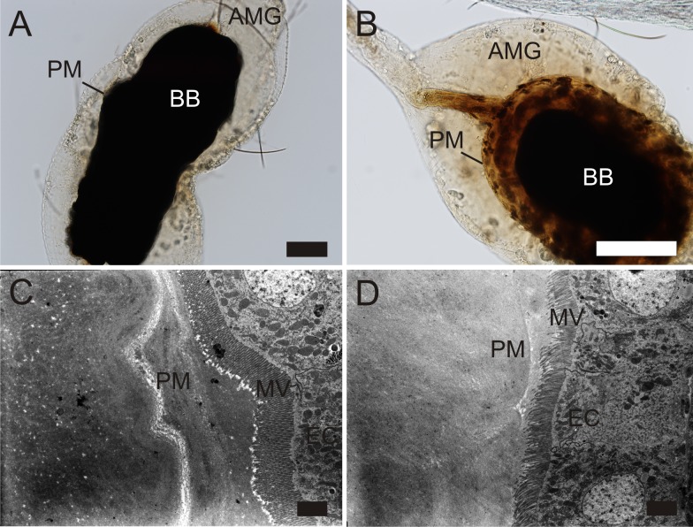
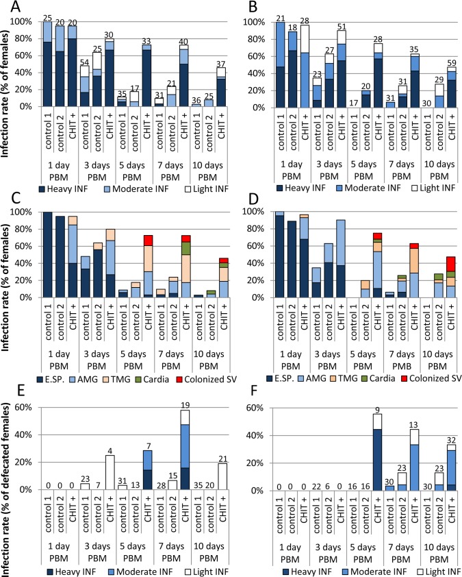
Methodology/principal findings: Morphology and ultrastructure of the PM as well the production of endogenous chitinase in S. schwetzi were compared with three sand fly species, which are natural vectors of Leishmania. Long persistence of the PM in S. schwetzi was not accompanied by different morphology or decreased production of chitinase. To confirm the role of the PM in refractoriness of S. schwetzi to Leishmania parasites, culture supernatant from the fungus Beauveria bassiana containing chitinase was added to the infective bloodmeal to disintegrate the PM artificially. In females treated with B. bassiana culture supernatants the PM was weakened and permeable, lacking multilayered inner structure; Leishmania colonized the midgut and the stomodeal valve and produced metacyclic forms. In control females Leishmania infections were lost during defecation.

Conclusions/significance: Persistence of the PM till defecation of the bloodmeal represents an important factor responsible for refractoriness of S. schwetzi to Leishmania development. Leishmania major as well as L. donovani promastigotes survived defecation and developed late-stage infections only in females with PM disintegrated artificially by B. bassiana culture supernatants containing exogenous chitinase.


Conflict of interest statement

The authors have declared that no competing interests exist.

Figures

Fig 1
Fig 1. Gross morphology of the S. schwetzi PM with detailed image of the posterior tail.
The complete PM dissected from the gut of S. schwetzi at 24 h PBM (A) and its enlarged posterior end with the transparent posterior tail showed in more detail (B). PT, posterior tail; AP, anterior plug; BB, blood bolus. Scale bars indicate 100 μm.
Fig 2
Fig 2. Ultrastructure of the PM in four sand fly species.
Electron micrographs of cross sections of the AMG in P. argentipes (A, B); P. orientalis (C, D); P. papatasi (E, F) and S. schwetzi (G, H) dissected at 24 h (P. argentipes) or 48 h PBM (P. orientalis, P. papatasi, S. schwetzi). PM, peritrophic matrix; MV, microvilli; EC, epithelial cells of the AMG. Scale bars indicate 2 μm.
Fig 3
Fig 3. Chitinase activity in midguts of the four sand fly species.
The exochitinase activity was quantified using the Chitinase Assay Kit (Sigma) in dissected sand fly midguts at 24, 48 and 72 h PBM. One arbitrary unit of chitinase releases 1 μmole of 4-nitrophenol per minute from the appropriate substrate at pH 4.8 at 37 °C. The values are arithmetic means from two experiments.
Fig 4
Fig 4
Mortality of L. donovani (A) and L. major (B) promastigotes exposed in vitro to culture supernatant of B. bassiana or chitinase from S. griseus. The mortality assessed as the ratio of the number of dead cells showing both green and blue fluorescence (GFP and DAPI) to the number of live cells showing green fluorescence (GFP). Control 1, culture medium for Leishmania with 0.025 V of PBS; Control 2, culture medium for Leishmania mixed at 1:1 with culture medium for B. bassiana; Control 3, parasites killed by 1% solution of formaldehyde and permeabilised by 0.5% Triton X-100; B. ba., Beauveria bassiana; S. gr., Streptomyces griseus.
Fig 5
Fig 5. Differences in the PM between S. schwetzi females treated with B. bassiana culture supernatant and control.
A and B, light microscopy; C and D, electron micrographs of cross sections of the abdominal midgut. A, appearance of the PM in a control female on day 3 PBM; B, appearance of the PM in female fed on a mixture of rabbit blood with supernatant of B. bassiana containing chitinase on day 3 PBM; C, multilayered thick PM in the control female on day 2 PBM; D, thin PM without apparent inner structure in the female fed on a mixture of rabbit blood with supernatant of B. bassiana containing chitinase on day 2 PBM. AMG, abdominal midgut; BB, blood bolus; PM, peritrophic matrix; MV, microvilli; EC, epithelial cells of the AMG. Scale bars indicate 100 μm in A, B and 2 μm in C, D.
Fig 6
Fig 6. Light and fluorescent microscopy of S. schwetzi midguts.
A and B, the gut of a control female (infectious meal comprising L. donovani promastigotes suspended in a mixture of fresh B. bassiana medium and inactivated rabbit blood) on day 3 PBM with L. donovani enclosed inside the PM; C and D, the gut of a treated female (infectious meal comprising L. donovani promastigotes suspended in a mixture of B. bassiana culture supernatant and inactivated rabbit blood) on day 3 PBM showing escape of L. donovani from the endoperitrophic space; E and F, the gut of a treated female on day 10 PBM showing colonization of the thoracic midgut and the stomodeal valve by L. major promastigotes expressing GFP (green). Images A—B, C—D and E—F are the same guts photographed by light and fluorescent microscopy, respectively. Both Leishmania species were marked with GFP protein, the midgut epithelium shows a natural mild autofluorescence. AMG, abdominal midgut; TMG, thoracic midgut; BB, blood bolus; SV, stomodeal valve; PM, peritrophic matrix. Scale bars indicate 100 μm.
Fig 7
Fig 7. Effect of B. bassiana culture supernatant on the development of L. donovani and L. major in S. schwetzi.
Experimental infections of S. schwetzi with L. donovani (A, C, E) and L. major (B, D, F) with addition of culture supernatant containing exogenous chitinase (0.07 U/mL) from B. bassiana (CHIT +). In control females, either fresh medium for B. bassiana or heat-inactivated supernatant from B. bassiana culture was used (control 1 and control 2, respectively). A, B: Rates and intensities of infections. Numbers of dissected females are shown above bars. C, D: Location of L. donovani in infected sand flies. E.SP., endoperitrophic space; AMG, abdominal midgut; TMG, thoracic midgut; SV, stomodeal valve. E, F: Rates and intensities of infections in females post defecation. Numbers of dissected females post defecation are shown above bars.

References

    1. Maroli M, Feliciangeli MD, Bichaud L, Charrel RN, Gradoni L. Phlebotomine sandflies and the spreading of leishmaniases and other diseases of public health concern. Med Vet Entomol 2013; 27: 123–147. doi: 10.1111/j.1365-2915.2012.01034.x - DOI - PubMed
    1. Seblova V, Sadlova J, Vojtkova B, Votypka J, Carpenter S, Bates PA, Volf P. The Biting Midge Culicoides sonorensis (Diptera: Ceratopogonidae) Is Capable of Developing Late Stage Infections of Leishmania enriettii. PLoS Negl Trop Dis 2015; 9: e0004060 doi: 10.1371/journal.pntd.0004060 PNTD-D-15-00735 [pii]. - DOI - PMC - PubMed
    1. Ramalho-Ortigao M, Saraiva EM, Traub-Cseko YM. Sand fly- Leishmania interactions: long relationships are not necessarily easy. Open Parasitol J 2010; 4: 195–204. doi: 10.2174/1874421401004010195 - DOI - PMC - PubMed
    1. Kamhawi S. Phlebotomine sand flies and Leishmania parasites: friends or foes? Trends Parasitol 2006; 22: 439–445. doi: 10.1016/j.pt.2006年06月01日2 - DOI - PubMed
    1. Dostalova A, Volf P. Leishmania development in sand flies: parasite-vector interactions overview. Parasit Vectors 2012; 5: 276 1756-3305年5月27日6 [pii]; doi: 10.1186/1756-3305年5月27日6 - DOI - PMC - PubMed

Publication types

LinkOut - more resources

Cite

AltStyle によって変換されたページ (->オリジナル) /