This site needs JavaScript to work properly. Please enable it to take advantage of the complete set of features!
Skip to main page content
U.S. flag

An official website of the United States government

Dot gov

The .gov means it’s official.
Federal government websites often end in .gov or .mil. Before sharing sensitive information, make sure you’re on a federal government site.

Https

The site is secure.
The https:// ensures that you are connecting to the official website and that any information you provide is encrypted and transmitted securely.

NIH NLM Logo
Log in
Access keys NCBI Homepage MyNCBI Homepage Main Content Main Navigation
Review
. 2019 Dec;10(1):822-831.
doi: 10.1080/21505594.2018.1431087. Epub 2018 Aug 1.

The capsule of Cryptococcus neoformans

Affiliations
Review

The capsule of Cryptococcus neoformans

Arturo Casadevall et al. Virulence. 2019 Dec.

Abstract

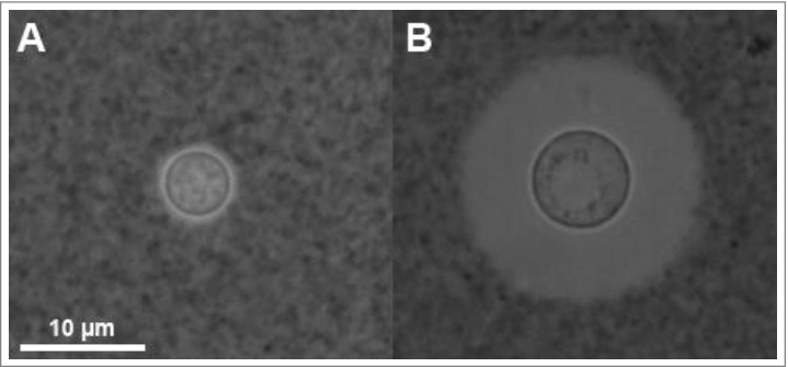
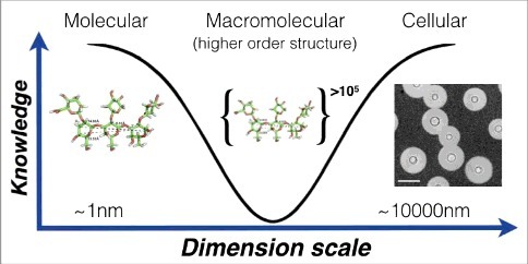
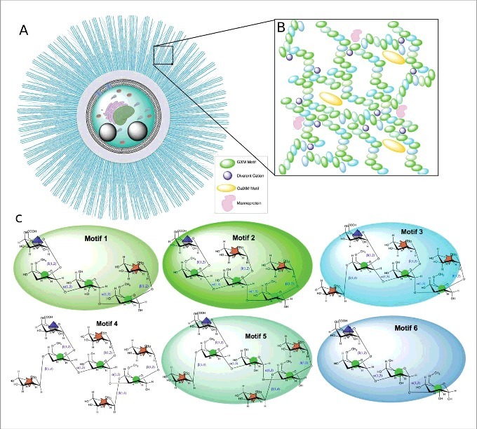
The capsule of Cryptococcus neoformans is its dominant virulence factor and plays a key role in the biology of this fungus. In this essay, we focus on the capsule as a cellular structure and note the limitations inherent in the current methodologies available for its study. Given that no single method can provide the structure of the capsule, our notions of what is the cryptococcal capsule must be arrived at by synthesizing information gathered from very different methodological approaches including microscopy, polysaccharide chemistry and physical chemistry of macromolecules. The emerging picture is one of a carefully regulated dynamic structure that is constantly rearranged as a response to environmental stimulation and cellular replication. In the environment, the capsule protects the fungus against desiccation and phagocytic predators. In animal hosts the capsule functions in both offensive and defensive modes, such that it interferes with immune responses while providing the fungal cell with a defensive shield that is both antiphagocytic and capable of absorbing microbicidal oxidative bursts from phagocytic cells. Finally, we delineate a set of unsolved problems in the cryptococcal capsule field that could provide fertile ground for future investigations.

Keywords: cryptococcal capsule; polysaccharide structure; virulence factor.


Figures

Figure 1.
Figure 1.
Cryptococcus neoformans yeast cells visualized by India Ink under phase contrast microscopy. India Ink particles are excluded from the dense PS capsule. A. Yeast cells cultured in an environment with optimal nutrition. B. Yeast cells cultured within mouse bone-marrow derived macrophages, experiencing starvation and oxidative stress from the phagolysosome for 24 h. Note the increased cell body, generation of what appears to be a large vacuole, and drastic increase in capsule size. Both images were obtained at 100x magnification with 2 ×ばつ 2 binning.
Figure 2.
Figure 2.
Knowledge of capsule structure as a function of dimension scale. Representative hyperbola of polysaccharide capsule structural understanding which is greatest at the smallest (molecular) and largest (cellular) levels. At the molecular level an in silico energy-minimized structure of the GXM M2 motif (see figure 3c) is estimated to cover a few nanometers in diameter. At the cellular level, capsule radii exist in the micrometer size range and are readily measured by negative staining and light microscopy (Scale bar in micrograph represents 10 μm). There is a knowledge gap in capsule structure at the macromolecular level or higher order structure (analogous to the secondary, tertiary and quaternary structural level of proteins) were many motifs are organized to form the overall capsular structure.
Figure 3.
Figure 3.
Illustration of the C. neoformans polysaccharide capsule structure. A. Cellular view of the polysaccharide capsule formed by a complex lattice-work of interconnected polymers that decreases in density, porosity and stiffness as it extends radially outward from the cell wall. B. Representative model of proposed polysaccharide lattice making up the capsular macromolecular structure involving interactions between GXM, GalXM, divalent cations, and mannoproteins. C. Structure of the six major motifs making up the GXM polysaccharide present in serotypes A, B, C, and D [35]. These tetrasaccharide to octasaccharide motifs fit together to make up the megadalton GXM polymer. Each motif is color coded to match the lattice illustration in B. In C. neoformans serotype A motif 4 is not present [35]. Motifs are represented using CFG nomenclature (mannose, green circles; xylose, orange 5-pointed start; glucuronic acid, blue/white diamond) along with chair pyranose rings and linkages.

References

    1. McClelland EE, Bernhardt P, Casadevall A. Estimating the relative contribution s of virulence factors for pathogenic microbes. Infect Immun. 2006;74:1500–4. doi:10.1128/IAI.74.3.1500-1504.2006 - DOI - PMC - PubMed
    1. Chang YC, Kwon-Chung KJ. Complementation of a capsule-deficient mutation of Cryptococcus neoformans restores its virulence. Mol Cell Biol. 1994;14:4912–9. doi:10.1128/MCB.14.7.4912 - DOI - PMC - PubMed
    1. Chang YC, Penoyer LA, Kwon-Chung KJ. The second capsule gene of cryptococcus neoformans, CAP64, is essential for virulence. Infect Immun. 1996;64:1977–83. - PMC - PubMed
    1. Kabanda T, Siedner MJ, Klausner JD, et al.. Point-of-care diagnosis and prognostication of cryptococcal meningitis with the cryptococcal antigen lateral flow assay on cerebrospinal fluid. Clinical infectious diseases : an official publication of the Infectious Diseases Society of America. 2014;58:113–6. doi:10.1093/cid/cit641 - DOI - PMC - PubMed
    1. O'Meara TR, Alspaugh JA. The Cryptococcus neoformans capsule: a sword and a shield. Clin Microbiol Rev. 2012;25:387–408. doi:10.1128/CMR.00001-12 - DOI - PMC - PubMed

Publication types

LinkOut - more resources

Cite

AltStyle によって変換されたページ (->オリジナル) /