This site needs JavaScript to work properly. Please enable it to take advantage of the complete set of features!
Skip to main page content
U.S. flag

An official website of the United States government

Dot gov

The .gov means it’s official.
Federal government websites often end in .gov or .mil. Before sharing sensitive information, make sure you’re on a federal government site.

Https

The site is secure.
The https:// ensures that you are connecting to the official website and that any information you provide is encrypted and transmitted securely.

NIH NLM Logo
Log in
Access keys NCBI Homepage MyNCBI Homepage Main Content Main Navigation
. 2018 Feb;24(2):294-302.
doi: 10.3201/eid2402.171065.

Yersinia pestis Survival and Replication in Potential Ameba Reservoir

Yersinia pestis Survival and Replication in Potential Ameba Reservoir

David W Markman et al. Emerg Infect Dis. 2018 Feb.

Abstract

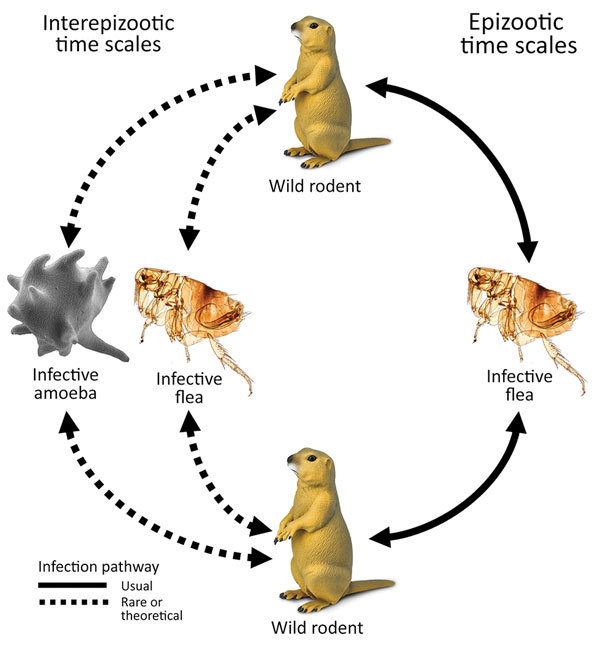
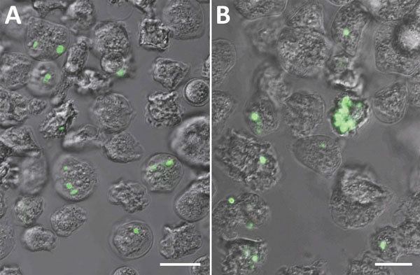
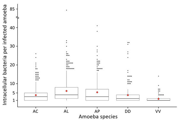
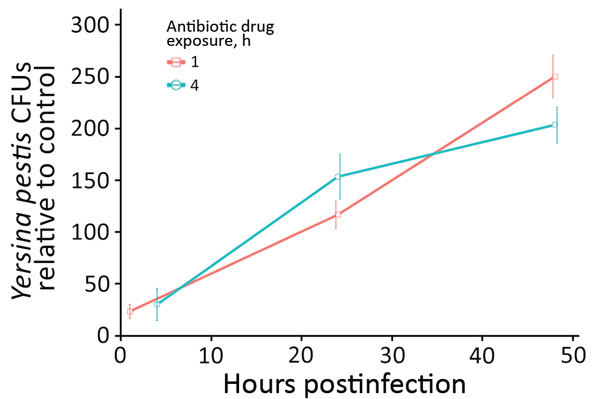
Plague ecology is characterized by sporadic epizootics, then periods of dormancy. Building evidence suggests environmentally ubiquitous amebae act as feral macrophages and hosts to many intracellular pathogens. We conducted environmental genetic surveys and laboratory co-culture infection experiments to assess whether plague bacteria were resistant to digestion by 5 environmental ameba species. First, we demonstrated that Yersinia pestis is resistant or transiently resistant to various ameba species. Second, we showed that Y. pestis survives and replicates intracellularly within Dictyostelium discoideum amebae for ˃48 hours postinfection, whereas control bacteria were destroyed in <1 hour. Finally, we found that Y. pestis resides within ameba structures synonymous with those found in infected human macrophages, for which Y. pestis is a competent pathogen. Evidence supporting amebae as potential plague reservoirs stresses the importance of recognizing pathogen-harboring amebae as threats to public health, agriculture, conservation, and biodefense.

Keywords: Dictyostelium discoideum; Plague; Yersinia pestis; ameba; amoeba; amoebae; biological evolution; coccobacillus; disease reservoir; ecology; endemic; epizootic; eukaryote; feral macrophage; phagocytosis; slime mold; trophozoite.


Figures

Figure 1
Figure 1
Infection pathways for plague. During plague epizootics, transmission occurs through flea vectors within meta-populations of ground-dwelling rodents. It is unknown by what route or mechanism Yersinia pestis is maintained during interepizootic periods of plague quiescence. Previous research on fleas has not strongly supported their reservoir potential across interepizootic periods (3). The experiment and analysis of this study test the hypothesis that amoeboid species demonstrate reservoir potential for Y. pestis. If Y. pestis is maintained within ameba reservoirs, we suspect that epizootic recrudescence may occur when infected soilborne amebae enter the bloodstream of naive rodent hosts (by entering wounds from antagonistic host-to-host interactions or burrowing activities). Amebae typically lyse when incubated at 37°C and simultaneously release their intracellular cargo, potentially initiating an infection.
Figure 2
Figure 2
Representative fluorescent confocal images of (A) Acanthamoeba castellanii (B) and Dictyostelium discoideum after experimental co-culture with Yersinia pestis (CO92 pgm+, pCD1, pGFPuv, amp+) and removal of extracellular bacteria. After co-culture of ameba trophozoites and Y. pestis, we determined the prevalence and intensity of bacterial uptake by manual counting of amebae by using z-stack fluorescent confocal microscopy and averaging across 15 fields per replicate of each ameba species. Confocal count data represent the minimum prevalence/intensity values. Bacteria adherent to the outside of ameba or those with uncertain intracellular status were discarded. The minimum count threshold to reduce random count bias to accepted levels was determined to be 500 per ameba species. Scale bars indicate 30 μm.
Figure 3
Figure 3
Boxplots of infection intensity across ameba species after experimental infection with Yersinia pestis. Infection intensity frequencies followed a strong negative binomial distribution. Median infection intensities (horizontal lines inside boxes): AC = 3, AL = 4, AP = 3, DD = 2, VV = 1. Red diamonds denote mean infection intensity (Table). Each ameba species had several high-intensity outliers ranging up to a maximum of 84 intracellular bacteria observed in 1 A. lenticulata ameba (note broken y-axis). AC, Acanthamoeba castellanii (n = 1,441); AL, A. lenticulata (n = 1,156); AP, A. polyphaga (n = 737); DD, Dictyostelium discoideum (n = 624); VV, Vermamoeba vermiformis (n = 528).
Figure 4
Figure 4
Representative transmission electron micrographs (TEM) depict Acanthamoeba castellanii amebae during A) 10-minute, B) 30-minute, and C) 24-hour co-cultures (multiplicity of infection 100) with Yersinia pestis (CO92 pgm+, pCD1, pGFPuv, amp+). Red arrows in panel A indicate potential intraameba mitotic division of Y. pestis bacterium. Visual analysis of TEM micrographs proved inconclusive for identifying the bacterial division septum. Y. pestis resides within the potential replicative niche of a tight-fitting vacuolar membrane, similar to Yersinia-containing vacuoles observed in macrophages. YP, Y. pestis; CV, central vacuole; DV, digestive vacuole; M, mitochondria. Scale bars indicate 3 μm.
Figure 5
Figure 5
Intraameba Yersinia pestis abundance in Dictyostelium discoideum across 2 postinfection antibiotic drug exposure periods, 1 hour and 4 hour. In D. discoideum, the abundance of viable intracellular Y. pestis was significantly greater at each successive time point (24 and 48 hours) after the 1-hour antibiotic drug treatment (p = 0.01 and p = 0.002, respectively). After the 4-hour antibiotic drug treatment in D. discoideum, the abundance of viable intracellular Y. pestis at 24 and 48 hours was significantly greater than at 4 hours (p = 0.008 and p = 0.001, respectively). The abundance of viable Y. pestis within D. discoideum at 48 hours postinfection was not significantly different between the 1-hour and 4-hour antibiotic drug treatments (p = 0.1624). Viable intracellular Y. pestis abundance was significantly greater in D. discoideum compared with all other species at 48 hours postinfection (p<0.001).

References

    1. Perry RD, Fetherston JD. Yersinia pestis—etiologic agent of plague. Clin Microbiol Rev. 1997;10:35–66. - PMC - PubMed
    1. Bertherat E; World Health Organization. Plague around the world, 2010–2015. Wkly Epidemiol Rec. 2016;91:89–93. - PubMed
    1. Webb CT, Brooks CP, Gage KL, Antolin MF. Classic flea-borne transmission does not drive plague epizootics in prairie dogs. Proc Natl Acad Sci U S A. 2006;103:6236–41. 10.1073/pnas.0510090103 - DOI - PMC - PubMed
    1. Girard JM, Wagner DM, Vogler AJ, Keys C, Allender CJ, Drickamer LC, et al. Differential plague-transmission dynamics determine Yersinia pestis population genetic structure on local, regional, and global scales. Proc Natl Acad Sci U S A. 2004;101:8408–13. 10.1073/pnas.0401561101 - DOI - PMC - PubMed
    1. Snäll T, O’Hara RB, Ray C, Collinge SK. Climate-driven spatial dynamics of plague among prairie dog colonies. Am Nat. 2008;171:238–48. 10.1086/525051 - DOI - PubMed

Publication types

Cite

AltStyle によって変換されたページ (->オリジナル) /