This site needs JavaScript to work properly. Please enable it to take advantage of the complete set of features!
Skip to main page content
U.S. flag

An official website of the United States government

Dot gov

The .gov means it’s official.
Federal government websites often end in .gov or .mil. Before sharing sensitive information, make sure you’re on a federal government site.

Https

The site is secure.
The https:// ensures that you are connecting to the official website and that any information you provide is encrypted and transmitted securely.

NIH NLM Logo
Log in
Access keys NCBI Homepage MyNCBI Homepage Main Content Main Navigation
doi: 10.1038/s41598-017-17765-5.

In situ immune response and mechanisms of cell damage in central nervous system of fatal cases microcephaly by Zika virus

Affiliations

In situ immune response and mechanisms of cell damage in central nervous system of fatal cases microcephaly by Zika virus

Raimunda S S Azevedo et al. Sci Rep. .

Abstract

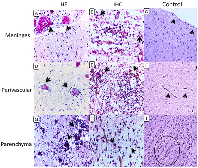
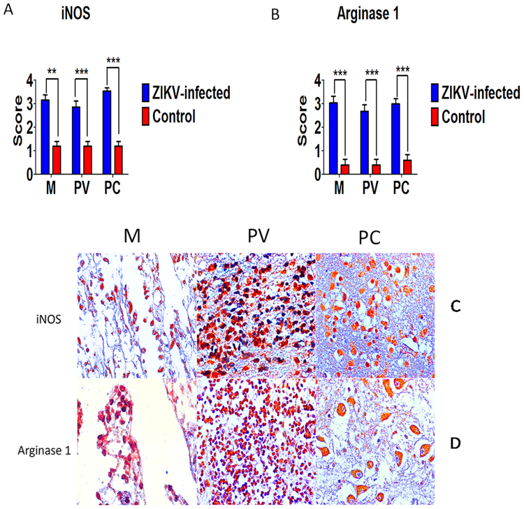
Zika virus (ZIKV) has recently caused a pandemic disease, and many cases of ZIKV infection in pregnant women resulted in abortion, stillbirth, deaths and congenital defects including microcephaly, which now has been proposed as ZIKV congenital syndrome. This study aimed to investigate the in situ immune response profile and mechanisms of neuronal cell damage in fatal Zika microcephaly cases. Brain tissue samples were collected from 15 cases, including 10 microcephalic ZIKV-positive neonates with fatal outcome and five neonatal control flavivirus-negative neonates that died due to other causes, but with preserved central nervous system (CNS) architecture. In microcephaly cases, the histopathological features of the tissue samples were characterized in three CNS areas (meninges, perivascular space, and parenchyma). The changes found were mainly calcification, necrosis, neuronophagy, gliosis, microglial nodules, and inflammatory infiltration of mononuclear cells. The in situ immune response against ZIKV in the CNS of newborns is complex. Despite the predominant expression of Th2 cytokines, other cytokines such as Th1, Th17, Treg, Th9, and Th22 are involved to a lesser extent, but are still likely to participate in the immunopathogenic mechanisms of neural disease in fatal cases of microcephaly caused by ZIKV.


Conflict of interest statement

The authors declare that they have no competing interests.

Figures

Figure 1
Figure 1
Histopathological and immunohistochemical (IHC) aspects of fatal ZIKV-associated microcephaly cases and controls. (A) (Case 6): Viral meningoencephalitis with vasocongestion and surrounding inflammatory infiltrate (arrow). (B) (Case 6): Immunostaining of ZIKV antigens in cells of the inflammatory infiltrate in the meninges (arrow). (C) (Case 15): Preserved meninges and IHC-negative to control (arrow). (D) (Case 6): Infiltration of mononuclear cells in the perivascular space (arrow). (E) (Case 8): Gliosis and immunostaining inflammatory cell infiltrate (arrow). (F) (Case 15): Preserved capillaries without inflammatory infiltrate and IHC-negative to control (arrow). (G) (Case 8): Calcification (arrow) in the neural parenchyma. (H) (Case 5): Neuronal necrosis (arrow) with ZIKV antigens in the neural parenchyma. (I) (Case 15): Parenchymal neural preservation and IHC-negative to control (circle) (magnification: ×ばつ).
Figure 2
Figure 2
Semiquantitative analysis and representative immunohistochemical pattern in meninges (M), perivascular region (PV) and parenchyma (PC) in fatal Zika microcephaly cases and control. (A) Expression of caspase 3. (B) Immunolabeling for caspase 3 (B-M and B-PV: Case 7; B-PC: Case 2). (C) Negative control (C-M, C-PV, and C-PC: Case 15) (magnification: ×ばつ) (**p < 0.005, ***p < 0.0005).
Figure 3
Figure 3
Semiquantitative analysis and Immunohistochemical pattern in meninges (M), perivascular region (PV), and parenchyma (PC) in fatal Zika microcephaly cases. (A) Expression of S100. H Immunolabeling for S100 (H-M and H-PV: Case 6; H-PC: Case 5). (B) Expression of CD57. I Immunolabeling for CD57 (I-M: Case 6; I-PV and I-PC: Case 7). (C) Expression of CD68. J Immunolabeling for CD68 (J-M: Case 6; J-PV and J-PC: Case 5). (D) Expression of CD163. K Immunolabeling for CD163 (K-M: Case 8; K-PV and K-PC: Case 5). (E) Expression of CD8. L Immunolabeling for CD8 (L-M: Case 6; L-PV and L-PC: Case 7). (F) Expression of CD4. M Immunolabeling for CD4 (M-M: Case 6; M-PV and M-PC: Case 7). (G) Expression of Foxp3. N Immunolabeling for Foxp3 (N-M: Case 8; N-PV and N-PC: Case 7) (magnification: ×ばつ) (ns: not statistically significant, *p < 0.05, **p < 0.005, ***p < 0.0005).
Figure 4
Figure 4
Semiquantitative analysis and Immunohistochemical pattern in meninges (M), perivascular region (PV), and parenchyma (PC) in fatal Zika microcephaly cases. (A) Expression of IFN-γ. H Immunolabeling for IFN-γ (H-M and H-PV: Case 8; H-PC: Case 9). (B) Expression of IFN-α. I Immunolabeling for IFN-α (I-M: Case 8; I-PV: Case 9; I-PC: Case 7). (C) Expression of IFN-β. J Immunolabeling for IFN-β (J-M and J-PV: Case 8; J-PC: Case 9). (D) Expression of IL-6. K Immunolabeling for IL-6 (K-M: Case 9; K-PV: Case 8; K-PC: Case 7) (E) Expression of IL-12A. L Immunolabeling for IL-12A (L-M and L-PV: Case 8; L-PC: Case 7). (F) Expression of IL-1β. M Immunolabeling for IL-1β (M-M: Case 8; M-PV and M-PC: Case 7). (G) Expression of TNF-α. N Immunolabeling for TNF-α (N-M and N-PV: Case 8 N-PC: Case 9) (magnification: ×ばつ) (ns: not statistically significant, *p < 0.05, **p < 0.005, ***p < 0.0005).
Figure 5
Figure 5
Semiquantitative analysis and immunohistochemical staining in meninges (M), perivascular region (PV), and parenchyma (PC) in fatal Zika microcephaly cases. (A) Expression of IL-4. F Immunolabeling for 4 (F-M and F-PV: Case 8; F-PC: Case 9). (B) Expression of IL-10. G Immunolabeling for 10 (G-M and G-PV: Case 8; G-PC: Case 4). (C) Expression of IL-33. H Immunolabeling for IL-33 (H-M: Case 7; H-PV: Case 8; H-PC: Case 9). (D) Expression of IL-37. I Immunolabeling for IL-37 (I-M and I-PV: Case 8; I-PC: Case 7). (E) Expression TGF-β1. J Immunolabeling for TGF-β1 (J-M: Case 8; J-PV: Case 7; J-PC: Case 9) (magnification: ×ばつ) (**p < 0.05, ***p < 0.0005).
Figure 6
Figure 6
Semiquantitative analysis and immunohistochemical staining in meninges (M), perivascular region (PV), and parenchyma (PC) in fatal Zika microcephaly cases. (A) Expression of IL-9. F Immunolabeling for IL-9 (F-M and F-PV: Case 8 F-PC: Case 4). (B) Expression of IL-17. G Immunolabeling for IL-17 (G-M: Case 8; G-PV and G-PC: Case 7). (C) Expression of IL-23. H Immunolabeling for IL-23 (H-M: Case 8 and H-PV: H-PC: Case 9), (D) Expression of IL-22. H Immunolabeling for IL-22 (H-M: Case 8; H-PV: Case 9; H-PC: Case 2) (magnification: ×ばつ) (**p < 0.005, ***p < 0.0005).
Figure 7
Figure 7
Semiquantitative analysis and immunohistochemical staining in meninges (M), perivascular region (PV), and parenchyma (PC) in fatal Zika microcephaly cases. (A) Expression of iNOS. (C) Immunolabeling for iNOS (C-M: Case 8; C-PV: Case 7; C-PC: Case 1). (B) Expression of arginase 1. (D) Immunolabeling for arginase 1 (D-M and D-PV: Case 8; D-PC: Case 9) (magnification: ×ばつ) (**p < 0.05, ***p < 0.0005).
Figure 8
Figure 8
Integrated view of the in situ immune response in fatal cases of microcephaly caused by ZIKV. In view of the complexity of the immune response in the central nervous system, initially dendritic cells (DC) may trigger the development of neuroinflammation or present the viral antigen to naive TCD4 lymphocytes that can differentiate into several subpopulations of T helper lymphocytes (Th1, Th2, Th9, Th17, Th22, and Treg) that produce a series of cytokines modulating the activity of M1 and M2 microglia, astrocytes (AS), and neurons (NE). The production of pro-inflammatory (IL-1β, IL-6, IL-9, IL-17, IL-22, IL23, TNF-α, IFN-γ, IFN-α, and IFN-β) and anti-inflammatory cytokines (IL-4, IL-10, IL-33 IL-37, and TGF-β1) by these cells induces the neuroinflammatory or immunosuppressive response by increasing the production of iNOS or arginase 1. In the context of cellular stress, cytokines such as IL-4, IL-10, IL-33, IL-37, and TNF-α as well as other immune factors contribute directly to necrosis and apoptosis, regulating the production of caspase 3. With regard to the inflammasome, probably IL-1β and IL-33 induce neuroinflammation. NK cells participate in the apoptotic response and neuroinflammation, inducing IFN-γ production. In negative regulation, the immunosuppressive effect of anti-inflammatory cytokines inhibits the activity of M1 microglia as well as NK cells. In the cytotoxic response produced by TCD8 lymphocytes, these cells participate in mechanisms that lead to neuroinflammation as well as apoptosis.

References

    1. Barzon, L., Trevisan, M., Sinigaglia, A., Lavezzo, E. & Palù, G. Zika virus pathogenesis to disease control. FEMS Microbiol Lett. 363, 10.1093/femsle/fnw202 (2016). - PubMed
    1. Azevedo RS, et al. Zika virus epidemic in Brazil. I. Fatal disease in adults: clinical and laboratorial aspects. J Clin Virol. 2016;85:56–64. doi: 10.1016/j.jcv.2016年10月02日4. - DOI - PMC - PubMed
    1. Cao-Lormeau VM, et al. Guillain-Barré syndrome outbreak associated with Zika virus infection in French Polynesia: a case-control study. Lancet. 2016;387:1531–1539. doi: 10.1016/S0140-6736(16)00562-6. - DOI - PMC - PubMed
    1. Thangamani S, Huang J, Hart CE, Guzman H, Tesh RB. Vertical Transmission of Zika Virus in Aedes aegypti Mosquitoes. Am J Trop Med Hyg. 2016;95:1169–1173. doi: 10.4269/ajtmh.16-0448. - DOI - PMC - PubMed
    1. Cugola FR, et al. The Brazilian Zika virus strain causes birth defects in experimental models. Nature. 2015;534:267–71. - PMC - PubMed

Publication types

Cite

AltStyle によって変換されたページ (->オリジナル) /