This site needs JavaScript to work properly. Please enable it to take advantage of the complete set of features!
Skip to main page content
U.S. flag

An official website of the United States government

Dot gov

The .gov means it’s official.
Federal government websites often end in .gov or .mil. Before sharing sensitive information, make sure you’re on a federal government site.

Https

The site is secure.
The https:// ensures that you are connecting to the official website and that any information you provide is encrypted and transmitted securely.

NIH NLM Logo
Log in
Access keys NCBI Homepage MyNCBI Homepage Main Content Main Navigation
. 2017 Dec 18;11(12):e0006123.
doi: 10.1371/journal.pntd.0006123. eCollection 2017 Dec.

Human Neutrophil Peptide 1 as immunotherapeutic agent against Leishmania infected BALB/c mice

Affiliations

Human Neutrophil Peptide 1 as immunotherapeutic agent against Leishmania infected BALB/c mice

Zahra Abdossamadi et al. PLoS Negl Trop Dis. .

Abstract

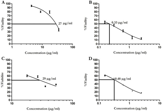
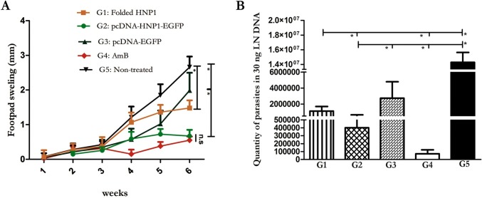
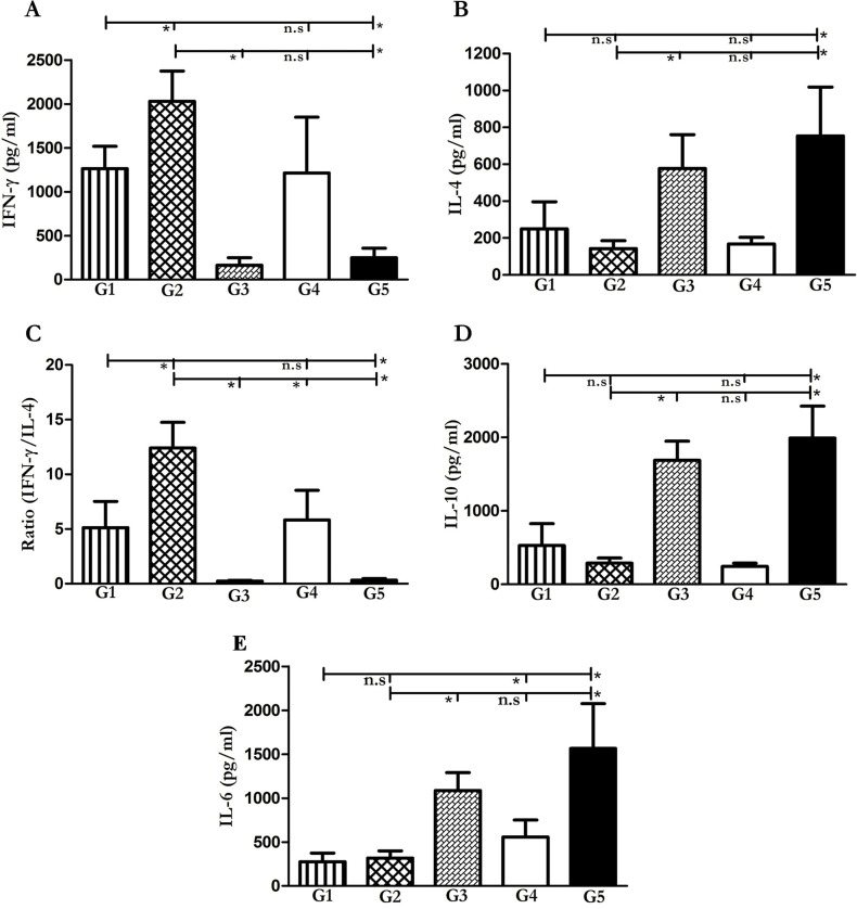
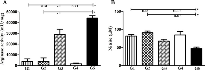
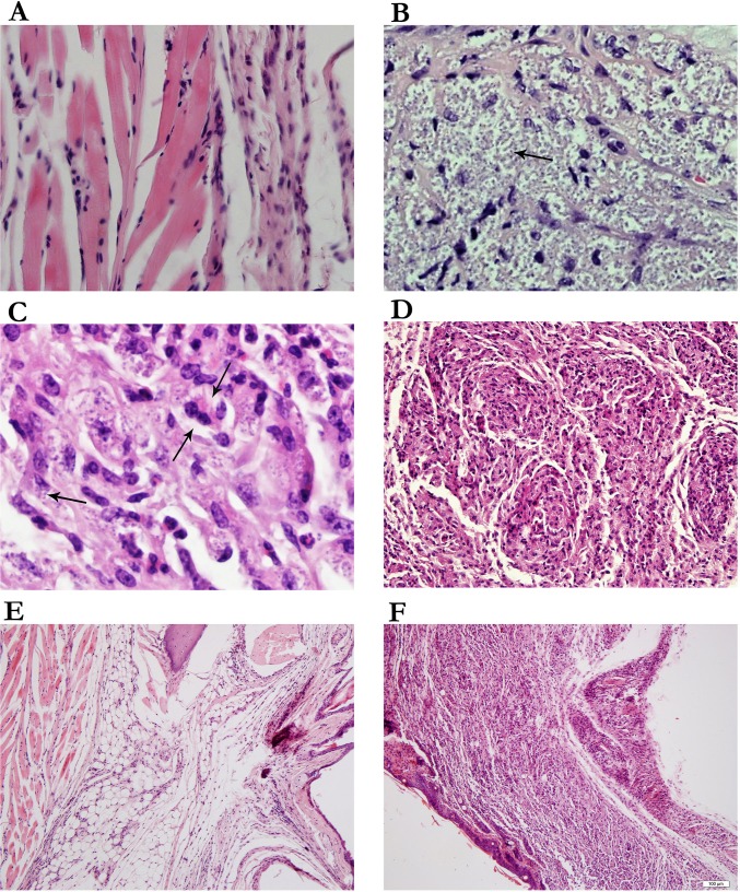
Human Neutrophil Peptide 1 (HNP1) produced by neutrophils, is a well-known antimicrobial peptide which plays a role both in innate as well as in adaptive immunity and is under intensive investigation as a potential therapeutic agent. Previous in vitro experiments have indicated the leishmaniacidal effect of recombinant HNP1 on Leishmania major (L. major) promastigotes and amastigotes. In the current study, we further extended the idea to explore the remedial effect of HNP1 in the two modalities of peptide therapy (folded HNP1) and gene therapy in L. major infected BALB/c mice. To this end, mice in five different groups received synthetic folded HNP1 (G1), pcDNA-HNP1-EGFP (G2), pcDNA-EGFP (G3), Amphotericin B (G4) and PBS (G5), which was started three weeks after infection for three consecutive weeks. Footpad swelling was monitored weekly and a day after the therapy ended, IFN-γ, IL-4, IL-10, IL-6 and nitric oxide produced by splenocytes were analyzed together with the parasite load in draining lymph nodes. Arginase activity and dermal histopathological changes were also analyzed in the infected footpads. We demonstrated that both therapeutic approaches effectively induced Th1 polarization and restricted parasite burden. It can control disease progression in contrast to non-treated groups. However, pcDNA-HNP1-EGFP is more promising in respect to parasite control than folded HNP1, but less effective than AmB treatment. We concluded with the call for a future approach, that is, a DNA-based expression of HNP1 combined with AmB as it can improve the leishmaniacidal efficacy.


Conflict of interest statement

The authors have declared that no competing interests exist.

Figures

Fig 1
Fig 1. Confirmation of HNP1-EGFP transfection in COS-7 cells by fluorescence microscopy and western blot analysis.
(A) COS-7 transfection confirmation by fluorescent microscopy 48h after transfection. A-1; Untransfected COS-7, A-2; COS-7 transfected with pcDNA-EGFP, A3; COS-7 transfected with pcDNA-HNP1-EGFP. (B) Confirmation of EGFP expression by western blot analysis in COS-7 transfected with pcDNA-HNP1-EGFP and pcDNA-EFGP; lane 1: untransfected COS-7, lane 2: COS-7 transfected with pcDNA-EFGP, lane 3: COS-7 transfected with pcDNA-HNP1-EFGP.
Fig 2
Fig 2. Anti promastigote and amastigote effects of folded HNP1.
After HNP1 folding by oxido-shuffling method, the leishmaniacidal effect of folded HNP1 was analyzed (A); the inhibitory effect of folded HNP1 on L. major promastigotes (B); the inhibitory effect of AmB on L. major promastigotes (C); effective dose of folded HNP1 on infected THP1 with L. major (D); effective dose of AmB on infected THP-1 with L. major.
Fig 3
Fig 3. Footpad swelling measurement and determination of L. major parasite quantification in lymph node of treated and control groups.
Footpad swelling measurement, started one week after challenge, was fulfilled weekly by metric capilar (B) Real time PCR was performed in 30 ng of DNA (n = 5). The G1-G5 represents the results from folded HNP1 (G1), pcDNA-HNP1-EGFP (G2), pcDNA-EGFP (G3), AmB (G4) and non-treated group (G5), respectively. The stars indicate significant difference and n.s displays not significant.
Fig 4
Fig 4. Cytokine production by splenocytes stimulated with F/T L. major antigen in treated and control mice.
Five mice per group were sacrificed and their spleens were dissected and stimulated by F/T antigen in order to measure IFN-γ (A), IL-4 (B), IFN-γ/IL-4 ratio (C), IL-10 (D) and IL-6 (E) cytokine production by ELISA system. The G1-G5 represents the results from folded HNP1 (G1), pcDNA-HNP1-EGFP (G2), pcDNA-EGFP (G3), AmB (G4) and non-treated group (G5), respectively. The star represents significant difference and n.s illustrates not significant.
Fig 5
Fig 5. Evaluation of arginase activity and NO production in mice treated with different HNP1 modalities.
Arginase activity (mU/ml) and nitrite production were determined in footpad and spleen (n = 5), respectively. Arginase activity (A) was performed by micro method test and NO (B) was fulfilled by Griess test. In both tests, the G1-G5 represents the results from folded HNP1 (G1), pcDNA-HNP1-EGFP (G2), pcDNA-EGFP (G3), AmB (G4) and non-treated group (G5), respectively. The star displays significant difference and n.s indicates not significant.
Fig 6
Fig 6. Histopathological changes in different mice groups.
Histopathological changes were analyzed directly on the infected footpads (A and B) Presence of parasites in the footpad lesion which represent; A: No parasite, B: parasite 4(+) (C) Presence of plasma cell in non-treated group (G5) (D) Immature granuloma reaction. Inflammation assessment (E): restricted area inflammation in treated group with HNP-1 and (F): diffuse inflammation in non-treated group was observed. (H&E, 400 HPF for Fig A, B, C. 250 for Fig D and 40 for Fig E and F).

References

    1. Bethony JM, Cole RN, Guo X, Kamhawi S, Lightowlers MW, et al. (2011) Vaccines to combat the neglected tropical diseases. Immunol Rev 239: 237–270. doi: 10.1111/j.1600-065X.2010.00976.x - DOI - PMC - PubMed
    1. Rafati S, Modabber F (2014) Cutaneous Leishmaniasis in Middle East and North Africa Neglected Tropical Diseases-Middle East and North Africa: Springer; pp. 117–139.
    1. Zandieh M, Kashi T, Taheri T, Zahedifard F, Taslimi Y, et al. (2015) Assessment of protection induced by DNA and live vaccine encoding Leishmania MHC class I restricted epitopes against L. major challenge in Balb/c mice model. Journal of Microbial & Biochemical Technology 2015.
    1. Abdossamadi Z, Seyed N, Rafati S (2016) Mammalian host defense peptides and their implication on combating Leishmania infection. Cell Immunol 309: 23–31. doi: 10.1016/j.cellimm.201610001 - DOI - PubMed
    1. Musa AM, Noazin S, Khalil EA, Modabber F (2010) Immunological stimulation for the treatment of leishmaniasis: a modality worthy of serious consideration. Trans R Soc Trop Med Hyg 104: 1–2. doi: 10.1016/j.trstmh.2009年07月02日6 - DOI - PubMed

Publication types

MeSH terms

Cite

AltStyle によって変換されたページ (->オリジナル) /