This site needs JavaScript to work properly. Please enable it to take advantage of the complete set of features!
Skip to main page content
U.S. flag

An official website of the United States government

Dot gov

The .gov means it’s official.
Federal government websites often end in .gov or .mil. Before sharing sensitive information, make sure you’re on a federal government site.

Https

The site is secure.
The https:// ensures that you are connecting to the official website and that any information you provide is encrypted and transmitted securely.

NIH NLM Logo
Log in
Access keys NCBI Homepage MyNCBI Homepage Main Content Main Navigation
Review
. 2017 Sep 18;9(9):290.
doi: 10.3390/toxins9090290.

A Review and Database of Snake Venom Proteomes

Affiliations
Review

A Review and Database of Snake Venom Proteomes

Theo Tasoulis et al. Toxins (Basel). .

Abstract

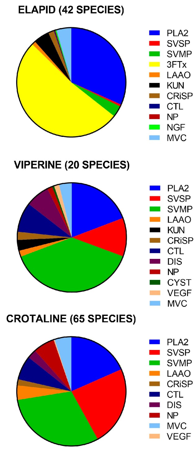
Advances in the last decade combining transcriptomics with established proteomics methods have made possible rapid identification and quantification of protein families in snake venoms. Although over 100 studies have been published, the value of this information is increased when it is collated, allowing rapid assimilation and evaluation of evolutionary trends, geographical variation, and possible medical implications. This review brings together all compositional studies of snake venom proteomes published in the last decade. Compositional studies were identified for 132 snake species: 42 from 360 (12%) Elapidae (elapids), 20 from 101 (20%) Viperinae (true vipers), 65 from 239 (27%) Crotalinae (pit vipers), and five species of non-front-fanged snakes. Approximately 90% of their total venom composition consisted of eight protein families for elapids, 11 protein families for viperines and ten protein families for crotalines. There were four dominant protein families: phospholipase A2s (the most common across all front-fanged snakes), metalloproteases, serine proteases and three-finger toxins. There were six secondary protein families: cysteine-rich secretory proteins, l-amino acid oxidases, kunitz peptides, C-type lectins/snaclecs, disintegrins and natriuretic peptides. Elapid venoms contained mostly three-finger toxins and phospholipase A2s and viper venoms metalloproteases, phospholipase A2s and serine proteases. Although 63 protein families were identified, more than half were present in <5% of snake species studied and always in low abundance. The importance of these minor component proteins remains unknown.

Keywords: elapid; proteomics; snakes; toxins; venom; viper.


Conflict of interest statement

The authors declare no conflict of interest. Any conclusions drawn are dependent on the accuracy of the original papers.

Figures

Figure 1
Figure 1
The relative proportions of different protein families for the venoms of: elapids (upper); viperines (middle); and crotalines (lower), averaged from the number of species noted in the brackets. PLA2, phospholipase A2; SVSP, snake venom serine protease; SVMP, snake venom metalloprotease; LAAO, l-amino acid oxidase; 3FTx, three-finger toxin; KUN, kunitz peptide; CRiSP, cysteine-rich secretory protein; CTL, C-type lectin; DIS, disintegrin; NP, natriuretic peptide; NGF, nerve growth factor; CYS, cystatin; VEGF, vascular endothelial growth factor; MVC, minor venom component.
Figure 2
Figure 2
Differences in the venom composition among the family elapidae, averaged from the number of species noted in the brackets. The 3FTx/PLA2 dichotomy is shown for New World coral snakes (upper pair), Australian elapids (middle pair) and Afro-Asian cobras and kraits (lower pair). The lowermost pie chart shows the unique venom composition of African black mamba. PLA2, phospholipase A2; SVSP, snake venom serine protease; SVMP, snake venom metalloprotease; LAAO, l-amino acid oxidase; 3FTx, three-finger toxin; KUN, kunitz peptide; CRiSP, cysteine-rich secretory protein; NP, natriuretic peptide; VEGF, vascular endothelial growth factor; NGF, nerve growth factor; MVC, minor venom component. Mildly venomous Australian species: Drysdalia coronoides, Austrelaps labialis and Toxicocalamus longissimus. Medically significant Australian elapids: Oxyuranus scutellatus, Notechis scutatus, Pseudechis papuanus and Micropechis ikaheka.
Figure 3
Figure 3
Differences in venom composition in different genera of viperids, showing the less extreme variation in individual protein families compared to elapids. The majority of the venoms are made up of SVMP, PLA2 and SVSP. Differences include the presence of KUNs in viperines and the greater importance of NPs in some crotalines. Abbreviations: SVMP, snake venom metalloprotease; PLA2, phospholipase A2; SVSP, snake venom serine protease; LAAO, l-amino acid oxidase; CRiSP, cysteine rich secretory protein; CTL, C-type lectin/snaclec; DIS, disintegrin; NP, natriuretic peptide; VEGF, vascular endothelial growth factor; MVC, minor venom components.
Figure 4
Figure 4
African black mamba Dendroaspis polylepis (left) and Australian coastal taipan Oxyuranus scutellatus (right): These elapids represent evolutionary parallels on different continents in terms of their morphology, ecology and biology, but the pharmacological effects caused by their venoms are the result of different protein families. Some of these protein families have convergently evolved to cause potent neurotoxicity. Photo credits: Nick Evans (black mamba), and Brendan Schembri (coastal taipan).

References

    1. Jackson T.N.W., Young B., Underwood G., McCarthy C., Kochva E., Vidal N., van der Weerd L., Nabuurs R., Dobson J., Whitehead D., et al. Endless forms most beautiful: The evolution of ophidian oral glands, including the venom system, and the use of appropriate terminology for homologous structures. Zoomorphology. 2017;136:107–130. doi: 10.1007/s00435-016-0332-9. - DOI
    1. Vonk F.J., Admiraal J.F., Jackson K., Reshef R., de Bakker M.A., Vanderschoot K., van den Berge I., van Atten M., Burgerhout E., Beck A. Evolutionary origin and development of snake fangs. Nature. 2008;454:630. doi: 10.1038/nature07178. - DOI - PubMed
    1. Doley R., Nguyen Ngoc Bao T., Reza M.A., Kini R.M. Unusual accelerated rate of deletions and insertions in toxin genes in the venom glands of the pygmy copperhead (Austrelaps labialis) from kangaroo island. BMC Evol. Biol. 2008;8:1–13. doi: 10.1186/1471-2148-8-70. - DOI - PMC - PubMed
    1. Chatrath S.T., Chapeaurouge A., Lin Q., Lim T.K., Dunstan N., Mirtschin P., Kumar P.P., Kini R.M. Identification of Novel Proteins from the Venom of a Cryptic Snake Drysdalia coronoides by a Combined Transcriptomics and Proteomics Approach. J. Proteome Res. 2011;10:739–750. doi: 10.1021/pr1008916. - DOI - PubMed
    1. Paiva O., Pla D., Wright C.E., Beutler M., Sanz L., Gutiérrez J.M., Williams D.J., Calvete J.J. Combined venom gland cDNA sequencing and venomics of the New Guinea small-eyed snake, Micropechis ikaheka. J. Proteom. 2014;110:209–229. doi: 10.1016/j.jprot.2014年07月01日9. - DOI - PubMed

Publication types

Cite

AltStyle によって変換されたページ (->オリジナル) /