Mycetoma laboratory diagnosis: Review article
- PMID: 28837657
- PMCID: PMC5570215
- DOI: 10.1371/journal.pntd.0005638
Mycetoma laboratory diagnosis: Review article
Abstract
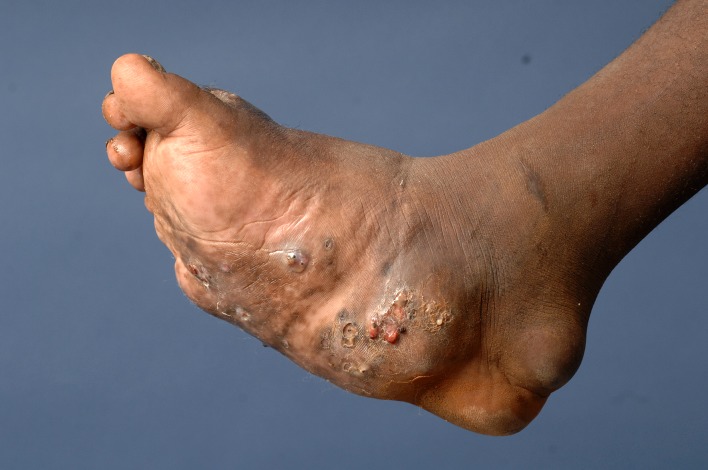
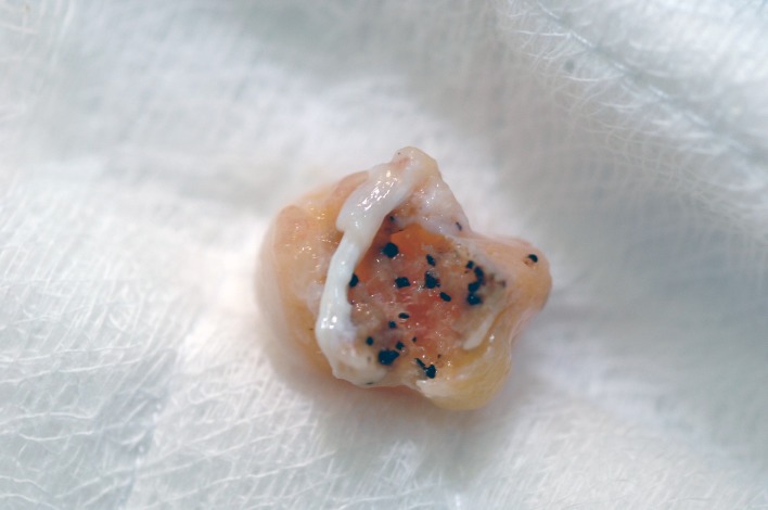
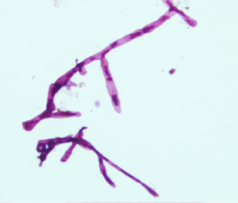
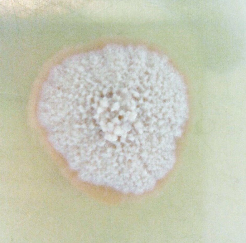
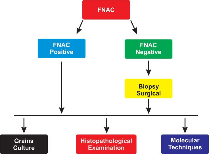
Mycetoma is a unique neglected tropical disease caused by a substantial number of microorganisms of fungal or bacterial origins. Identification of the causative organism and the disease extension are the first steps in the management of the affected patients and predicting disease treatment outcome and prognosis. Different laboratory-based diagnostic tools and techniques were developed over the years to determine and identify the causative agents. These include direct microscopy and cytological, histopathological, and immunohistochemical techniques in addition to the classical grain culture. More recently, various molecular-based techniques have joined the mycetoma diagnostic armamentarium. The available mycetoma diagnostic techniques are of various specificity and sensitivity rates. Most are invasive, time consuming, and operator dependent, and a combination of them is required to reach a diagnosis. In addition, they need a well-equipped laboratory and are therefore not field friendly. This review aims to provide an update on the laboratory investigations used in the diagnosis of mycetoma. It further aims to assist practising health professionals dealing with mycetoma by outlining the guidelines developed by the Mycetoma Research Centre, University of Khartoum, WHO collaborating centre on mycetoma following a cumulative experience of managing more than 7,700 mycetoma patients.
Conflict of interest statement
The authors have declared that no competing interests exist.
Figures
References
-
- Fahal A, Mahgoub ES, Hassan AME, Abdel-Rahman ME. (2015) Mycetoma in the Sudan: An Update from the Mycetoma Research Centre, University of Khartoum, Sudan. PLoS Negl Trop Dis 9(3): e0003679 doi: 10.1371/journal.pntd.0003679 - DOI - PMC - PubMed
-
- Bonifaz A, Tirado-Sánchez A, Calderón L, Saúl A, Araiza J, Hernández M, et al. (2014) Mycetoma: Experience of 482 Cases in a Single Center in Mexico. PLoS Negl Trop Dis 8(8): e3102 doi: 10.1371/journal.pntd.0003102 - DOI - PMC - PubMed
-
- Fahal A, Mahgoub ES, EL Hassan AM, Jacoub AO, Hassan D. (2015) Head and Neck Mycetoma: The Mycetoma Research Centre Experience. PLoS Negl Trop Dis 9(3): e0003587 doi: 10.1371/journal.pntd.0003587 - DOI - PMC - PubMed
-
- Fahal AH, Hassan MA. (1991) Mycetoma. Br J Surg. 79(11): 1138–1141. - PubMed
-
- Fahal AH. (2004) Mycetoma: a thorn in the flesh. Trans R Soc Trop Med Hyg. 98(1):3–11. - PubMed
Publication types
MeSH terms
Substances
LinkOut - more resources
Full Text Sources
Other Literature Sources
Medical