This site needs JavaScript to work properly. Please enable it to take advantage of the complete set of features!
Skip to main page content
U.S. flag

An official website of the United States government

Dot gov

The .gov means it’s official.
Federal government websites often end in .gov or .mil. Before sharing sensitive information, make sure you’re on a federal government site.

Https

The site is secure.
The https:// ensures that you are connecting to the official website and that any information you provide is encrypted and transmitted securely.

NIH NLM Logo
Log in
Access keys NCBI Homepage MyNCBI Homepage Main Content Main Navigation
. 2017 Aug 17;12(8):e0183198.
doi: 10.1371/journal.pone.0183198. eCollection 2017.

Cross-reactivity between apical membrane antgen 1 and rhoptry neck protein 2 in P. vivax and P. falciparum: A structural and binding study

Affiliations

Cross-reactivity between apical membrane antgen 1 and rhoptry neck protein 2 in P. vivax and P. falciparum: A structural and binding study

Brigitte Vulliez-Le Normand et al. PLoS One. .

Abstract

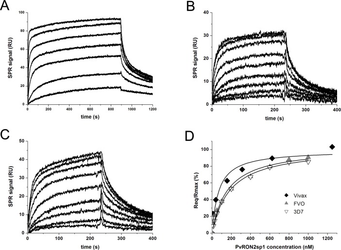
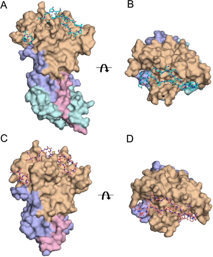
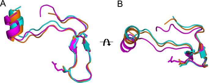
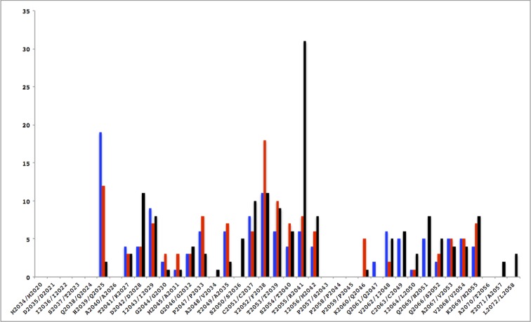
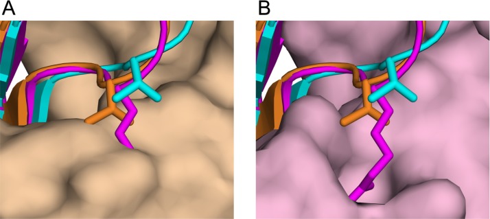
Malaria, a disease endemic in many tropical and subtropical regions, is caused by infection of the erythrocyte by the apicomplexan parasite Plasmodium. Host-cell invasion is a complex process but two Plasmodium proteins, Apical Membrane Antigen 1 (AMA1) and the Rhoptry Neck protein complex (RON), play a key role. AMA1, present on the surface of the parasite, binds tightly to the RON2 component of the RON protein complex, which is inserted into the erythrocyte membrane during invasion. Blocking the AMA1-RON2 interaction with antibodies or peptides inhibits invasion, underlining its importance in the Plasmodium life cycle and as a target for therapeutic strategies. We describe the crystal structure of the complex formed between AMA1 from P. vivax (PvAMA1) and a peptide derived from the externally exposed region of P. vivax RON2 (PvRON2sp1), and of the heterocomplex formed between P. falciparum AMA1 (PfAMA1) and PvRON2sp1. Binding studies show that the affinity of PvRON2sp1 for PvAMA1 is weaker than that previously reported for the PfRON2sp1-PfAMA1 association. Moreover, while PvRON2sp1 shows strong cross-reactivity with PfAMA1, PfRON2sp1 displays no detectable interaction with PvAMA1. The structures show that the equivalent residues PvRON2-Thr2055 and PfRON2-Arg2041 largely account for this pattern of reactivity.


Conflict of interest statement

Competing Interests: The authors have declared that no competing interests exist.

Figures

Fig 1
Fig 1. Binding measurements of PvRON2sp1 to PvAMA1 and PfAMA1 using surface plasmon resonance.
Sensograms for the binding of PvRON2sp1 to (A) PvAMA1, (B) PfAMA1 (FVO strain), and (C) PfAMA1 (3D7 strain). (D) Steady state binding curves showing the fraction of bound sites as a function of PvRON2sp1 concentration. The following peptide concentrations were injected: 10, 39, 156, 313, 625, 1250, 2500 nM for PvAMA1, and 12.5, 25, 50, 100, 200, 400, 600, 800 and 1000 nM for 3D7 and FVO.
Fig 2
Fig 2. Comparison between the homo-complex PvAMA1-PvRON2sp1 and the hetero-complex PfAMA1-PvRON2sp1.
Two orthogonal views of the complexes are shown: (A), (B) The PvAMA1-PvRON2sp1 complex is shown in surface representation with the N-terminal extension in pink (residues 43 to 62), Domain I in light brown (residues 63 to 248), Domain II in mauve (residues 249 to 385) and Domain III in cyan (residues 386 to 474); PvRON2sp1 is shown in stick representation. (C), (D) The PfAMA1-PvRON2sp1 complex is shown with the same relative orientations as in A and B, respectively. PfAMA1 is shown in surface representation with the N-terminal extension in pink (residues 102 to 123), Domain I in light brown (residues 124 to 303), Domain II in mauve (residues 304 to 440); PvRON2sp1 is shown in stick representation.
Fig 3
Fig 3. Comparison of the PvRON2sp1 peptides in the homo- and hetero-complexes, and PfRON2sp1 in the homo-complex PfAMA1-PfRON2sp1.
PvRON2sp1 is shown in cyan for the homo-complex and in orange for the hetero-complex; PfRON2sp1 from the PfAMA1-PfRON2sp1 homo-complex (PDB entry 3ZWZ) is shown in magenta. (A) The three ligands, shown in ribbon representation, were superimposed using the main-chain coordinates of the disulphide-bridged β-hairpin (Cys2051-Cys2063 in PvRON2sp1, Cys2037-Cys2049 in PfRON2sp1). The side chains of Arg2041 of PfRON2sp1, which makes critical contacts with PfAMA1 in the P. falciparum homo-complex, and of the equivalent residue in PvRON2sp1, Thr2055, are also shown. The superimposed ligands are oriented such that the N-terminal helix of PvRON2sp1 in the homo-complex (cyan) is vertical. (B) View of the superimposed ligands rotated by 90° about the horizontal axis.
Fig 4
Fig 4. Comparison of contacts between AMA1 and the RON2 peptide in the homo- and hetero-complexes.
The number of interatomic contacts < 3.8 Å (ordinate) is given as a function of the RON2 sequence (abscissa; PvRON2/PfRON2). In the histogram, the PvAMA1-PvRON2sp1 complex is shown in blue, the PfAMA1(FVO)-PvRON2sp1 complex is in red and the PfAMA1(3D7)-PfRON2sp1 complex [10] is in black.
Fig 5
Fig 5. The critical role of Arg2041 in specific binding to PfAMA1.
View of the disulphide-linked β-hairpin of the peptides in ribbon representation after superposition of PfAMA1-PvRON2sp1 and PfAMA1-PfRON2sp1 onto PvAMA1-PvRON2sp1 using the AMA1 coordinates only. PvRON2sp1 is shown in cyan for the homo-complex and in orange for the hetero-complex; PfRON2sp1 from the PfAMA1-PfRON2sp1 homo-complex is in magenta. The side chains of Arg2041 of PfRON2sp1 and the equivalent residue in PvRON2sp1, Thr2055, are also shown. (A) View of the superimposed peptides and PfAMA1 from the homo-complex in surface representation (pink). (B) The equivalent view with PvAMA1 from the hetero-complex in surface representation (light brown).

References

    1. Chitnis CE, Blackman MJ. Host cell invasion by malaria parasites. Parasitol Today. 2000;16:411–5. - PubMed
    1. Aikawa M, Miller LH, Johnson J, Rabbege J. Erythrocyte entry by malarial parasites. A moving junction between erythrocyte and parasite. J Cell Biol. 1978;77:72–82. - PMC - PubMed
    1. Cowman AF, Crabb BS. Invasion of red blood cells by malaria parasites. Cell; (2006)124:755–66. doi: 10.1016/j.cell.200602006 - DOI - PubMed
    1. Srinivasan P, Beatty WL, Diouf A, Herrera R, Ambroggio X, Moch JK, et al. Binding of Plasmodium merozoite proteins RON2 and AMA1 triggers commitment to invasion. Proc Natl Aca Sci USA. (2011);108:13275–80. - PMC - PubMed
    1. Tyler JS, Treeck M, Boothroyd JC. Focus on the ringleader: the role of AMA1 in apicomplexan invasion and replication. Trends Parasitol. 2011;27:410–20. doi: 10.1016/j.pt.201104002 - DOI - PMC - PubMed
Cite

AltStyle によって変換されたページ (->オリジナル) /