This site needs JavaScript to work properly. Please enable it to take advantage of the complete set of features!
Skip to main page content
U.S. flag

An official website of the United States government

Dot gov

The .gov means it’s official.
Federal government websites often end in .gov or .mil. Before sharing sensitive information, make sure you’re on a federal government site.

Https

The site is secure.
The https:// ensures that you are connecting to the official website and that any information you provide is encrypted and transmitted securely.

NIH NLM Logo
Log in
Access keys NCBI Homepage MyNCBI Homepage Main Content Main Navigation
. 2017 Jul 25:7:341.
doi: 10.3389/fcimb.2017.00341. eCollection 2017.

Toxoplasma Co-infection Prevents Th2 Differentiation and Leads to a Helminth-Specific Th1 Response

Affiliations

Toxoplasma Co-infection Prevents Th2 Differentiation and Leads to a Helminth-Specific Th1 Response

Norus Ahmed et al. Front Cell Infect Microbiol. .

Abstract

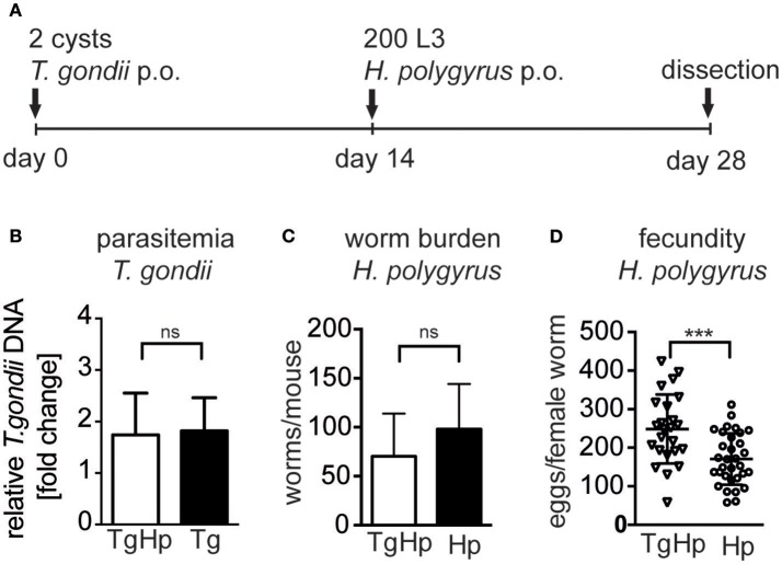
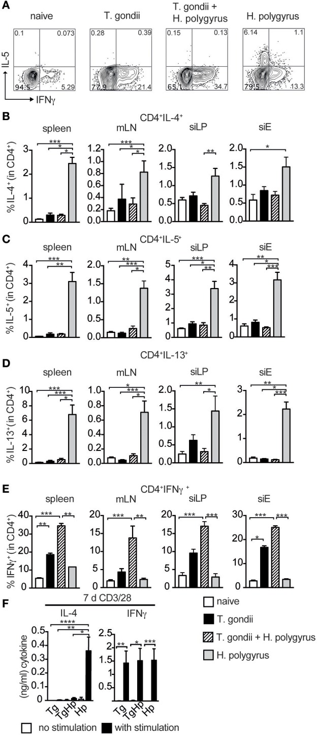
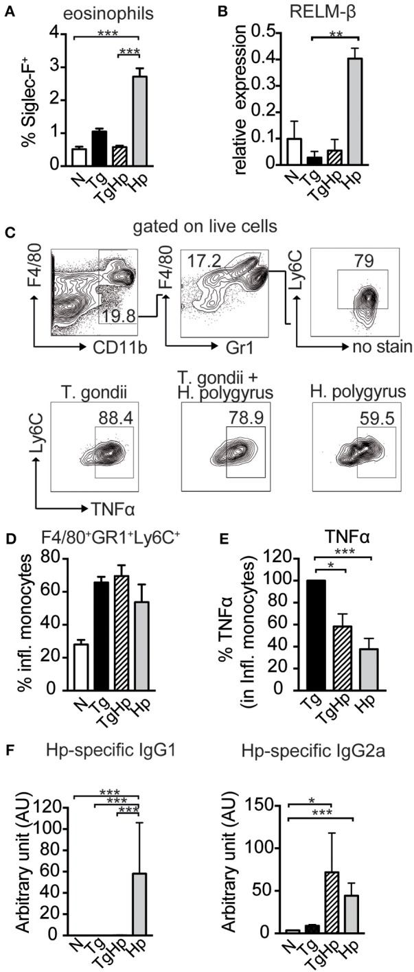
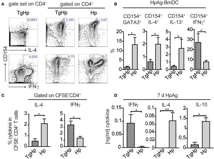
Nematode infections, in particular gastrointestinal nematodes, are widespread and co-infections with other parasites and pathogens are frequently encountered in humans and animals. To decipher the immunological effects of a widespread protozoan infection on the anti-helminth immune response we studied a co-infection with the enteric nematode Heligmosomoides polygyrus in mice previously infected with Toxoplasma gondii. Protective immune responses against nematodes are dependent on parasite-specific Th2 responses associated with IL-4, IL-5, IL-13, IgE, and IgG1 antibodies. In contrast, Toxoplasma gondii infection elicits a strong and protective Th1 immune response characterized by IFN-γ, IL-12, and IgG2a antibodies. Co-infected animals displayed significantly higher worm fecundity although worm burden remained unchanged. In line with this, the Th2 response to H. polygyrus in co-infected animals showed a profound reduction of IL-4, IL-5, IL-13, and GATA-3 expressing T cells. Co-infection also resulted in the lack of eosinophilia and reduced expression of the Th2 effector molecule RELM-β in intestinal tissue. In contrast, the Th1 response to the protozoan parasite was not diminished and parasitemia of T. gondii was unaffected by concurrent helminth infection. Importantly, H. polygyrus specific restimulation of splenocytes revealed H. polygyrus-reactive CD4+ T cells that produce a significant amount of IFN-γ in co-infected animals. This was not observed in animals infected with the nematode alone. Increased levels of H. polygyrus-specific IgG2a antibodies in co-infected mice mirrored this finding. This study suggests that polarization rather than priming of naive CD4+ T cells is disturbed in mice previously infected with T. gondii. In conclusion, a previous T. gondii infection limits a helminth-specific Th2 immune response while promoting a shift toward a Th1-type immune response.

Keywords: Heligmosomoides polygyrus; Th1; Th2; Toxoplasma gondii; co-infection; helminth.


Figures

Figure 1
Figure 1
Experimental design (A), C57BL/6 mice were infected with 2 ME49 T. gondii tissue cysts and/or 200 H. polygyrus L3 larvae. (B) Parasitemia of T. gondii in the heart, pooled from two independent experiments, fold change compared to uninfected, data shown as mean ± SEM, n = 6–7 (C) H. polygyrus worm burden. (D) Fecundity of female worms. Panels (C,D) are representative of three independent experiments with n = 4. Data shown as mean ± SD; statistical analysis was performed using the Mann-Whitney test, ***P ≤ 0.001.
Figure 2
Figure 2
Restricted Th2 responses in co-infected mice. Cells from spleen, mesenteric lymph nodes (mLN), small intestinal lamina propria (siLP) and small intestinal epithelium (siE) were isolated and stimulated with PMA and ionomycin in the presence of Brefeldin A followed by intranuclear staining for the lineage transcription factors GATA3 and T-bet. Gating strategy shown in (A), Bar graphs showing frequencies of CD4+ T cells expressing GATA3 (B), T-bet (C), and T-bet expression in CD8+ T cells (D). GATA3 expression in the duodeunum of the small intestine with scale bar of 100 μm (E), data shown as mean ± SEM pooled from 2 independent experiments n = 6, statistical analysis was performed using the Mann-Whitney test. (B–D) shown as mean ± SEM, pooled from two independent experiments with n = 8–10. Statistical analysis was performed using the Kruskal-Wallis with Dunn's multiple comparison test, *P ≤ 0.05, **P ≤ 0.01, and ***P ≤ 0.001.
Figure 3
Figure 3
Th2 but not Th1 immune responses are absent in co-infected mice. Cells from spleen, mLN, siLP, and siE of single and co-infected animals were isolated and stimulated with PMA and ionomycin in the presence of Brefeldin A followed by intracellular cytokine staining. Gating strategy for the cytokines IL-5 and IFN-γ in CD4+ cells isolated from spleen (A). Bar graphs showing frequencies of CD4+ T cells expressing IL-4 (B), IL-5 (C), IL-13 (D), and IFN-γ (E) in spleen, mLN, siLP, and siE. (F) IL-4 and IFN-γ production detected by ELISA in supernatant from 3 ×ばつ 105 splenocytes stimulated with (black bars) and without (white bars) anti-CD3/CD28 antibodies. (B–F) Data shown as mean ± SEM, pooled from two independent experiments with n = 8–9 Statistical analysis was performed using the Kruskal-Wallis with Dunn's multiple comparison test, *P ≤ 0.05, **P ≤ 0.01, and ***P ≤ 0.001.
Figure 4
Figure 4
Impact of co-infection on Th1- and Th2-dependent effector cells, effector molecules and antibody isotypes. (A) Bar graph showing the frequency of Siglec-F+ eosinophils in the spleen, pooled from 2 independent experiments n = 7–8. (B) Relative Gene Expression of RELM-β compared to housekeeping gene β-actin (pooled from two experiments, n = 5–8). (C,D) Gating strategy and bar graphs showing frequency of inflammatory monocytes (F4/80+GR1+Ly6C+) in spleen stimulated with LPS (pooled from two experiments, n = 8). (E) Percentage of TNFα, with the response of the T. gondii infected group represented as 100%, stimulated with LPS, pooled from two experiments, n = 9. (F) H. polygyrus specific IgG1 and IgG2a detected by ELISA, n = 7–8 pooled from two independent experiments. Data shown as mean ± SEM. Statistical analysis was performed using the Kruskal-Wallis with Dunn's multiple comparison test, *P ≤ 0.05, **P ≤ 0.01, and ***P ≤ 0.001.
Figure 5
Figure 5
Helminth-reactive CD4+ T cells from co-infected mice show enhanced production of IFN-γ. BmDC (1 ×ばつ 105) were pulsed with 50 μg/ml H. polygyrus antigen (HpAg) and cultured with 1 ×ばつ 106 splenocytes from single or co-infected animals for 5 h followed by intracellular staining for CD154 and cytokines. Gating strategy is shown in (A) whereby the gate for cells coexpressing CD154 and IL-4, IL-13, or IFN-γ was set using CD4 cells. (B) Bar graphs showing frequencies of CD154+GATA3+, CD154+IL-4+, CD154+IL-13+, and CD154+IFN-γ+ in CD4+ T cells. Data shown as mean ± SD and representative of two independent experiments with n = 4–5. (C) CFSE labeled CD4+ spleen cells were stimulated for 6 days with 20 μg/ml HpAg. Proliferating spleen cells were gated on (CFSE-CD4+). CD4+ cells were re-stimulated with PMA/Ionomycin and stained for intracellular cytokines IL-4, and IFN-γ; data shown as mean ± SD and is representative for two independent experiments n = 4. (D) Bar graphs show detection of IFN-γ, IL-4 and IL-10 by ELISA from collected supernatant of 3 ×ばつ 105 splenocytes stimulated with H. polygyrus antigen for 6 days; representative of two independent experiments n = 4–5, mean ± SD. Statistical analysis was performed using the Mann-Whitney test, *P ≤ 0.05, **P ≤ 0.01.

References

    1. Artis D., Wang M. L., Keilbaugh S. A., He W., Brenes M., Swain G. P., et al. . (2004). RELMbeta/FIZZ2 is a goblet cell-specific immune-effector molecule in the gastrointestinal tract. Proc. Natl. Acad. Sci. U.S.A. 101, 13596–13600. 10.1073/pnas.0404034101 - DOI - PMC - PubMed
    1. Bahia-Oliveira L. M., da Silva J. A., Peixoto-Rangel A. L., Boechat M. S. B., Oliveira A. M. W. A., Massara C. L., et al. . (2009). Host immune response to Toxoplasma gondii and Ascaris lumbricoides in a highly endemic area: evidence of parasite co-immunomodulation properties influencing the outcome of both infections. Mem. Inst. Oswaldo Cruz 104, 273–280. 10.1590/S0074-02762009000200021 - DOI - PubMed
    1. Bansemir A. D., Sukhdeo M. V. (1994). The food resource of adult Heligmosomoides polygyrus in the small intestine. J. Parasitol. 80, 24–28. 10.2307/3283340 - DOI - PubMed
    1. Besnard A.-G., Togbe D., Guillou N., Erard F., Quesniaux V., Ryffel B. (2011). IL-33-activated dendritic cells are critical for allergic airway inflammation. Eur. J. Immunol. 41, 1675–1686. 10.1002/eji.201041033 - DOI - PubMed
    1. Biswas A., Bruder D., Wolf S. A., Jeron A., Mack M., Heimesaat M. M., et al. . (2015). LY6C(high) monocytes control cerebral toxoplasmosis. J. Immunol. 194, 3223–3235. 10.4049/jimmunol.1402037 - DOI - PubMed

MeSH terms

Cite

AltStyle によって変換されたページ (->オリジナル) /