This site needs JavaScript to work properly. Please enable it to take advantage of the complete set of features!
Skip to main page content
U.S. flag

An official website of the United States government

Dot gov

The .gov means it’s official.
Federal government websites often end in .gov or .mil. Before sharing sensitive information, make sure you’re on a federal government site.

Https

The site is secure.
The https:// ensures that you are connecting to the official website and that any information you provide is encrypted and transmitted securely.

NIH NLM Logo
Log in
Access keys NCBI Homepage MyNCBI Homepage Main Content Main Navigation
. 2017 Sep 6;45(15):9068-9084.
doi: 10.1093/nar/gkx519.

Polypyrimidine tract-binding protein (PTB) and PTB-associated splicing factor in CVB3 infection: an ITAF for an ITAF

Affiliations

Polypyrimidine tract-binding protein (PTB) and PTB-associated splicing factor in CVB3 infection: an ITAF for an ITAF

Pratik Dave et al. Nucleic Acids Res. .

Abstract

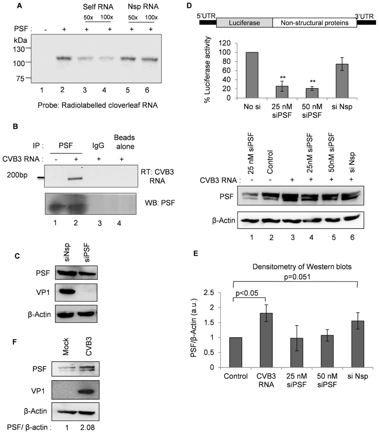
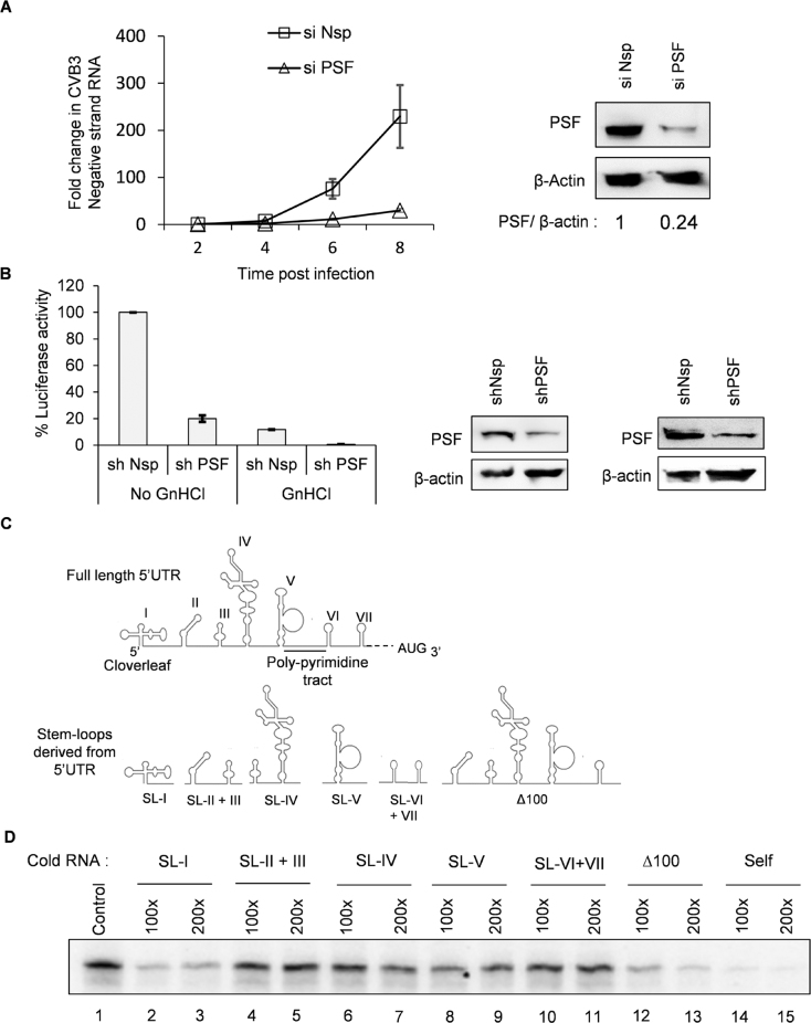
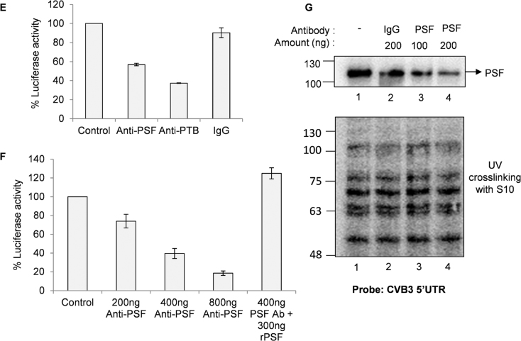
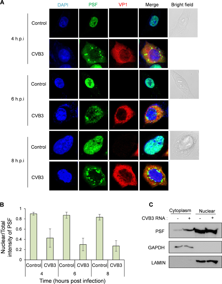
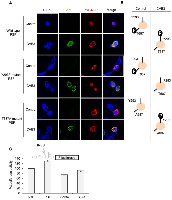
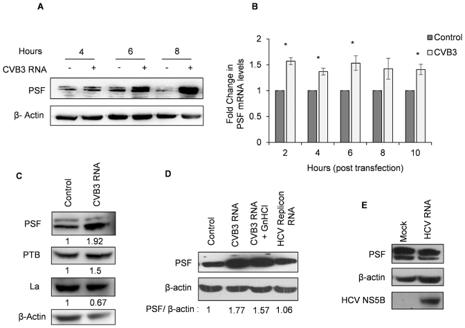
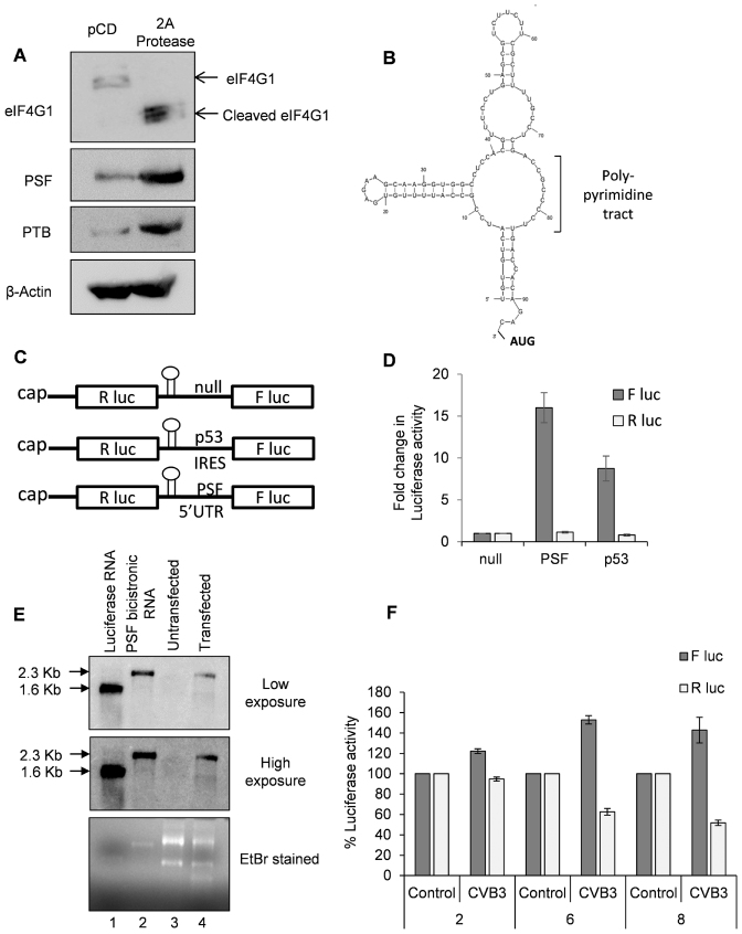
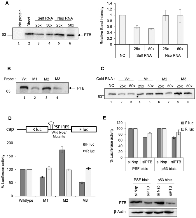
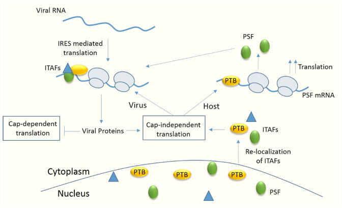
The 5' UTR of Coxsackievirus B3 (CVB3) contains internal ribosome entry site (IRES), which allows cap-independent translation of the viral RNA and a 5'-terminal cloverleaf structure that regulates viral replication, translation and stability. Here, we demonstrate that host protein PSF (PTB associated splicing factor) interacts with the cloverleaf RNA as well as the IRES element. PSF was found to be an important IRES trans acting factor (ITAF) for efficient translation of CVB3 RNA. Interestingly, cytoplasmic abundance of PSF protein increased during CVB3 infection and this is regulated by phosphorylation status at two different amino acid positions. Further, PSF protein was up-regulated in CVB3 infection. The expression of CVB3-2A protease alone could also induce increased PSF protein levels. Furthermore, we observed the presence of an IRES element in the 5'UTR of PSF mRNA, which is activated during CVB3 infection and might contribute to the elevated levels of PSF. It appears that PSF IRES is also positively regulated by PTB, which is known to regulate CVB3 IRES. Taken together, the results suggest for the first time a novel mechanism of regulations of ITAFs during viral infection, where an ITAF undergoes IRES mediated translation, sustaining its protein levels under condition of translation shut-off.


Figures

Figure 1.
Figure 1.
PSF interacts with cloverleaf RNA and influences CVB3 lifecycle. (A) UV cross-linking assay using recombinant PSF and radio-labelled cloverleaf RNA. Lane 1 corresponds to no protein and only RNA probe and lane 2 shows interaction between RNA and recombinant PSF protein. For competition, 50- and 100-fold molar excess of unlabelled cloverleaf RNA (lanes 3 and 4) or non-specific RNA (lanes 5 and 6) was also included in the reaction. Non-specific RNA of equivalent length was derived from pGEM-T vector MCS region. (B) CVB3 replicon RNA was transfected in HeLa S3 cells, followed by immuno-precipitation of PSF using anti-PSF antibody or IgG isotype antibody (as a control). RNA was purified from the immuno-precipitated complexes and viral positive strand RNA was detected by semi-quantitative PCR. (C) HeLa S3 cells were either transfected with siPSF or siNsp, and 24 h later, CVB3 infection was carried out. Cells were harvested at 8 h post-infection and lysates were subjected to immunoblot analysis. (D) Schematic of CVB3 sub-genomic replicon RNA adapted from Lanke et al. (25).Hela S3 cells were either transfected with siPSF or siNsp, and 24 h later CVB3 sub-genomic replicon RNA was transfected. Cells were harvested at 10 h post-transfection and firefly luciferase activity was measured. Error bars represent standard deviation in three independent experiments. **P value < 0.01. PSF protein levels as determined by western blotting are indicated compared to β-actin protein levels. (E) Densitometry of three independent western blots from (D) are represented in graphical format. Error bars represent standard deviation in three independent experiments. (F) HeLa S3 cells were infected with CVB3 and cells were harvested at 8 h post-infection. Western blot indicating the PSF protein level in presence and absence of CVB3 infection. PSF levels are quantified by densitometric analysis of band intensities and are represented by fold change in PSF/β-actin.
Figure 2.
Figure 2.
Role of PSF in CVB3 replication and translation. (A) HeLa S3 cells, priorly transfected with siNsp or siPSF, were infected with CVB3 and cells were harvested in trizol at indicated time points post-infection. At each time point, RNA was isolated and negative strand RNA was detected by qPCR. Fold change in negative strand RNA level was calculated in both control cells and PSF knockdown cells and is indicated in the graph. Error bars indicate standard deviation in three independent experiments. (B) Effect of partial knockdown of PSF on CVB3 translation was studied in presence and absence of viral replication inhibitor GnHCl (concentration = 2 mM), using CVB3 sub-genomic replicon RNA as described earlier. Luciferase activity was found to be significantly reduced even in presence of GnHCl. ** indicates P < 0.005. Error bars indicate the standard deviation in three independent experiments. PSF and β-actin protein levels are indicated in the immunoblots. (C) Schematic diagram of CVB3 5′UTR. Different stem-loops derived from the full length 5′UTR for competition UV induced cross-linking assay are indicated. Cloverleaf RNA is indicated by SL-I. (D) UV-crosslinking assay with 32-P labelled full length 5′UTR of CVB3 genomic RNA alone or in presence of indicated un-labelled stem-loop RNAs. Δ1–100 indicates the entire CVB3 5′UTR without the cloverleaf RNA or SL-1. (E) in vitro translation of CVB3 replicon RNA carried out using rabbit reticulocyte system supplemented with S10 extract from HeLa S3 cells and resultant luciferase activities are indicated. Anti-PSF and anti-PTB antibodies were added in the reaction to sequester these proteins. Anti-Rabbit IgG antibody was used as control. Error bars indicate standard deviation in three independent experiments. (F) In vitro translation carried out using rabbit reticulocyte system supplemented with S10 extract from HeLa S3 cells in presence of indicated amounts of anti-PSF antibody. Indicated amount of bacterially expressed recombinant PSF (rPSF) was supplemented in one of the reactions to rescue the inhibitory effects of PSF antibody. Error bars indicate standard deviation in three independent experiments. (G) UV cross-linking assay carried out using radio-labelled CVB3 5′UTR with either recombinant PSF protein or S10 extract from HeLa S3 cells (lane 1). The assay was carried out in presence of PSF antibody (lanes 3 and 4) or IgG antibody (lane 2) as indicated.
Figure 2.
Figure 2.
Role of PSF in CVB3 replication and translation. (A) HeLa S3 cells, priorly transfected with siNsp or siPSF, were infected with CVB3 and cells were harvested in trizol at indicated time points post-infection. At each time point, RNA was isolated and negative strand RNA was detected by qPCR. Fold change in negative strand RNA level was calculated in both control cells and PSF knockdown cells and is indicated in the graph. Error bars indicate standard deviation in three independent experiments. (B) Effect of partial knockdown of PSF on CVB3 translation was studied in presence and absence of viral replication inhibitor GnHCl (concentration = 2 mM), using CVB3 sub-genomic replicon RNA as described earlier. Luciferase activity was found to be significantly reduced even in presence of GnHCl. ** indicates P < 0.005. Error bars indicate the standard deviation in three independent experiments. PSF and β-actin protein levels are indicated in the immunoblots. (C) Schematic diagram of CVB3 5′UTR. Different stem-loops derived from the full length 5′UTR for competition UV induced cross-linking assay are indicated. Cloverleaf RNA is indicated by SL-I. (D) UV-crosslinking assay with 32-P labelled full length 5′UTR of CVB3 genomic RNA alone or in presence of indicated un-labelled stem-loop RNAs. Δ1–100 indicates the entire CVB3 5′UTR without the cloverleaf RNA or SL-1. (E) in vitro translation of CVB3 replicon RNA carried out using rabbit reticulocyte system supplemented with S10 extract from HeLa S3 cells and resultant luciferase activities are indicated. Anti-PSF and anti-PTB antibodies were added in the reaction to sequester these proteins. Anti-Rabbit IgG antibody was used as control. Error bars indicate standard deviation in three independent experiments. (F) In vitro translation carried out using rabbit reticulocyte system supplemented with S10 extract from HeLa S3 cells in presence of indicated amounts of anti-PSF antibody. Indicated amount of bacterially expressed recombinant PSF (rPSF) was supplemented in one of the reactions to rescue the inhibitory effects of PSF antibody. Error bars indicate standard deviation in three independent experiments. (G) UV cross-linking assay carried out using radio-labelled CVB3 5′UTR with either recombinant PSF protein or S10 extract from HeLa S3 cells (lane 1). The assay was carried out in presence of PSF antibody (lanes 3 and 4) or IgG antibody (lane 2) as indicated.
Figure 3.
Figure 3.. Cytoplasmic abundance of PSF increases in presence of CVB3. (A) Immunofluorescence microscopy of HeLa S3 cells infected with CVB3 or mock infection at different time points post-infection. Green represents PSF, Red represents viral protein VP1 indicating infected cells, blue represents nucleus of the cell. (B) Quantification of the confocal images. Mean intensities of PSF were calculated in total cell and in nucleus using ImageJ software. Ratio of nuclear intensity by total cell intensity is indicated in graph. For 4 h.p.i., 6 h.p.i. and 8 h.p.i. 20, 25 and 40 cells were quantified from three independent experiments. Error bars represent standard deviation in three independent experiments. (C) Western blot analysis of nuclear and cytoplasmic extracts of HeLa S3 cells with and without CVB3 infection. Cells were harvested at 8 hours post infection processed for fractionation. Lamin and GAPDH were used as nuclear and cytoplasmic markers respectively.
Figure 4.
Figure 4.
Regulation of PSF localization by phosphorylation. (A) Plasmids harbouring PSF gene fused with RFP were used for immunofluorescence assay. Mutations were incorporated individually in the same plasmid to generate Y293F and T687A phosphodead mutants. HeLa cells were transfected with plasmids expressing wild type PSF fused with RFP or the phosphodead mutants (as described before) and 24 h later cells were infected with CVB3. Eight hours post-infection; cells were processed for immunofluorescence microscopy. (B) A schematic summary of the information obtained from (A). Phosphorylation status of wild type PSF and phosphodead mutants in control cells and upon CVB3 infection is indicated. (C) Luciferase assay carried out using CVB3 monocistronic construct as represented by the schematic. HeLa cells were transfected with plasmids expressing either wild type PSF or Y293F or T687A phosphodead mutants. Twenty four hours post-transfection, cells were transfected with CVB3 monocistronic RNAs and 8 hours post transfection cells were processed for luciferase assay. Graphs indicates the % luciferase activity as compared to pCD vector control. Error bars indicates the standard deviation in three independent experiments.
Figure 5.
Figure 5.. PSF is specifically up-regulated in CVB3 infection. (A) PSF levels at different time points post CVB3 infection in HeLa S3 cells. (B) PSF mRNA levels at indicated time points post CVB3 RNA transfection as determined by qPCR. At each time point, mRNA level in un-transfected cells were used as control. * indicates P < 0.05. (C) Western blots of HeLa S3 cells transfected with CVB3 replicon RNA indicating levels of PSF, PTB and La. Cells were harvested at 10 h post-transfection. (D) Western blots of HeLa S3 cells transfected with CVB3 replicon RNA in presence or absence of 2 mM GnHCL and HCV replicon RNA. Cells were harvested at 10 h post-transfection. Densitometry of the represented western blot is indicated as PSF/β-actin below the lanes. (E) Western blot to check the levels of PSF upon HCV infection. Huh 7.5 cells were infected with HCV and cells were harvested at 48 h post-infection. NS5B blot is to indicate viral infection.
Figure 6.
Figure 6.
PSF mRNA undergoes IRES mediated translation. (A) Western blots indicating the levels of PSF and PTB upon expression of 2A protease. Both PSF and PTB appear to be up-regulated upon 2A protease expression. 2A protease activity is represented by the cleavage of eIF4G1. (B) Structure of PSF 5′UTR as predicted by mfold server. The presence of hairpin loop followed by poly-pyrimidine tract suggests a potential PTB dependent IRES (33). (C) Schematic of different bicistronic construct used in this study. (D) Capped RNAs were prepared from the constructs described in Figure 5C. These capped RNAs were transfected in cells and cells were harvested 8 h post-transfection, followed by dual luciferase assay. Fold change in luciferase activities from PSF 5′UTR and p53 5′UTR (positive control) compared to La ORF as negative control (null) is represented as firefly luciferase activity while renilla luciferase activity represents the cap-dependent translation. (E) Integrity of capped PSF bicistronic RNA in the cell as assessed by northern blotting. Cells were transfected with in vitro transcribed capped PSF bicistronic RNA and 8 h post-transfection cells were harvested in trizol and RNA was isolated. 2 μg of total RNA from untransfected and transfected cells was used for formaldehyde agarose gel electrophoresis (lanes 3 and 4) along with luciferase RNA (lane 1) and in vitro transcribed PSF bicistronic RNA (lane 2). RNAs were transferred to nitrocellulose membrane by capillary transfer method and the membrane was probed for luciferase gene using complementary radio-labelled primer. Two different exposures after northern blotting are indicated, along with the ethidium bromide stained agarose gel. In both the exposures, PSF bicistronic RNA was found to be intact in the cell with negligible degradation. (F) PSF IRES activity upon CVB3 infection was measured. HeLa S3 cells previously transfected with PSF bicistronic construct were infected with CVB3 or mock. Cells were harvested at 2, 6 and 8 h.p.i. and luciferase activity was measured. Error bars indicate the standard deviation in three independent experiments.
Figure 7.
Figure 7.
PTB interacts with PSF IRES and positively influences its IRES activity. (A) UV-crosslinking assay carried out with recombinant PTB with radio-labelled PSF IRES in presence of un-labelled PSF IRES or non-specific RNA. Graph represents the average intensities of bands visible in the autoradiograph of UV-crosslinking gel from 3 independent experiments. (B) UV-crosslinking assay carried out using recombinant PTB with wild type and mutant radio-labelled PSF IRESs. M1, M2 and M3 mutants were generated as represented in Supplementary Figure S6A. (C) Competition UV crosslinking assay carried out using radio-labelled wild type PSF 5′UTR with recombinant PTB, in presence of un-labelled wild type PSF IRES or mutant PSF IRESs as indicated. (D) Ex vivo IRES activity of wild type and mutant PSF IRESs. Capped RNAs were prepared from bicistronic constructs harbouring mutations in PSF IRES and wild type PSF IRES and were transfected in the cells. Cells were harvested at 8 h post-transfection and firefly luciferase activity indicating the IRES activity of wild type and mutant RNAs and renilla luciferase activity indicating cap-dependent translation are indicated. M1 and M3 mutants showed lower IRES activity as compared to wild type. * indicates P value < 0.05. Error bars indicate standard deviation from three independent experiments. (E) Effect of knockdown of PTB on PSF IRES activity. p53 IRES activity is used as positive control. Cells previously transfected with si-PTB or si-NSP were transfected with the PSF IRES. Luciferase assay was carried out after harvesting the cells in PLB buffer at 8 hours post transfection. * indicates P value < 0.05.
Figure 8.
Figure 8.
Model showing the regulation of host cell translation under CVB3 infection. Upon entry of CVB3 RNA in cells, host factors known as ITAFs relocalize from nucleus to cytoplasm and these proteins stimulate viral IRES mediated translation. Viral proteins inhibit cap-dependent translation and favour cap-independent translation. ITAFs like PSF also have an IRES in their mRNA and are stimulated by PTB, also an ITAF for viral RNA. Thus, level of PSF in cell is maintained even under condition where cap-dependent translation is shut off and this is beneficial for viral IRES mediated translation.

References

    1. Jackson R.J., Hellen C.U.T., Pestova T.V.. The mechanism of eukaryotic translation initiation and principles of its regulation. Nat. Rev. Mol. Cell. Biol. 2010; 11:113–127. - PMC - PubMed
    1. Andino R., Rieckhof G.E., Baltimore D.. A functional ribonucleoprotein complex forms around the 5′ end of poliovirus RNA. Cell. 1990; 63:369–380. - PubMed
    1. Ray P.S., Das S.. La autoantigen is required for the internal ribosome entry site-mediated translation of Coxsackievirus B3 RNA. Nucleic Acids Res. 2002; 30:4500–4508. - PMC - PubMed
    1. Bedard K.M., Daijogo S., Semler B.L.. A nucleo-cytoplasmic SR protein functions in viral IRES-mediated translation initiation. EMBO J. 2007; 26:459–467. - PMC - PubMed
    1. Verma B., Bhattacharyya S., Das S.. Polypyrimidine tract-binding protein interacts with coxsackievirus B3 RNA and influences its translation. J. Gen. Virol. 2010; 91:1245–1255. - PubMed

MeSH terms

Cite

AltStyle によって変換されたページ (->オリジナル) /