This site needs JavaScript to work properly. Please enable it to take advantage of the complete set of features!
Skip to main page content
U.S. flag

An official website of the United States government

Dot gov

The .gov means it’s official.
Federal government websites often end in .gov or .mil. Before sharing sensitive information, make sure you’re on a federal government site.

Https

The site is secure.
The https:// ensures that you are connecting to the official website and that any information you provide is encrypted and transmitted securely.

NIH NLM Logo
Log in
Access keys NCBI Homepage MyNCBI Homepage Main Content Main Navigation
. 2017 Jun 1;11(6):e0005654.
doi: 10.1371/journal.pntd.0005654. eCollection 2017 Jun.

Mosquito co-infection with Zika and chikungunya virus allows simultaneous transmission without affecting vector competence of Aedes aegypti

Affiliations

Mosquito co-infection with Zika and chikungunya virus allows simultaneous transmission without affecting vector competence of Aedes aegypti

Giel P Göertz et al. PLoS Negl Trop Dis. .

Abstract

Background: Zika virus (ZIKV) and chikungunya virus (CHIKV) are highly pathogenic arthropod-borne viruses that are currently a serious health burden in the Americas, and elsewhere in the world. ZIKV and CHIKV co-circulate in the same geographical regions and are mainly transmitted by Aedes aegypti mosquitoes. There is a growing number of case reports of ZIKV and CHIKV co-infections in humans, but it is uncertain whether co-infection occurs via single or multiple mosquito bites. Here we investigate the potential of Ae. aegypti mosquitoes to transmit both ZIKV and CHIKV in one bite, and we assess the consequences of co-infection on vector competence.

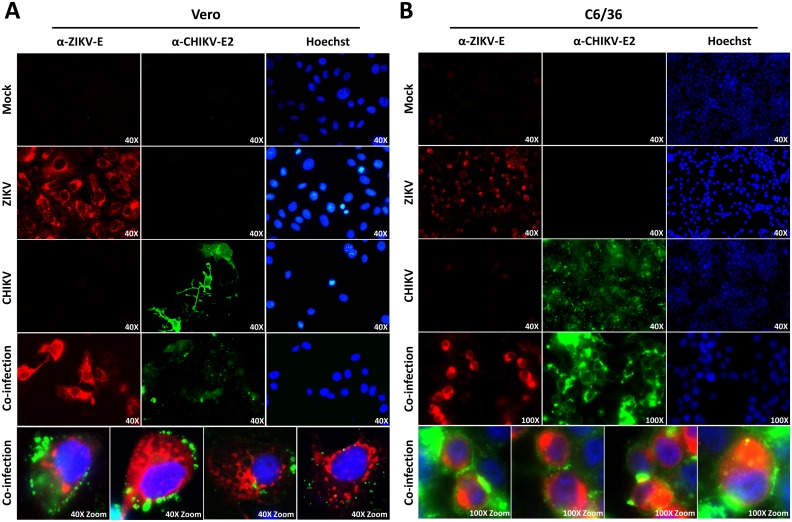
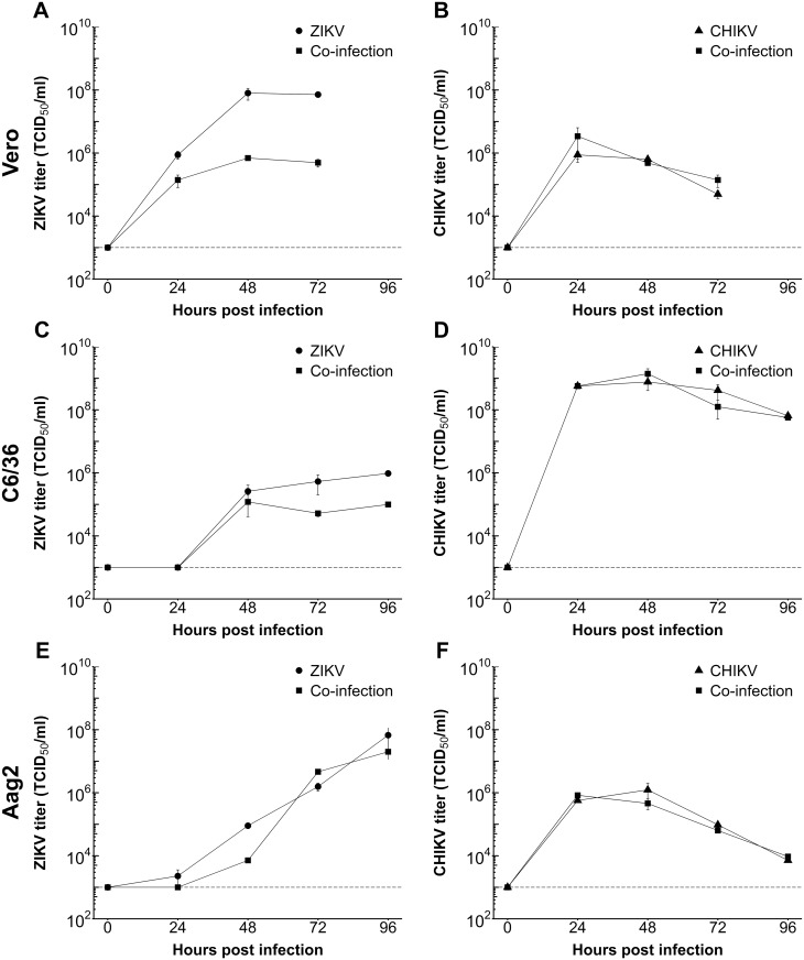
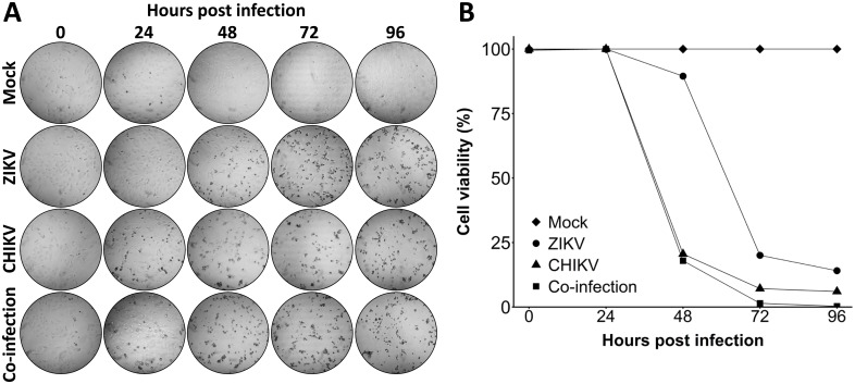
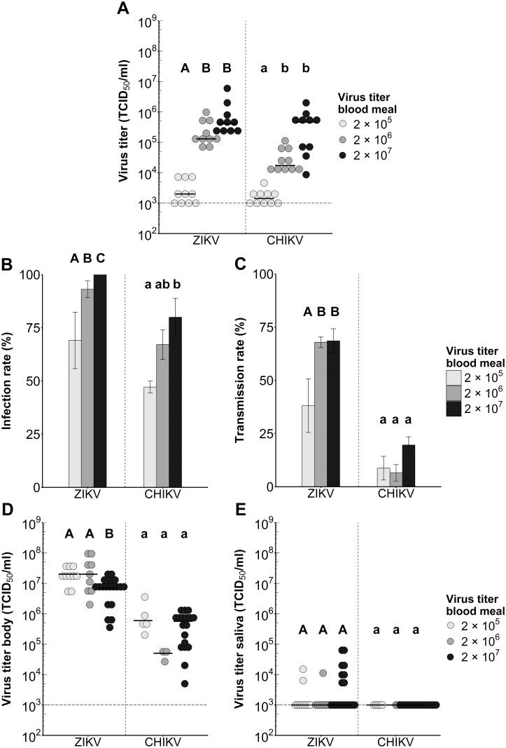
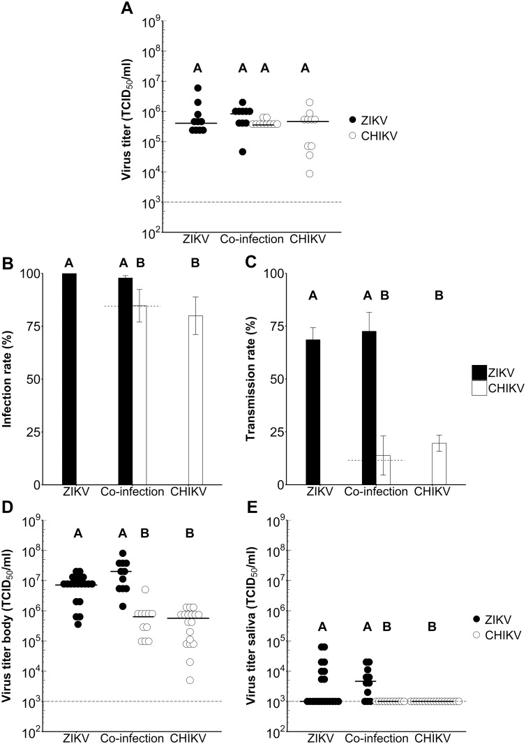
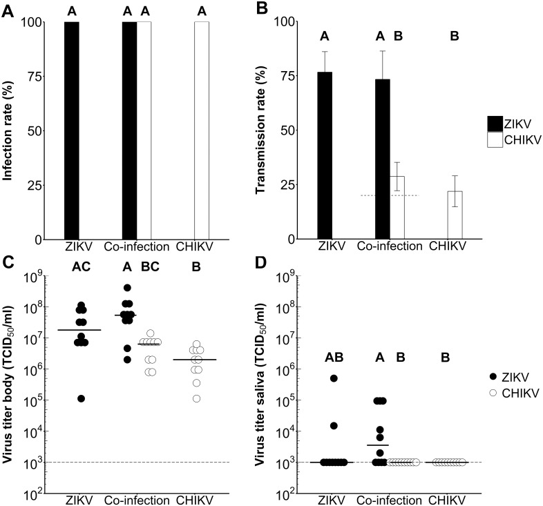
Methodology/principal findings: First, growth curves indicated that co-infection with CHIKV negatively affects ZIKV production in mammalian, but not in mosquito cells. Next, Ae. aegypti mosquitoes were infected with ZIKV, CHIKV, or co-infected via an infectious blood meal or intrathoracic injections. Infection and transmission rates, as well as viral titers of positive mosquitoes, were determined at 14 days after blood meal or 7 days after injection. Saliva and bodies of (co-)infected mosquitoes were scored concurrently for the presence of ZIKV and/or CHIKV using a dual-colour immunofluorescence assay. The results show that orally exposed Ae. aegypti mosquitoes are highly competent, with transmission rates of up to 73% for ZIKV, 21% for CHIKV, and 12% of mosquitoes transmitting both viruses in one bite. However, simultaneous oral exposure to both viruses did not change infection and transmission rates compared to exposure to a single virus. Intrathoracic injections indicate that the selected strain of Ae. aegypti has a strong salivary gland barrier for CHIKV, but a less profound barrier for ZIKV.

Conclusions/significance: This study shows that Ae. aegypti can transmit both ZIKV and CHIKV via a single bite. Furthermore, co-infection of ZIKV and CHIKV does not influence the vector competence of Ae. aegypti.


Conflict of interest statement

The authors have declared that no competing interests exist.

Figures

Fig 1
Fig 1. Immunofluorescence of ZIKV and CHIKV single- and co-infections in mammalian and mosquito cells.
(A) Vero and (B) C6/36 cells were infected with ZIKVSUR, CHIKV37997 or both. At 48 hpi the monolayers were fixed, permeabilized, stained with antibodies for ZIKV-E (pan-Flavivirus α-E (4G2)), CHIKV-E2 and Hoechst33258 (details in Materials and methods) and visualized by immunofluorescence. Magnifications are indicated in each picture. Bottom panels indicate zoomed and merged images of co-infected cells.
Fig 2
Fig 2. Growth curves of ZIKV and CHIKV during single- and co-infections.
(A-B) Vero, (C-D) C6/36 and (E-F) Aag2 cells were infected with ZIKVSUR, CHIKV37997 or co-infected at an MOI of 0.1. The virus titers during single- and co-infection were determined by EPDA at the indicated time points for (A,C,E) ZIKV and (B,D,F) CHIKV. Shown are the mean virus titers ± SEM from duplicate samples. The detection limit of the EPDA is indicated by a dashed line.
Fig 3
Fig 3. Cell viability of Vero cells during ZIKV, CHIKV single- or co-infections.
(A-B) Vero cells were infected with ZIKVSUR, CHIKV37997, or co-infected at an MOI of 0.1. At 0, 24, 48, 72 and 96 hpi cells were visualized by (A) bright field microscopy and (B) lysed before measuring the cell viability by CellTiter-Glo assay. Cell viability is presented as percentage normalized to the mock.
Fig 4
Fig 4. Infection and transmission rates of Ae. aegypti orally infected with different doses of ZIKV or CHIKV.
Mosquitoes were inoculated with an infectious blood meal containing a dose of 2.0 ×ばつ 105, 2.0 ×ばつ 106, or 2.0 ×ばつ 107 TCID50/ml ZIKVSUR or CHIKV37997. (A) Ingested virus titers of Ae. aegypti immediately after blood feeding. Each dot represents one mosquito body, and horizontal bars indicate median titers. The detection limit of the EPDA is indicated by the dashed line. Results were evaluated with a Kruskal-Wallis test. Significant differences between viral doses (P < 0.05) are indicated by different letters. (B) Infection and (C) transmission rates of Ae. aegypti mosquitoes at 14 dpi presented as the percentage of the total number of engorged mosquitoes. Shown are the mean percentages from three independent replicates. Error bars show the standard error of the mean. Sample size ranged between 48–101 female mosquitoes per treatment. Results were evaluated with Chi-squared test. Significant differences between viral doses (P < 0.05) are indicated by different letters. (D-E) Virus titers of CHIKV and ZIKV mosquito (D) bodies and (E) saliva. Each dot represents one mosquito body or saliva sample, and the horizontal bars indicate median titers. The detection limit of the EPDA is indicated by a dashed line. Results were evaluated with a Kruskal-Wallis test. Significant differences between viral doses (P < 0.05) are indicated by different letters.
Fig 5
Fig 5. Infection and transmission rates of Ae. aegypti mosquitoes orally inoculated with ZIKV, CHIKV, or both viruses.
Mosquitoes were orally infected with an infectious blood meal containing a dose of 2.0 ×ばつ 107 TCID50/ml ZIKVSUR, CHIKV37997, or both. (A) Ingested virus titers of Ae. aegypti immediately after blood feeding. Each dot represents one mosquito body, and the horizontal bars indicate median titers. The detection limit of the EPDA is indicated by a dashed line. Results were evaluated with a Kruskal-Wallis test. Significant differences between treatments (P < 0.05) are indicated by different letters. (B) Infection and (C) transmission rates of Ae. aegypti mosquitoes at 14 dpi presented as the percentage of the total number of engorged mosquitoes. The percentage of co-infected mosquitoes and saliva samples is indicated by the dashed line. Shown are the mean percentages from three independent replicates. Error bars show the standard error of the mean. Sample size ranged between 85–101 female mosquitoes per treatment. Results were evaluated with Chi-squared tests. Significant differences between treatments (P < 0.05) are indicated by different letters. (D-E) Virus titers of CHIKV and ZIKV mosquito (D) bodies and (E) saliva. Each dot represents one mosquito body or saliva sample, and the horizontal bars indicate median titers. The detection limit of the EPDA is indicated by a dashed line. Results were evaluated with a Kruskal-Wallis test. Significant differences between treatments (P < 0.05) are indicated by different letters.
Fig 6
Fig 6. Infection and transmission rates of Ae. aegypti intrathoracically injected with ZIKV, CHIKV, or both viruses.
Mosquitoes were infected through intrathoracic injections with a dose of 2.8 ×ばつ 103 TCID50 units of ZIKVSUR, CHIKV37997, or both. (A) Infection and (B) transmission rates of Ae. aegypti mosquitoes at 7 dpi presented as percentage of the total number of injected mosquitoes. The percentage of co-infected mosquito bodies and saliva samples is indicated by the dashed line. Shown are the mean percentages from three independent replicates. Error bars show the standard error of the mean. Sample size ranged between 48–49 female mosquitoes per treatment. Results were evaluated with Chi-squared tests, and corrected for multiple comparisons with the Bonferroni correction. Significant differences between treatments (P < 0.05) are indicated by different letters.(C-D). Virus titers of CHIKV and ZIKV mosquito (C) bodies and (D) saliva. Each dot represents one mosquito body, and the horizontal bars indicate median titers. The detection limit of the EPDA is indicated by a dashed line. Results were evaluated with a Kruskal-Wallis test, and Dunn’s test for multiple comparisons, corrected with the Bonferroni correction. Significant differences between treatments (P < 0.05) are indicated by different letters.

References

    1. Johansson MA, Reefhuis LMJ, Gilboa SM, Hills Susan L., Mlakar J, Korva M, et al. Zika virus associated with microcephaly. N Engl J Med. 2016;374: 951–958. 10.1056/NEJMoa1600651 - DOI - PubMed
    1. Hayes EB. Zika virus outside Africa. Emerg Infect Dis. 2009;15: 1347–1350. 10.3201/eid1509.090442 - DOI - PMC - PubMed
    1. Musso D, Nilles EJ, Cao-Lormeau VM. Rapid spread of emerging Zika virus in the Pacific area. Clin Microbiol Infect. 2014;20: O595–O596. 10.1111/1469-0691.12707 - DOI - PubMed
    1. PAHO, WHO. Zika cases and congenital syndrome associated with Zika virus reported by countries and territories in the Americas Cumulative cases, 2015–2016. PAHO/WHO. 2016;
    1. Pas SD, Raj VS, Haagmans BL, Koopmans MPG. Miscarriage associated with Zika virus infection. N Engl J Med. 2016;375: 1002–1004. 10.1056/NEJMc1605898 - DOI - PubMed
Cite

AltStyle によって変換されたページ (->オリジナル) /