This site needs JavaScript to work properly. Please enable it to take advantage of the complete set of features!
Skip to main page content
U.S. flag

An official website of the United States government

Dot gov

The .gov means it’s official.
Federal government websites often end in .gov or .mil. Before sharing sensitive information, make sure you’re on a federal government site.

Https

The site is secure.
The https:// ensures that you are connecting to the official website and that any information you provide is encrypted and transmitted securely.

NIH NLM Logo
Log in
Access keys NCBI Homepage MyNCBI Homepage Main Content Main Navigation
. 2017 May 18;11(5):e0005537.
doi: 10.1371/journal.pntd.0005537. eCollection 2017 May.

Neotropical bats that co-habit with humans function as dead-end hosts for dengue virus

Affiliations

Neotropical bats that co-habit with humans function as dead-end hosts for dengue virus

Amanda Vicente-Santos et al. PLoS Negl Trop Dis. .

Abstract

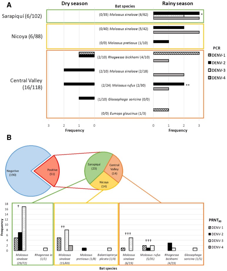
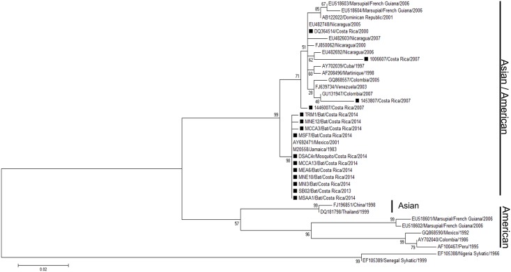
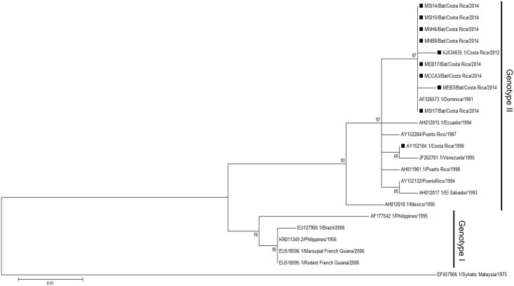
Several studies have shown Dengue Virus (DENV) nucleic acids and/or antibodies present in Neotropical wildlife including bats, suggesting that some bat species may be susceptible to DENV infection. Here we aim to elucidate the role of house-roosting bats in the DENV transmission cycle. Bats were sampled in households located in high and low dengue incidence regions during rainy and dry seasons in Costa Rica. We captured 318 bats from 12 different species in 29 households. Necropsies were performed in 205 bats to analyze virus presence in heart, lung, spleen, liver, intestine, kidney, and brain tissue. Histopathology studies from all organs showed no significant findings of disease or infection. Sera were analyzed by PRNT90 for a seroprevalence of 21.2% (51/241), and by PCR for 8.8% (28/318) positive bats for DENV RNA. From these 28 bats, 11 intestine samples were analyzed by RT-PCR. Two intestines were DENV RNA positive for the same dengue serotype detected in blood. Viral isolation from all positive organs or blood was unsuccessful. Additionally, viral load analyses in positive blood samples by qRT-PCR showed virus concentrations under the minimal dose required for mosquito infection. Simultaneously, 651 mosquitoes were collected using EVS-CO2 traps and analyzed for DENV and feeding preferences (bat cytochrome b). Only three mosquitoes were found DENV positive and none was positive for bat cytochrome b. Our results suggest an accidental presence of DENV in bats probably caused from oral ingestion of infected mosquitoes. Phylogenetic analyses suggest also a spillover event from humans to bats. Therefore, we conclude that bats in these urban environments do not sustain DENV amplification, they do not have a role as reservoirs, but function as epidemiological dead end hosts for this virus.


Conflict of interest statement

The authors have declared that no competing interests exist.

Figures

Fig 1
Fig 1. Detection of DENV RNA and antibodies against DENV in the captured bats in the 3 sites of study in Costa Rica (Nicoya, Sarapiquí and Central Valley, S1 Fig) during the dry and rainy season, 2013–2014.
(A) Prevalence of DENV RNA obtained by PCR from each serotype of DENV in positive bat species. *Two individuals with joint detection of DENV-2 and DENV-4. **One individual with joint detection of DENV-2 and DENV-3. (B) Seroprevalence against each dengue serotype obtained from serum diluted 1:20 by PRNT90 from positive bat species. Twelve individuals presenting antibodies against more than one serotype: † DENV-1 & DENV-3 (3) and DENV-2 & DENV-3 (4); † † DENV-1 & DENV-3 (2) and DENV-1 & DENV-4 (1); † † † DENV-1 & DENV-3 (2).
Fig 2
Fig 2. Maximum likelihood tree of 38 DENV-2 C/prM gene sequences (390 bp) including 10 from bats, 1 from a mosquito, and 3 from humans from former outbreaks in Costa Rica.
Black rectangles (しかく) indicate the strains from Costa Rica. Bootstrap values are indicated at the respecting nodes. The sequences were named according to reference number/country/year of collection or detection.
Fig 3
Fig 3. Maximum likelihood tree of 24 DENV-4 C/prM gene sequences (390 bp) including 8 from bats.
Black rectangles (しかく) indicate the strains from Costa Rica. Bootstrap values are indicated at the respecting nodes. The sequences were named according to reference number/country/year of collection or detection.

References

    1. Murray NEA, Quam MB, Wilder-Smith A. Epidemiology of dengue: past, present and future prospects. Clin Epidemiol. 2013;5: 299–309. 10.2147/CLEP.S34440 - DOI - PMC - PubMed
    1. OMS, TDR. Dengue: guias para el diagnóstico, tratamiento, prevención y control. 20091 OPS, OMS, editors. Bolivia: OMS; 2009.
    1. Weaver SC, Vasilakis N. Molecular evolution of dengue viruses: contributions of phylogenetics to understanding the history and epidemiology of the preeminent arboviral disease. Infect Genet Evol. 2009;9: 523–40. 10.1016/j.meegid.200902003 - DOI - PMC - PubMed
    1. Platt KB, Mangiafico JA, Rocha OJ, Zaldivar ME, Mora J, Trueba G, et al. Detection of dengue virus neutralizing antibodies in bats from Costa Rica and Ecuador. J Med Entomol. 2000;37: 965–7. Available: http://www.ncbi.nlm.nih.gov/pubmed/11126559 - PubMed
    1. de Thoisy B, Lacoste V, Germain A, Muñoz-Jordán J, Colón C, Mauffrey J-F, et al. Dengue infection in neotropical forest mammals. Vector Borne Zoonotic Dis. 2009;9: 157–70. 10.1089/vbz.2007.0280 - DOI - PubMed

LinkOut - more resources

Cite

AltStyle によって変換されたページ (->オリジナル) /