This site needs JavaScript to work properly. Please enable it to take advantage of the complete set of features!
Skip to main page content
U.S. flag

An official website of the United States government

Dot gov

The .gov means it’s official.
Federal government websites often end in .gov or .mil. Before sharing sensitive information, make sure you’re on a federal government site.

Https

The site is secure.
The https:// ensures that you are connecting to the official website and that any information you provide is encrypted and transmitted securely.

NIH NLM Logo
Log in
Access keys NCBI Homepage MyNCBI Homepage Main Content Main Navigation
doi: 10.1038/srep44141.

Leishmania (Leishmania) amazonensis induces macrophage miR-294 and miR-721 expression and modulates infection by targeting NOS2 and L-arginine metabolism

Affiliations

Leishmania (Leishmania) amazonensis induces macrophage miR-294 and miR-721 expression and modulates infection by targeting NOS2 and L-arginine metabolism

Sandra Marcia Muxel et al. Sci Rep. .

Abstract

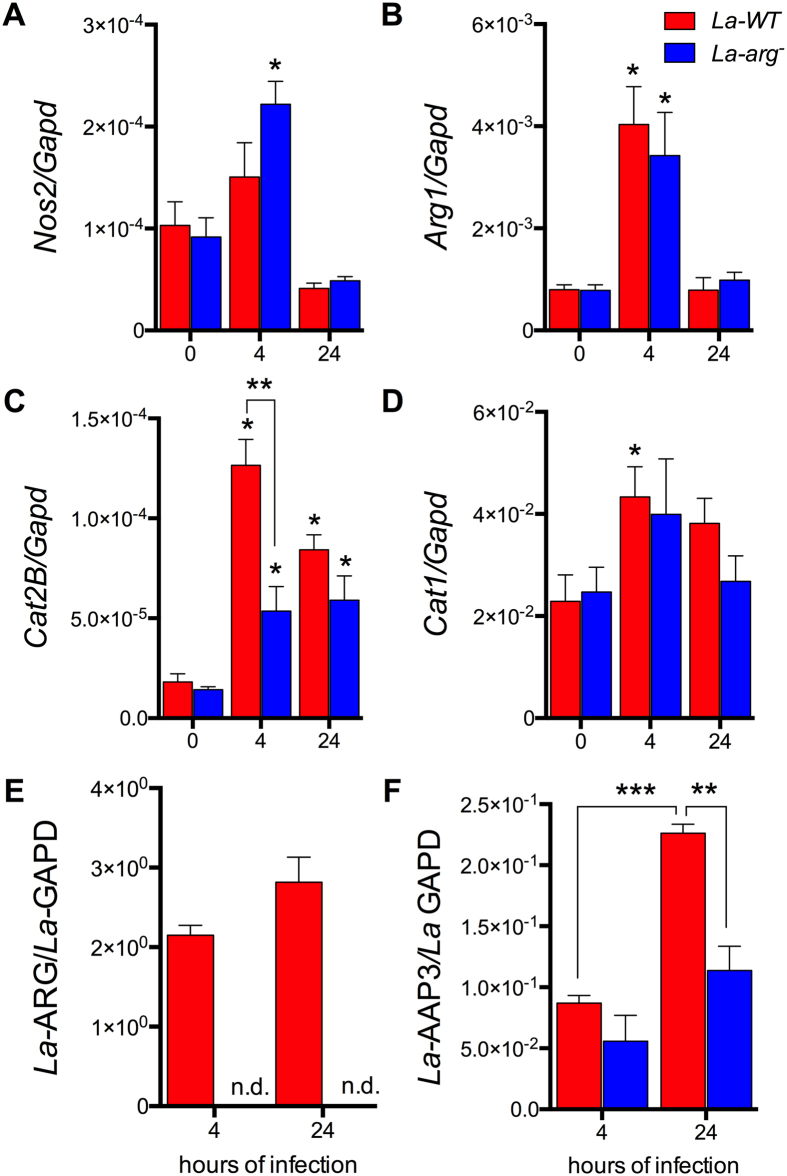
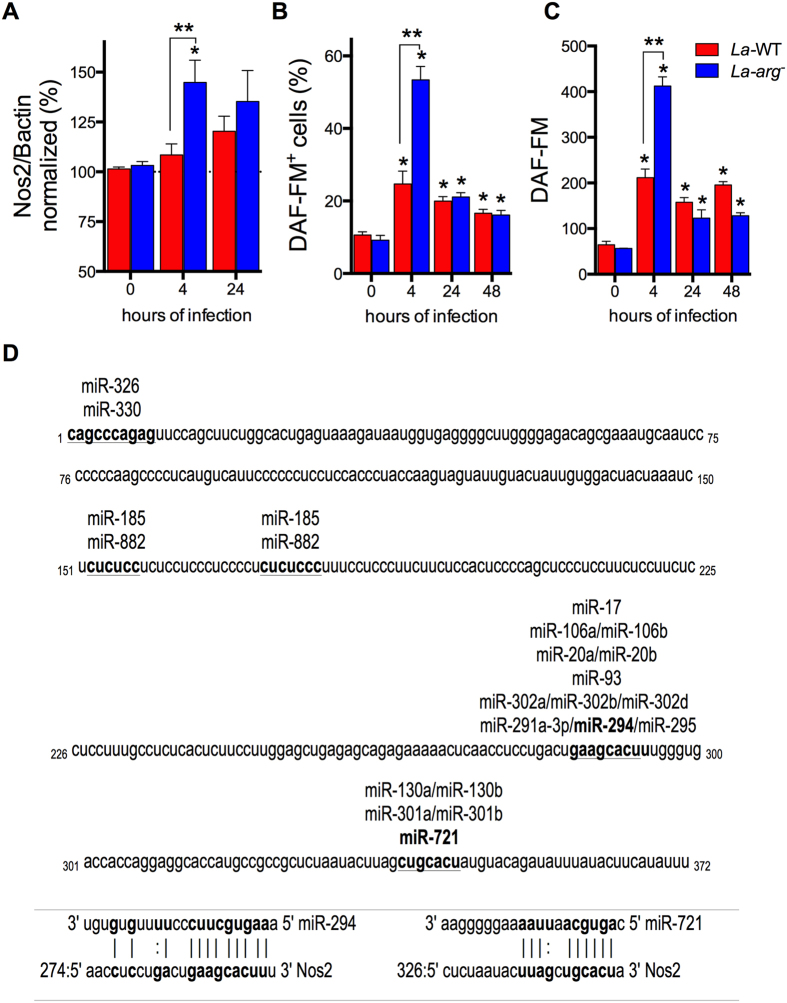
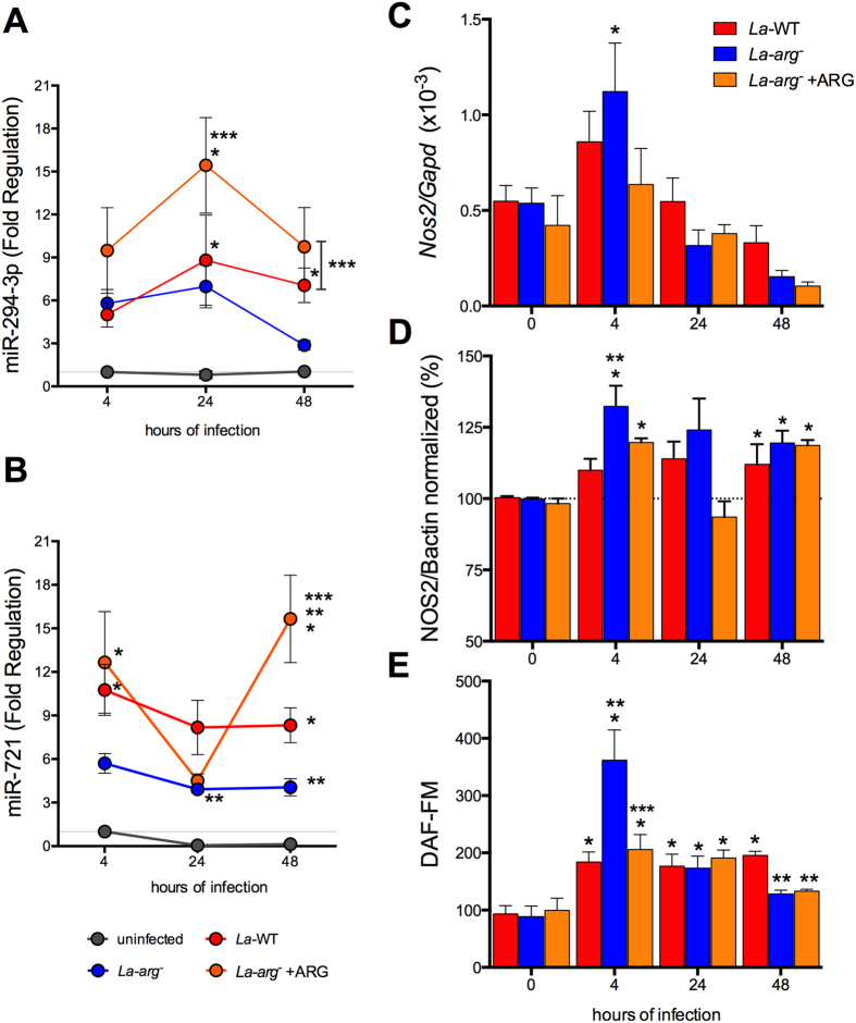
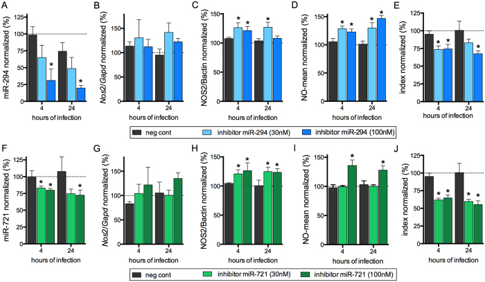
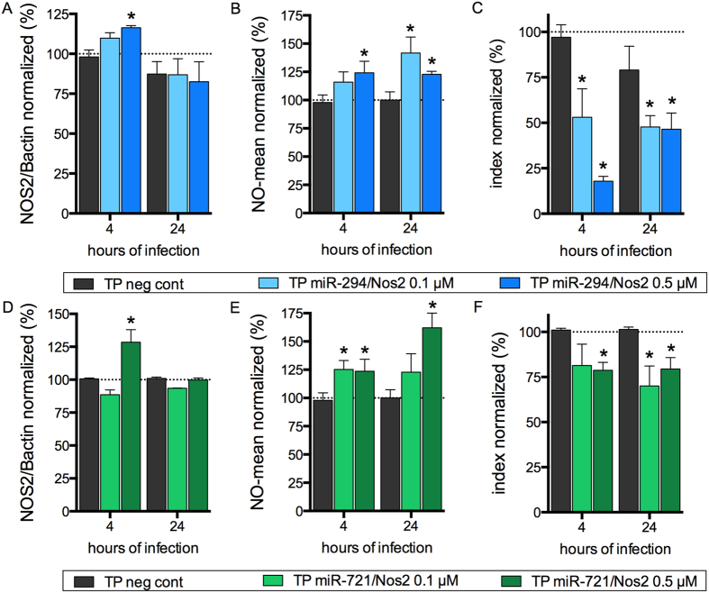
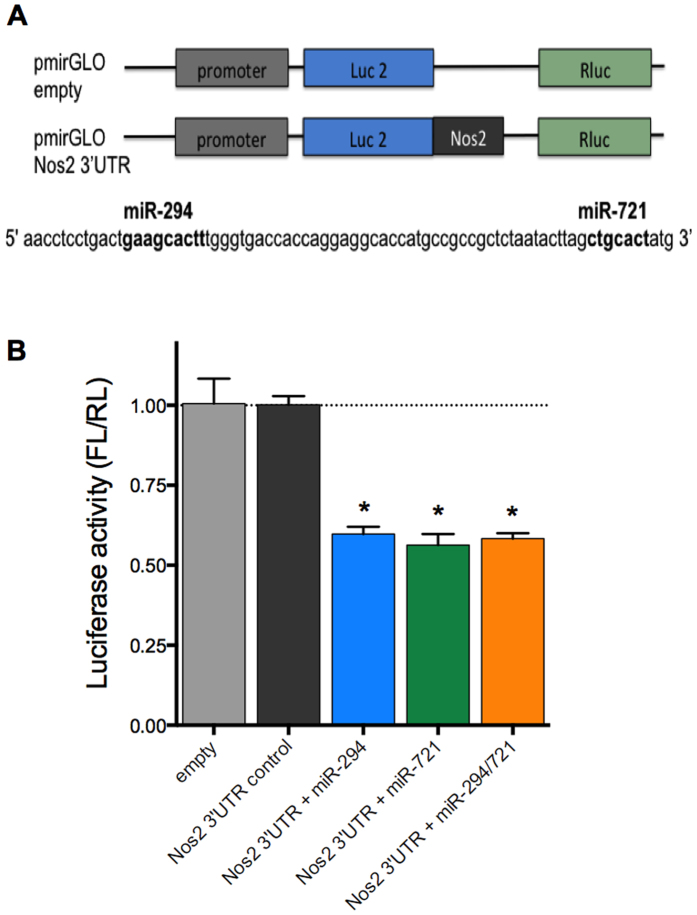
Leishmania (Leishmania) amazonensis is an intracellular protozoan parasite responsible for the cutaneous leishmaniasis. The parasite replicates inside mammalian macrophage to establish infection. Host-pathogen interactions result in microRNA-mediated post-transcriptional regulation of host genes involved in inflammatory immune response. We analyzed macrophage miRNA profiles during L. (L.) amazonensis infection. The regulation of macrophage miRNA expression by the parasite correlates with/depends on parasite arginase activity during infection. L. (L.) amazonensis (La-WT) presented significant miRNA profile alteration (27%) compared to L. (L.) amazonensis arginase knockout (La-arg-) (~40%) in relation to uninfected-macrophages. We observed that 78% of the altered miRNAs were up-regulated in La-WT infection, while only 32% were up-regulated in La-arg--infected macrophages. In contrast to La-WT, the lack of L. (L.) amazonensis arginase led to the inhibition of miR-294 and miR-721 expression. The expression of miR-294 and miR-721 was recovered to levels similar to La-WT in La-arg- addback mutant. The inhibition of miR-294/Nos2 and miR721/Nos2 interactions increased NOS2 expression and NO production, and reduced L. (L.) amazonensis infectivity, confirming Nos2 as target of these miRNAs. The role of miR-294 and miR-721 in the regulation of NOS2 expression during Leishmania replication in infected macrophages pointing these miRNAs as potential new targets for drug development.


Conflict of interest statement

The authors declare no competing financial interests.

Figures

Figure 1
Figure 1. Volcano plot of the miRNA profiles of BMDMs infected with La-WT and La-arg L. (L.) amazonensis.
Each dot represents one miRNA of BMDMs infected for 4, 12, 24 and 48 h with La-WT or La-arg L. (L.) amazonensis. The red dot indicated up-regulated miRNA and green dot indicated down-regulated miRNAs. Blue dotted line corresponds to p = 0.05, log 10. The relative up- and down-regulation of miRNAs, expressed as boundaries of 2 or −2 of Fold Regulation, respectively. P-value was determined based on two-tailed Student’s t test. The data are representative of three independent experiments.
Figure 2
Figure 2. Lack of L. (L.) amazonensis arginase leads to differential modulation of expression of genes involved in the polyamine and NO production pathways.
The BMDMs (1 ×ばつ 106) were infected with La-WT (red) or La-arg (blue) L. (L.) (MOI 5:1). After 4 and 24 h of infection, the copy numbers of mRNAs of Nos2 (A), Arg1 (B), Cat2B (C), and Cat1 (D) from macrophages and La-ARG (E) and the La-AAP3 5.1 (F) from Leishmania were quantified by real-time RT-qPCR. Each bar represents the average ± SEM of the values obtained in 3 independent experiments (n = 6). Statistical significance was determined based on two-tailed Student’s t test. *p < 0.05, compared to uninfected macrophages (0 hours of infection). **p < 0.05, La-WT compared to La-arg. n.d. (not detected).
Figure 3
Figure 3. NOS2 protein amount and NO production in BMDMs infected with La-WT and La-arg L. (L.) amazonensis.
BMDMs (1 ×ばつ 106) were infected with La-WT (red) or La-arg (blue) L. (L.) amazonensis (MOI 5:1). After 4 and 24 h of infection, the protein levels of NOS2 (A), the frequency of cells producing NO (B) and the average of fluorescence intensity reflecting NO production (C) were determined by in-cell western blotting and flow cytometry analysis of DAF-FM staining. Each bar represents the average ± SEM of the values obtained in 3 independent experiments, n = 6. Statistical significance was determined based on two-tailed Student’s t test. *p < 0.05, compared to uninfected macrophages (0 hours of infection). **p < 0.05, La-WT compared to La-arg. (D) Sequence alignment of miR-294 and miR-721 and its target site in the 3′UTR of Nos2 using the microRNA.org database. Mmu-miR-294/Nos2 Alignment: mirSVR score −0.3992, PhastCons: 0.5607; Mmu-miR-721/Nos2 Alignment: mirSVR score −0.1183, PhastCons: 0.6227. Target sites of conserved miRNAs with good mirSVR scores.
Figure 4
Figure 4. Levels of miR-294-3p, miR-721, Nos2 mRNA, NOS2 and NO in BMDMs infected with La-WT, La-arg or La-arg + ARG L. (L.) amazonensis.
BMDMs (1 ×ばつ 106) were infected with La-WT (red) or La-arg (blue) or La-arg + ARG (orange) L. (L.) amazonensis (MOI 5:1). After 4, 24 and 48 h of infection, miR-294-3p (A), miR-721 (B) and Nos2 mRNA levels were quantified via qPCR (C), NOS2 protein levels was quantified via in-cell western blotting (D) and the average of fluorescence intensity reflecting NO production (E) was determined by flow cytometry analysis of DAF-FM staining and values was normalized by uninfected macrophages (100%). Each bar or dot represents the average ± SEM of the values obtained in 3 independent experiments (n = 4–6). Statistical significance was determined based on two-tailed Student’s t test. *p < 0.05, compared to uninfected macrophages (0 hours of infection). **p < 0.05, La-arg or La-arg + ARG compared to La-WT. ***p < 0.05, La-WT or La-arg + ARG compared to La-arg.
Figure 5
Figure 5. Inhibition of miR-294 and miR-721 functions increased NO production and reduced the infectivity of L. (L.) amazonensis.
BMDMs (5 ×ばつ 105) were transiently transfected with the negative control, 30 or 100 nM of miR-294-5p (AE) or miR-721 (FJ) inhibitors, or left non-transfected (untreated, black bars). After 24 h of incubation, the cells were co-cultivated with La-WT L. (L.) amazonensis (MOI 5:1) for 4 h, and the cultures were then washed. After 4 and 24 h of infection, the samples were analyzed for miR-294 (A), miR-721 (F) and Nos2 mRNA (B,G) levels via RT-qPCR of total RNA, for NOS2 expression (C,H) via in-cell western, for NO production (D,I) via flow cytometry DAF-FM fluorescence analysis, and for infectivity (E,J) via microscopy analysis, counting of the numbers of infected macrophages and amastigotes per macrophage (n = 1,000 macrophages/treatment). The values were normalized based on the average values of untreated infected macrophages. Each bar represents the average ± SEM of the values obtained in 3 independent experiments (n = 4–6). Statistical significance was determined based on two-tailed Student’s t test. *p < 0.05, compared to negative control infected macrophages.
Figure 6
Figure 6. miR-294 and miR-721 bind to Nos2 mRNA and regulating NOS2 expression and L. (L.) amazonensis infectivity.
BMDMs (5 ×ばつ 105) were plated into chamber slides overnight and transfected with negative control, 0.1 or 0.5 μM miScript Target Protector for miR-294/Nos2 or miR-721/Nos2 or negative control for 24 h. Then, the BMDMs were co-cultivated with La-WT L. (L.) amazonensis (MOI 5:1) for 4 h, and the cultures were washed. After 4 h and 24 h of infection, the samples were analyzed for NOS2 protein levels (A,D) via in-cell western, NO production (B,E) via flow cytometry DAF-FM fluorescence analysis, and for infectivity (C,F) via microscopy analysis, counting of the numbers of infected macrophages and amastigotes per macrophage (n = 1,000 macrophages/treatment). The values were normalized based on the average of untreated infected macrophages. Each bar represents the average ± SEM of the values obtained in 3 independent experiments (n = 4–6). Statistical significance was determined based on two-tailed Student’s t test. *p < 0.05, compared to negative control infected macrophages.
Figure 7
Figure 7. miR-294 and miR-721 bind to Nos2 3′UTR.
RAW 264.7 (5 ×ばつ 105) were transfected with 5 μg of pmiRGLO, empty or with the regions containing miR-294 or miR-721 binding sites to Nos2 3′UTR, plus 100 nM of miR-294 or/and miR-721 mimics (A). After 24 h, the cells lysates were analyzed for luciferase activity (B). The values of firefly luciferase (FL) were firstly normalized by renilla luciferase (RL), and then normalized by the values obtained with cells transfected with pmiRGLO empty construct. Each bar represents the average ± SEM of the values obtained in 3 independent experiments (n = 5). Statistical significance was determined based on two-tailed Student’s t test. *p < 0.05, compared to pmiRGLO empty vector.

References

    1. Bogdan C. Mechanisms and consequences of persistence of intracellular pathogens: leishmaniasis as an example. Cellular microbiology 10, 1221–1234, doi: 10.1111/j.1462-5822.2008.01146.x (2008). - DOI - PubMed
    1. Liese J., Schleicher U. & Bogdan C. The innate immune response against Leishmania parasites. Immunobiology 213, 377–387, doi: 10.1016/j.imbio.200712005 (2008). - DOI - PubMed
    1. Nathan C. & Shiloh M. U. Reactive oxygen and nitrogen intermediates in the relationship between mammalian hosts and microbial pathogens. Proc Natl Acad Sci USA 97, 8841–8848 (2000). - PMC - PubMed
    1. Gregory D. J. & Olivier M. Subversion of host cell signalling by the protozoan parasite Leishmania. Parasitology 130 Suppl, S27–35, doi: 10.1017/S0031182005008139 (2005). - DOI - PubMed
    1. Green S. J., Crawford R. M., Hockmeyer J. T., Meltzer M. S. & Nacy C. A. Leishmania major amastigotes initiate the L-arginine-dependent killing mechanism in IFN-gamma-stimulated macrophages by induction of tumor necrosis factor-alpha. J Immunol 145, 4290–4297 (1990). - PubMed

Publication types

Cite

AltStyle によって変換されたページ (->オリジナル) /