This site needs JavaScript to work properly. Please enable it to take advantage of the complete set of features!
Skip to main page content
U.S. flag

An official website of the United States government

Dot gov

The .gov means it’s official.
Federal government websites often end in .gov or .mil. Before sharing sensitive information, make sure you’re on a federal government site.

Https

The site is secure.
The https:// ensures that you are connecting to the official website and that any information you provide is encrypted and transmitted securely.

NIH NLM Logo
Log in
Access keys NCBI Homepage MyNCBI Homepage Main Content Main Navigation
Comparative Study
. 2017 Jan 23;11(1):e0005310.
doi: 10.1371/journal.pntd.0005310. eCollection 2017 Jan.

Diagnosing Polyparasitism in a High-Prevalence Setting in Beira, Mozambique: Detection of Intestinal Parasites in Fecal Samples by Microscopy and Real-Time PCR

Affiliations
Comparative Study

Diagnosing Polyparasitism in a High-Prevalence Setting in Beira, Mozambique: Detection of Intestinal Parasites in Fecal Samples by Microscopy and Real-Time PCR

Lynn Meurs et al. PLoS Negl Trop Dis. .

Abstract

Background: Many different intestinal parasite species can co-occur in the same population. However, classic diagnostic tools can only frame a particular group of intestinal parasite species. Hence, one or two tests do not suffice to provide a complete picture of infecting parasite species in a given population. The present study investigated intestinal parasitic infections in Beira, Mozambique, i.e. in the informal settlement of Inhamudima. Diagnostic accuracy of five classical microscopy techniques and real-time PCR for the detection of a broad spectrum of parasites was compared.

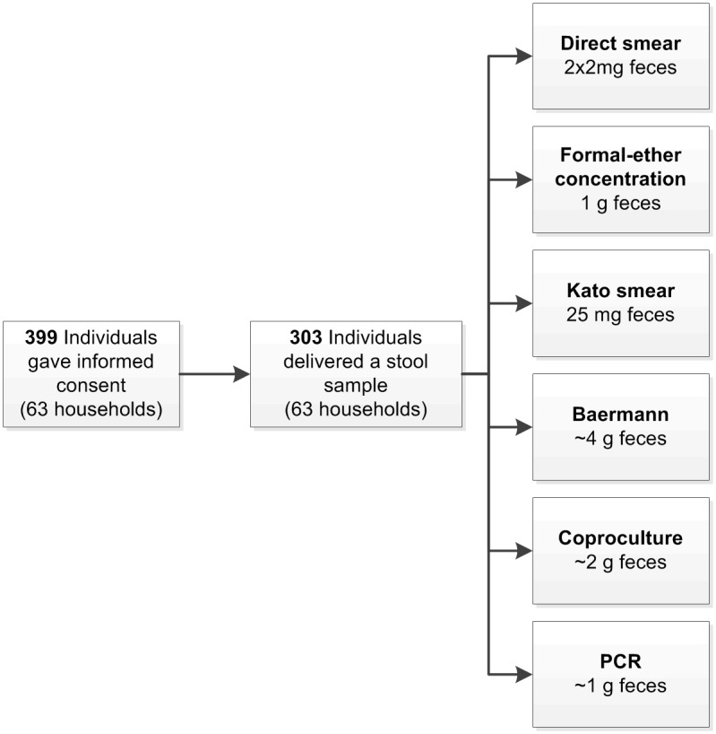
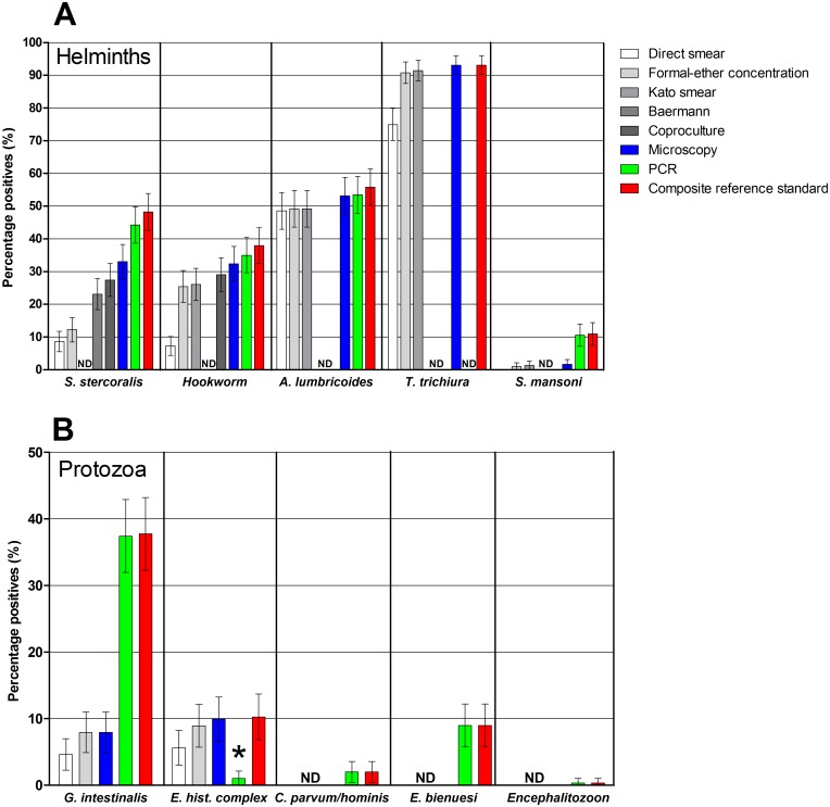
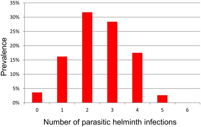
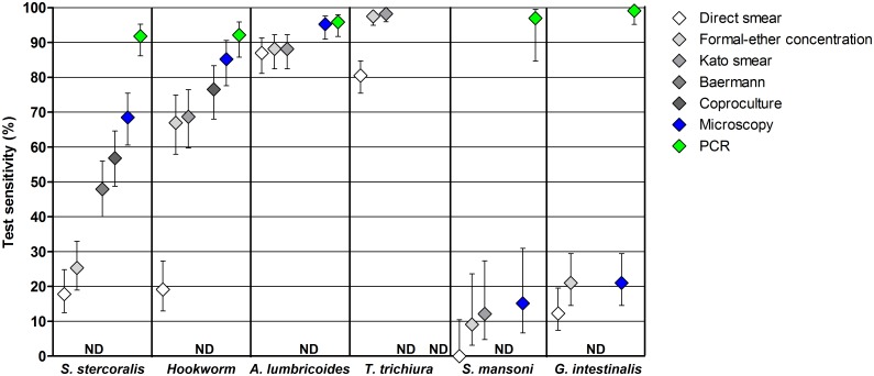
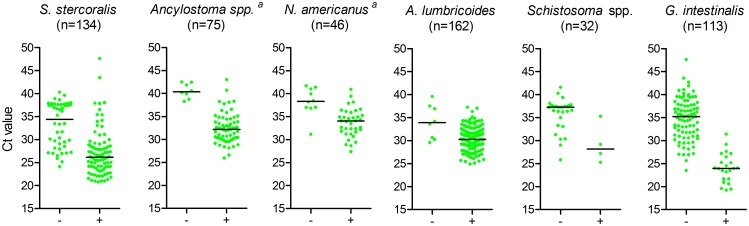
Methodology/principal findings: A cross-sectional population-based survey was performed. One stool sample per participant (n = 303) was examined by direct smear, formal-ether concentration (FEC), Kato smear, Baermann method, coproculture and real-time PCR. We found that virtually all people (96%) harbored at least one helminth, and that almost half (49%) harbored three helminths or more. Remarkably, Strongyloides stercoralis infections were widespread with a prevalence of 48%, and Ancylostoma spp. prevalence was higher than that of Necator americanus (25% versus 15%), the hookworm species that is often assumed to prevail in East-Africa. Among the microscopic techniques, FEC was able to detect the broadest spectrum of parasite species. However, FEC also missed a considerable number of infections, notably S. stercoralis, Schistosoma mansoni and G. intestinalis. PCR outperformed microscopy in terms of sensitivity and range of parasite species detected.

Conclusions/significance: We showed intestinal parasites-especially helminths-to be omnipresent in Inhamudima, Beira. However, it is a challenge to achieve high diagnostic sensitivity for all species. Classical techniques such as FEC are useful for the detection of some intestinal helminth species, but they lack sensitivity for other parasite species. PCR can detect intestinal parasites more accurately but is generally not feasible in resource-poor settings, at least not in peripheral labs. Hence, there is a need for a more field-friendly, sensitive approach for on-the-spot diagnosis of parasitic infections.


Conflict of interest statement

The authors have declared that no competing interests exist.

Figures

Fig 1
Fig 1. Flow diagram of the selection of the study population and diagnostic procedures.
Fig 2
Fig 2. Prevalence of intestinal parasitic infections in the study population according to different diagnostic methods.
Whiskers indicate 95% confidence intervals of the observed prevalence. Percentages are based on observations in 303 individuals. (A) Prevalence of helminth infections. Strongyloides stercoralis infection was not determined (ND) in Kato smears, hookworm was not determined by the Baermann method, while A. lumbricoides, T. trichiura, and S. mansoni were not determined by the Baermann method or coproculture. Trichuris trichiura was not determined by PCR either, and the composite reference standard (CRS) for this infection was consequently based on microscopic results only. (B) Prevalence of intestinal protozoan infections. Feces were examined by both microscopy and PCR for G. intestinalis, and E. histolytica complex spp. (one observation was missing for PCR, and consequently for the CRS). Only PCR data was used for C. parvum/C. hominis (one observation missing) and for, E. bieneusi and Encephalitozoon spp. (two observations missing).* While microscopy cannot differentiate between the pathogenic species Entamoeba histolytica and the nonpathogenic species of the E. histolytica complex, PCR is specific for the pathogenic species (E. histolytica).
Fig 3
Fig 3. Number of helminth species found per person.
Prevalence of infection is based on the composite reference standard for S. stercoralis, A. lumbricoides, T. trichiura, and S. mansoni, and on PCR for hookworm—Ancylostoma spp. and N. americanus (n = 303).
Fig 4
Fig 4. Sensitivities of the different diagnostic methods for the detection of intestinal parasitic infections.
Whiskers indicate 95% confidence intervals of observed sensitivities (n = 303). Strongyloides stercoralis infection was not determined (ND) in Kato smears, hookworm was not determined by the Baermann method, while A. lumbricoides, T. trichiura, and S. mansoni were not determined by the Baermann method or coproculture. Trichuris trichiura was not determined by PCR, and for this species the sensitivity was therefore based on microscopic results only. Giardia intestinalis was not determined by Kato smear, the Baermann method or coproculture (one observation missing).
Fig 5
Fig 5. Ct values in PCR-positives: microscopy-negative versus -positive samples.
Total number of PCR-positives per species is indicated between brackets. ‘-’ indicates microscopy-negative and ‘+’ microscopy-positive samples. Horizontal lines indicate median Ct values. Differences in Ct values between microscopy-positive and–negative samples were all significant p≤0.007, a microscopy cannot differentiate the two hookworm species.

References

    1. Guerrant RL, Oria RB, Moore SR, Oria MO, Lima AA. Malnutrition as an enteric infectious disease with long-term effects on child development. Nutr Rev 2008. September;66(9):487–505. 10.1111/j.1753-4887.2008.00082.x - DOI - PMC - PubMed
    1. Hall A, Hewitt G, Tuffrey V, de Silva N. A review and meta-analysis of the impact of intestinal worms on child growth and nutrition. Matern Child Nutr 2008. April;4 Suppl 1:118–236. - PMC - PubMed
    1. Hotez PJ, Kamath A. Neglected tropical diseases in sub-saharan Africa: review of their prevalence, distribution, and disease burden. PLoS Negl Trop Dis 2009;3(8):e412 10.1371/journal.pntd.0000412 - DOI - PMC - PubMed
    1. Bethony J, Brooker S, Albonico M, Geiger SM, Loukas A, Diemert D, et al. Soil-transmitted helminth infections: ascariasis, trichuriasis, and hookworm. Lancet 2006. May 6;367(9521):1521–32. 10.1016/S0140-6736(06)68653-4 - DOI - PubMed
    1. Colley DG, Bustinduy AL, Secor WE, King CH. Human schistosomiasis. Lancet 2014. March 31. - PMC - PubMed

Publication types

MeSH terms

Cite

AltStyle によって変換されたページ (->オリジナル) /