This site needs JavaScript to work properly. Please enable it to take advantage of the complete set of features!
Skip to main page content
U.S. flag

An official website of the United States government

Dot gov

The .gov means it’s official.
Federal government websites often end in .gov or .mil. Before sharing sensitive information, make sure you’re on a federal government site.

Https

The site is secure.
The https:// ensures that you are connecting to the official website and that any information you provide is encrypted and transmitted securely.

NIH NLM Logo
Log in
Access keys NCBI Homepage MyNCBI Homepage Main Content Main Navigation
Review
. 2016 Oct 21:6:129.
doi: 10.3389/fcimb.2016.00129. eCollection 2016.

Type Three Secretion System in Attaching and Effacing Pathogens

Affiliations
Review

Type Three Secretion System in Attaching and Effacing Pathogens

Meztlli O Gaytán et al. Front Cell Infect Microbiol. .

Abstract

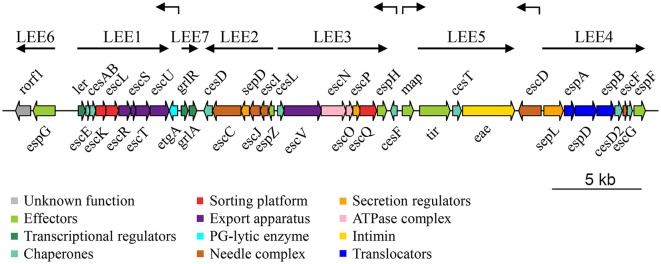
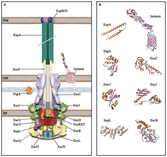
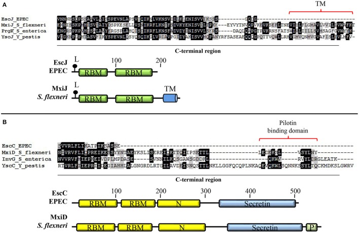
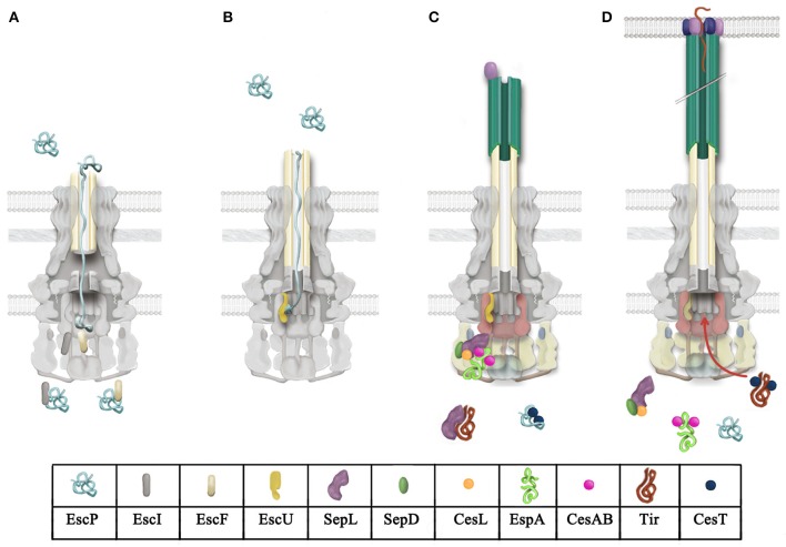
Enteropathogenic Escherichia coli and enterohemorrhagic E. coli are diarrheagenic bacterial human pathogens that cause severe gastroenteritis. These enteric pathotypes, together with the mouse pathogen Citrobacter rodentium, belong to the family of attaching and effacing pathogens that form a distinctive histological lesion in the intestinal epithelium. The virulence of these bacteria depends on a type III secretion system (T3SS), which mediates the translocation of effector proteins from the bacterial cytosol into the infected cells. The core architecture of the T3SS consists of a multi-ring basal body embedded in the bacterial membranes, a periplasmic inner rod, a transmembrane export apparatus in the inner membrane, and cytosolic components including an ATPase complex and the C-ring. In addition, two distinct hollow appendages are assembled on the extracellular face of the basal body creating a channel for protein secretion: an approximately 23 nm needle, and a filament that extends up to 600 nm. This filamentous structure allows these pathogens to get through the host cells mucus barrier. Upon contact with the target cell, a translocation pore is assembled in the host membrane through which the effector proteins are injected. Assembly of the T3SS is strictly regulated to ensure proper timing of substrate secretion. The different type III substrates coexist in the bacterial cytoplasm, and their hierarchical secretion is determined by specialized chaperones in coordination with two molecular switches and the so-called sorting platform. In this review, we present recent advances in the understanding of the T3SS in attaching and effacing pathogens.

Keywords: A/E pathogens; Citrobacter rodentium; EHEC; EPEC; injectisome; locus of enterocyte effacement; secretion hierarchy; type III secretion system.


Figures

Figure 1
Figure 1
Genetic structure of the LEE island of Escherichia coli E2348/69 O127:H6 strain (EPEC). Genes are depicted as filled arrows colored according to their proposed functional category (see enclosed box). The operon organization of the island (LEE1 to LEE7) is indicated by solid black arrows above the LEE genes, while individual transcriptional units (etgA, cesF, map, escD) are denoted by broken arrows. Schematic representation is drawn at scale (scale bar 5 kb). PG, peptidoglycan.
Figure 2
Figure 2
Schematic representation of the type III secretion system of A/E pathogens. (A) The T3SS is divided into three main parts, from top to bottom (i) extracellular appendages: translocation pore (inserted into the host membrane, HM), filament and needle; (ii) basal body: consisting of three membrane rings that span the inner and outer membrane (IM and OM, respectively) connected through a periplasmic inner rod. The IM rings house the export apparatus components; (iii) cytoplasmic components: the C-ring, the ATPase complex and the gatekeeper protein. The outer membrane protein intimin and the PG lytic enzyme EtgA are also illustrated. (B) Solved protein structures of the depicted T3SS components. Protein Data Bank (PDB) accession numbers: SepL, 5C9E; EscN, 2OBM; cytoplasmic C-terminal domain of EscU, 3BZL; periplasmic domain of EscC, 3GR5; EtgA, 4XP8; periplasmic domain of EscJ, 1YJ7; the EspA structure was obtained from that of the CesAB/EspA complex, 1XOU, chain A; transmembrane beta-domain of intimin, 4E1S and its C-terminal domain, 1F00. Protein structures are displayed as ribbon diagrams and were colored according to their secondary structure.
Figure 3
Figure 3
Basal body components of A/E pathogens possess distinctive features. (A) EscJ lacks the C-terminal transmembrane segment typically present in proteins of the SctJ family. Multiple protein alignment of the C-terminal domain of the lipoproteins EscJ (EPEC), MxiJ (S. flexneri), PrgK (S. enterica), and YscJ (Y. pestis) forming the inner membrane ring. The schematic comparison of EscJ and MxiJ domain organization is shown at the bottom. (B) The proposed pilotin-binding domain is absent in the EscC secretin. Multiple alignment of the C-terminal domain of secretins EscC (EPEC), MxiD (S. flexneri), InvG (S. enterica), and YscC (Y. pestis). The schematic comparison of EscC and MxiD domain organization is shown at the bottom. Even though only the basal body proteins from EPEC are depicted, all A/E pathogens share these features. TM, transmembrane helix; L, lipidation site; RBM, ring-building motif; Secretin, secretin domain (PF00263); N, secretin N domain (PF03958); P, pilotin-binding domain.
Figure 4
Figure 4
Model for T3 secretion regulation in A/E pathogens. (A) Rod and needle assembly occur simultaneously (Marlovits et al., ; Lefebre and Galán, 2014). EscP directly interacts with early substrates (EscI and EscF, rod and needle subunits, respectively), regulating its secretion. EscP is secreted occasionally during needle assembly. (B) Once rod and needle assembly is completed, EscP makes contact with the full-length needle, causing a pause in substrate secretion that allows the productive interaction between EscP and the pre-cleaved C-terminal domain of EscU (EscUcc) (Monjarás Feria et al., 2012). This interaction is proposed to promote a conformational change in EscUcc that flicks substrate specificity, probably generating a docking site for a different category of substrates (Zarivach et al., ; Thomassin et al., 2011). (C) Translocator secretion is now allowed. The SepL/SepD/CesL complex targets translocator/chaperone complexes to the sorting platform (formed by EscQ, EscL, and EscK; Lara-Tejero et al., 2011). SepL also interacts with the export gate component EscV and probably modifies its affinity for certain substrate classes; it has also been proposed that it might block access of effectors to the export gate (Lee et al., ; Shen and Blocker, 2016). SepL interacts with the effector Tir, preventing its secretion (Wang et al., 2008). (D) Upon host cell contact, the SepL/SepD/CesL complex might disengage from the export gate component and the sorting platform, alleviating the effector recognition blockade exerted on EscV and allowing effector translocation into host cells.

References

    1. Abby S. S., Rocha E. P. (2012). The non-flagellar type III secretion system evolved from the bacterial flagellum and diversified into host-cell adapted systems. PLoS Genet. 8:e1002983. 10.1371/journal.pgen.1002983 - DOI - PMC - PubMed
    1. Abe A., de Grado M., Pfuetzner R. A., Sánchez-Sanmartín C., Devinney R., Puente J. L., et al. . (1999). Enteropathogenic Escherichia coli translocated intimin receptor, Tir, requires a specific chaperone for stable secretion. Mol. Microbiol. 33, 1162–1175. - PubMed
    1. Abrusci P., Vergara-Irigaray M., Johnson S., Beeby M. D., Hendrixson D. R., Roversi P., et al. . (2013). Architecture of the major component of the type III secretion system export apparatus. Nat. Struct. Mol. Biol. 20, 99–104. 10.1038/nsmb.2452 - DOI - PMC - PubMed
    1. Agrain C., Sorg I., Paroz C., Cornelis G. R. (2005). Secretion of YscP from Yersinia enterocolitica is essential to control the length of the injectisome needle but not to change the type III secretion substrate specificity. Mol. Microbiol. 57, 1415–1427. 10.1111/j.1365-2958.2005.04758.x - DOI - PubMed
    1. Akeda Y., Galán J. E. (2005). Chaperone release and unfolding of substrates in type III secretion. Nature 437, 911–915. 10.1038/nature03992 - DOI - PubMed

Publication types

MeSH terms

Substances

LinkOut - more resources

Cite

AltStyle によって変換されたページ (->オリジナル) /