This site needs JavaScript to work properly. Please enable it to take advantage of the complete set of features!
Skip to main page content
U.S. flag

An official website of the United States government

Dot gov

The .gov means it’s official.
Federal government websites often end in .gov or .mil. Before sharing sensitive information, make sure you’re on a federal government site.

Https

The site is secure.
The https:// ensures that you are connecting to the official website and that any information you provide is encrypted and transmitted securely.

NIH NLM Logo
Log in
Access keys NCBI Homepage MyNCBI Homepage Main Content Main Navigation
. 2016 Sep 28:7:12766.
doi: 10.1038/ncomms12766.

Systematic functional analysis of kinases in the fungal pathogen Cryptococcus neoformans

Affiliations

Systematic functional analysis of kinases in the fungal pathogen Cryptococcus neoformans

Kyung-Tae Lee et al. Nat Commun. .

Abstract

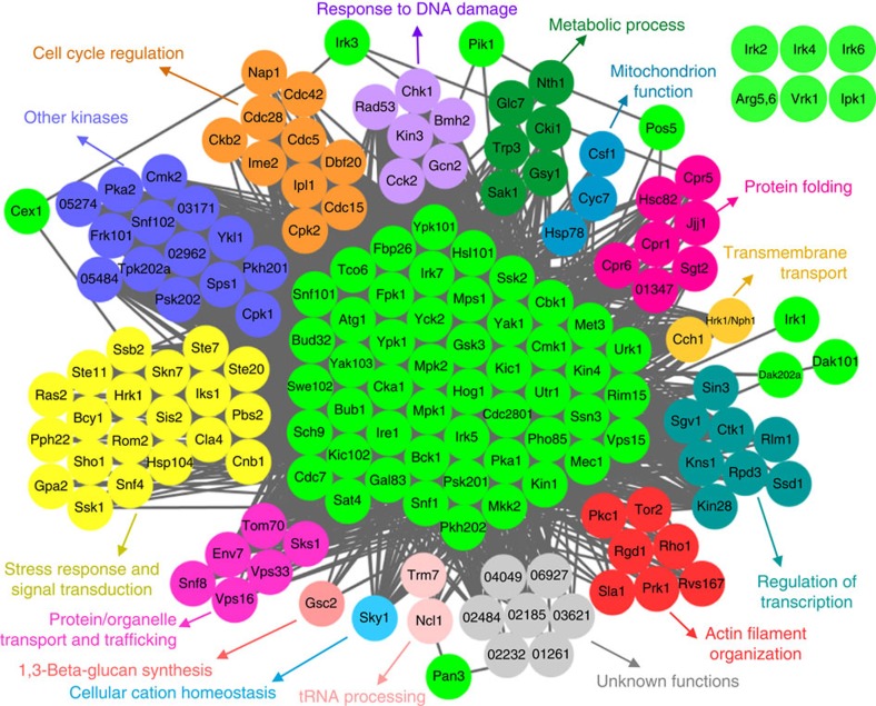
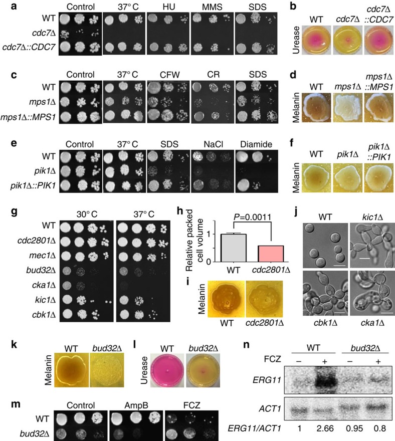
Cryptococcus neoformans is the leading cause of death by fungal meningoencephalitis; however, treatment options remain limited. Here we report the construction of 264 signature-tagged gene-deletion strains for 129 putative kinases, and examine their phenotypic traits under 30 distinct in vitro growth conditions and in two different hosts (insect larvae and mice). Clustering analysis of in vitro phenotypic traits indicates that several of these kinases have roles in known signalling pathways, and identifies hitherto uncharacterized signalling cascades. Virulence assays in the insect and mouse models provide evidence of pathogenicity-related roles for 63 kinases involved in the following biological categories: growth and cell cycle, nutrient metabolism, stress response and adaptation, cell signalling, cell polarity and morphology, vacuole trafficking, transfer RNA (tRNA) modification and other functions. Our study provides insights into the pathobiological signalling circuitry of C. neoformans and identifies potential anticryptococcal or antifungal drug targets.


Figures

Figure 1
Figure 1. Phylogenetic correlation among kinases in Cryptococcus neoformans and kinase distribution in human fungal pathogens.
(a) Protein sequence-based alignment was performed using ClustalX2 windows interface program run by University College Dublin. Using this alignment data, the phylogenetic tree was illustrated by a web-based drawing application named Interactive Tree Of Life (http://itol.embl.de). Among the 183 kinases found in C. neoformans, we constructed signature-tagged gene deletion strains for 129 kinases genes. Asterisks indicate the kinases that were first functionally characterized by this study and named based on the published nomenclature rules for C. neoformans genes. The different colour codes represent the different classes of protein kinases predicted by Kinomer 1.0 (http://www.compbio.dundee.ac.uk/kinomer). Red marked genes indicate the 63 pathogenicity-related kinases discovered in this study. (b) Pie-chart for the classification of 183 kinases in C. neoformans. The description of the Broad institute database was grouped by using previously reported classification methods. (c) Pie-chart for the kinase classes predicted by Kinomer 1.0 to reveal the relative portion of protein kinase classes in human fungal pathogens, C. neoformans, Candida albicans and Aspergillus fumigatus.
Figure 2
Figure 2. Phenotypic clustering of kinases in Cryptococcus neoformans.
The phenotypes were scored by seven grades (−3: strongly sensitive/reduced, −2: moderately sensitive/reduced, −1: weakly sensitive/reduced, 0: wild-type like, +1: weakly resistant/increased, +2: moderately resistant/increased, +3: strongly resistant/increased). The excel file containing the phenotype scores of each kinase mutant was loaded by Gene-E software (http://www.broadinstitute.org/cancer/software/GENE-E/) and then the kinase phenome clustering was drawn using one minus Pearson correlation. T25, 25 °C; T30, 30 °C; T37, 37 °C; T39, 39 °C; CAP, capsule production; MEL, melanin production; URE, urease production; MAT, mating; HPX, hydrogen peroxide; TBH, tert-butyl hydroperoxide; MD, menadione; DIA, diamide; MMS, methyl methanesulphonate; HU, hydroxyurea; 5FC, 5-flucytosine; AMB, amphotericin B; FCZ, fluconazole; FDX, fludioxonil; TM, tunicamycin; DTT, dithiothreitol; CDS, cadmium sulphate; SDS, sodium dodecyl sulphate; CR, Congo red; CFW, calcofluor white; KCR, YPD+KCl; NCR, YPD+NaCl; SBR, YPD+sorbitol; KCS, YP+KCl; NCS, YP+NaCl; SBS, YP+sorbitol.
Figure 3
Figure 3. Pathogenicity-related kinases in Cryptococcus neoformans.
(a) Results of the signature-tagged mutagenesis (STM)-based murine infectivity test. We used ste50Δ and hxl1Δ strains for the virulent (positive control, PC) and avirulent (negative control, NC) controls. STM scores were calculated by the quantitative PCR method, arranged numerically and coloured in gradient scales. Red marked letters show the novel infectivity-related kinases revealed by this study. Gene names for the 30 kinases that were co-identified by both insect killing and STM assays were depicted below the STM zero line. The P-value between control and mutant strains was determined by one-way analysis of variance employing Bonferroni correlation with three mice per each STM set. In addition, the STM score of the second independent strain was measured in another independent set with three mice. The y axis indicates the average value of the two independent STM scores for each kinase. This graph only shows kinases whose deletion reduces or increases STM score with statistically significant difference (P<0.05). The entire STM scoring was available in Supplementary Fig. 3. Error bars indicate s.e.m. (b) Five kinases were shown to be involved in virulence only by the wax moth killing assay. P values shown in the graph were calculated using the Log-rank test to measure statistical differences between the WT strain (H99S) and each kinase mutant strain.
Figure 4
Figure 4. Phylogenetic relationships between pathogenicity-related kinases in Cryptococcus neoformans and other eukaryotic kinases.
(a) BLAST matrix for the 63 pathogenicity-related kinases using the Comparative Fungal Genomics Platform (CFGP, http://cfgp.riceblast.snu.ac.kr) database. Using the pathogenicity-related 63 kinase protein sequence query, orthologue proteins were retrieved and matched from the genome database from the 35 eukaryotic species (Supplementary Data 7). (b) Correlation of the pathogenicity-related kinases in fungal pathogens. To determine the orthologue proteins among the indicated fungal pathogens, each protein sequence was analysed by BLAST and reverse-BLAST using genome databases (CGD; Candida genome database for C. albicans, Broad institute database for Fusarium graminearum and C. neoformans).
Figure 5
Figure 5. Network-based functional relationships among 63 pathogenicity-related kinases.
Gene-function network analysis of the 63 pathogenicity-related kinases in C. neoformans by CryptoNet (http://www.inetbio.org/cryptonet). The functional correlation network was drawn based on 100 candidate genes that are predicted to have functional relationships with the 63 pathogenicity-related kinases by CryptoNet. Genes in this pathogenicity network were classified by the information of their Gene Ontology (GO) term or predicted biological functions listed in Supplementary Data 5. Six kinases (Arg5,6, Ipk1, Irk2, Irk4, Irk6, Vrk1) did not have any functionally related genes in CryptoNet.
Figure 6
Figure 6. Kinases involved in growth and cell morphology of Cryptococcus neoformans.
Various phenotypic tests were performed using the WT strain (H99S) and the following kinase mutants and complemented strains: (a,b) cdc7Δ (YSB2912), cdc7Δ::CDC7 (YSB4356), (c,d) mps1Δ (YSB3632), mps1Δ::MPS1 (YSB4351), (e,f) pik1Δ (YSB1493), pik1Δ::PIK1 (YSB4360), (gn) cdc2801Δ (YSB2370), mec1Δ (YSB3063), cka1Δ (YSB3051), kic1Δ (YSB2915), cbk1Δ (YSB2941), and bud32Δ (YSB1968) strains. (a,c,e,m) Cells were spotted on YPD medium containing the following chemicals at the indicated concentrations: 110 mM hydroxyurea (HU), 0.04% methyl methanesulphonate (MMS), 0.03% sodium dodecyl sulphate (SDS), 4 mg ml−1 calcofluor-white (CFW), 0.8% Congo red (CR), 1.5 M NaCl, 2 mM diamide, 1 μg ml−1 amphotericin B (AmpB), or 14 μg ml−1 fluconazole (FCZ). The cells were incubated at 30 °C and photographed after 3 days. For testing thermotolerance, each strain was spotted on YPD medium, incubated at the indicated temperature, and photographed after 3 days. (b,l) Urease production assay. Each strain was spotted on Christensen's agar media, incubated at 30 °C, and photographed after 3 days. (d,i,k) Melanin production assay. Each strain was spotted on Niger seed media containing 0.1% glucose, incubated at 37 °C, and photographed after 2–3 days. (h) The relative packed cell volume of cdc2801Δ mutants. The ratio was calculated from three biological replicates with three technical replicates normalized to the WT strain. The statistical significance was calculated by the Bonferroni's test for multiple comparison. Error bars indicate the s.e.m. (j) Cell morphology picture of WT H99S, kic1Δ, cbk1Δ and cka1Δ strains as observed by differential interference contrast microscopy at 16 h after inoculation in liquid YPD medium. Scale bars, 10 μm. (n) WT and bud32Δ strains grown at 30 °C to the logarithmic phase were treated with (+) or without (−) 10 μg ml−1 FCZ for 90 min, and total RNA was extracted. The expression levels were visualized by northern blotting and quantified using a phosphorimager (Fujifilm BAS-1500). The whole gel and phosphorimages were displayed on Supplementary Fig. 6d.
Figure 7
Figure 7. Retrograde vacuole trafficking controls the pathogenicity of Cryptococcus neoformans.
Various tests were performed using WT strain (H99S) and vps15Δ mutants (YSB1500 and YSB1501) (a) Vps15 is required for virulence of C. neoformans. WT and PBS were used as positive and negative virulence controls, respectively. (b) vps15Δ mutants display enlarged vacuole morphology. Scale bars,10 μm. (c) vps15Δ mutants show significant growth defect under ER stresses. Overnight cultured cells were serially diluted tenfold (undiluted to 104-fold dilution), spotted on the solid YPD medium containing 15 mM dithiothreitol (DTT) or 0.3 μg ml−1 tunicamycin (TM), further incubated at 30 °C for 3 days, and photographed. (d) vps15Δ mutants show significant growth defects at high temperature and under cell membrane/wall stresses. Overnight cultured cells were spotted on the YPD medium and further incubated at indicated temperature (upper panel) or the YPD medium containing 0.03% SDS or 5 mg ml−1 calcofluor white (CFW) and further incubated at 30 °C (lower panel). Plates were photographed after 3 days. (e) Vps15 is not involved in the regulation of the calcineurin pathway in C. neoformans. For quantitative RT–PCR (qRT–PCR), RNA was extracted from three biological replicates with three technical replicates of WT and vps15Δ mutants. CNA1, CNB1, CRZ1, UTR2 expression levels were normalized by ACT1 expression levels as controls. Error bars represent s.e.m. (f) Vps15 negatively regulates the HXL1 splicing. For RT–PCR, total RNA was extracted from WT and vps15Δ mutants and cDNA was synthesized. HXL1 and ACT1-specific primer pairs were used for RT–PCR. This experiment was repeated twice and one representative experiment is presented. The whole-gel images were displayed on Supplementary Fig. 6e.
Figure 8
Figure 8. Phenotypic traits and phosphoproteomic analysis of the vrk1Δ mutant in Cryptococcus neoformans.
(a) WT strain (H99S) and vrk1Δ mutants (YSB2216 and YSB2217) grown overnight were serially diluted tenfold (102 to 104-fold dilution), spotted on solid YPD medium containing 2.5 mM H2O2 (HPX), 600 μg ml−1 flucytosine (5-FC) or 1 μg ml−1 fludioxonil (FDX), further incubated for 3 days at 30 °C and were photographed. (b) vrk1Δ mutants show increased capsule production. Their relative packed cell volume ratio was calculated from three biological replicates with three technical replicates with normalization to that of WT strain. Based on the Bonferroni's multiple comparison test, double and triple asterisks indicate P values of 0.0038 and 0.0004, respectively. Error bars mean s.e.m. (c) Functional network relationship among the Vrk1 phospho-target proteins using CryptoNet. Significant differences were observed in 23 proteins by statistical analysis of the phosphopeptides qualified in WT and/or vrk1Δ mutant strains listed in Supplementary Data 6. The functional relationship of each protein was connected by a line. (d) Multi-layered pie-chart for summarizing gene-function network analysis of the Vrk1 target proteins by CryptoNet. The network analysis was based on 100 candidate genes that are predicted to have functional relationship with the indicated eight Vrk1-regulated target proteins. Genes were classified by the information of their Gene Ontology (GO) term or predicted biological functions listed in Supplementary Data 5.

References

    1. Park B. J. et al. Estimation of the current global burden of cryptococcal meningitis among persons living with HIV/AIDS. AIDS 23, 525–530 (2009). - PubMed
    1. Lin X. & Heitman J. The biology of the Cryptococcus neoformans species complex. Annu. Rev. Microbiol. 60, 69–105 (2006). - PubMed
    1. Perfect J. R. et al. Clinical practice guidelines for the management of cryptococcal disease: 2010 update by the infectious diseases society of America. Clin. Infect. Dis. 50, 291–322 (2010). - PMC - PubMed
    1. Idnurm A. et al. Deciphering the model pathogenic fungus Cryptococcus neoformans. Nat. Rev. Microbiol. 3, 753–764 (2005). - PubMed
    1. Liu O. W. et al. Systematic genetic analysis of virulence in the human fungal pathogen Cryptococcus neoformans. Cell 135, 174–188 (2008). - PMC - PubMed
Cite

AltStyle によって変換されたページ (->オリジナル) /