This site needs JavaScript to work properly. Please enable it to take advantage of the complete set of features!
Skip to main page content
U.S. flag

An official website of the United States government

Dot gov

The .gov means it’s official.
Federal government websites often end in .gov or .mil. Before sharing sensitive information, make sure you’re on a federal government site.

Https

The site is secure.
The https:// ensures that you are connecting to the official website and that any information you provide is encrypted and transmitted securely.

NIH NLM Logo
Log in
Access keys NCBI Homepage MyNCBI Homepage Main Content Main Navigation
. 2016 Jun 28:7:12204.
doi: 10.1038/ncomms12204.

A rhesus macaque model of Asian-lineage Zika virus infection

Affiliations

A rhesus macaque model of Asian-lineage Zika virus infection

Dawn M Dudley et al. Nat Commun. .

Abstract

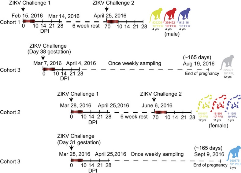
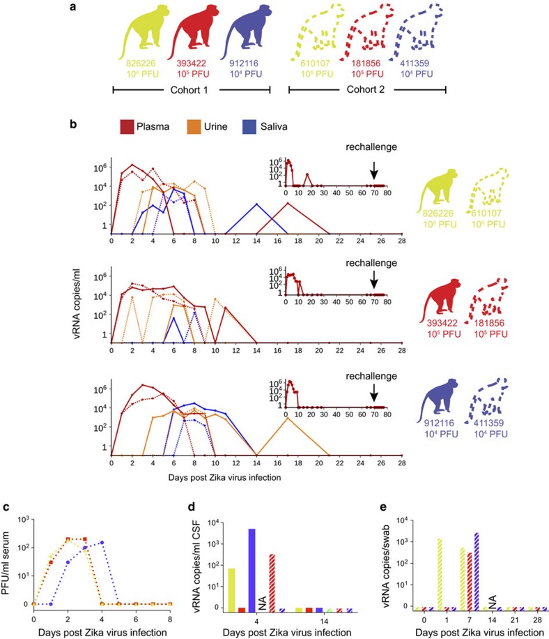
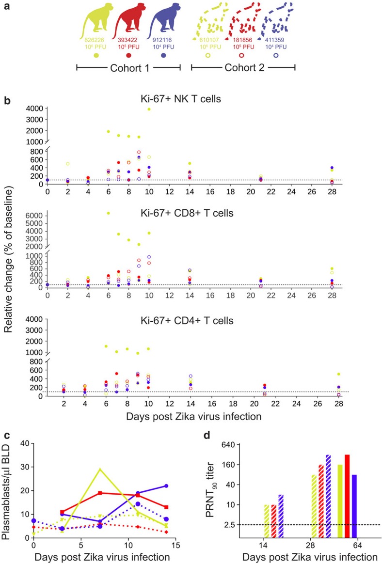
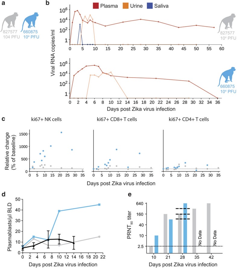
Infection with Asian-lineage Zika virus (ZIKV) has been associated with Guillain-Barré syndrome and fetal abnormalities, but the underlying mechanisms remain poorly understood. Animal models of infection are thus urgently needed. Here we show that rhesus macaques are susceptible to infection by an Asian-lineage ZIKV closely related to strains currently circulating in the Americas. Following subcutaneous inoculation, ZIKV RNA is detected in plasma 1 day post infection (d.p.i.) in all animals (N=8, including 2 pregnant animals), and is also present in saliva, urine and cerebrospinal fluid. Non-pregnant and pregnant animals remain viremic for 21 days and for up to at least 57 days, respectively. Neutralizing antibodies are detected by 21 d.p.i. Rechallenge 10 weeks after the initial challenge results in no detectable virus replication, indicating protective immunity against homologous strains. Therefore, Asian-lineage ZIKV infection of rhesus macaques provides a relevant animal model for studying pathogenesis and evaluating potential interventions against human infection, including during pregnancy.


Figures

Figure 1
Figure 1. Schematic representation of the timeline of infection and sampling for each animal in the presented studies.
Cohort 1 received the first ZIKV challenges and was then rested for 6 weeks before a rechallenge. For all studies, samples were collected daily for 10 days and then on 14, 21 and 28 d.p.i. as indicated by hashes in the timelines. Cohort 3 represents the two pregnant animals that were challenged on two different days. Both animals are currently in the once weekly sampling phase until the pregnancies come to term (∼165 gestational days). Cohort 2 was a repeat experiment of cohort 1 that allowed for additional experiments and sample collection (for example, serum plaque infectivity) that were not feasible when we initiated cohort 1 studies. These animals are currently in a 6-week rest period and will be rechallenged on 6 June 2016. Ages of all animals are indicated under each macaque identification number.
Figure 2
Figure 2. Animal cohort definitions and ZIKV viral load from rhesus macaque fluids.
(a) Animals included in this study and the ZIKV doses used to infect them. Solid lines and bars throughout the figure represent cohort 1 animals by colour, while stripped bars and dotted lines represent cohort 2 animals by colour. (b) Viral RNA loads measured in plasma, urine and saliva for the two animals challenged with each dose of virus through 28 d.p.i. Cohort 1 animals are represented by a solid line, while cohort 2 animals are represented by a dotted line for each fluid. Inset: vRNA loads from cohort 1 animals measured before and after rechallenge with homotypic Zika virus as indicated by an arrow. (c) Number of plaque-forming units per ml of serum for cohort 2 animals. (d) Viral RNA load per ml of CSF collected on 4 and 14 d.p.i. (e) Viral RNA load per vaginal swab collected on 0, 7, 14, 21 and 28 d.p.i. NA, sample not available.
Figure 3
Figure 3. Immune cell expansion and neutralizing antibody titres following ZIKV infection.
(a) Solid dots, lines and bars with corresponding colour represent cohort 1 animals and open circles, dotted lines or stripped bars represent cohort 2 animals throughout the figure. (b) Expansion of Ki-67+ (activated) NK cells, CD8+ T cells and CD4+ T cells were measured daily for 10 days and then on days 14, 21 and 28 post infection. Absolute numbers of activated cells per μl of blood are presented relative to the baseline value set to 100%. (c) Total number of plasmablast cells found in PBMCs collected at 0 (cohort 2 only), 3, 7, 11 and 14 d.p.i. for each animal. (d) PRNT90 titres for cohorts 1 and 2. The dotted line indicates the first dilution of serum tested.
Figure 4
Figure 4. Characterization of Zika virus infection in animals infected during the first trimester of pregnancy.
(a) Schematic of animals presented in this figure as cohort 3. Dots, lines and bars representing each animal match in colour throughout the figure. (b) vRNA copies of the plasma, urine and saliva from each pregnant animal. Oral swabs could not be obtained from 660875. (c) Absolute numbers of Ki-67+ NK, CD8+ T-cell and CD4+ T-cell populations presented as a percentage relative to baseline (×ばつ 100) over time in each animal. (d) Plasmablast expansion over time from each pregnant animal. The average plasmablast expansion of cohorts 1 and 2 animals infected with the 104 p.f.u. is presented by the black line. Error bars represent s.d. (e) PRNT90 titres over time for each animal. Lines representing the titres from cohort 2 animals are overlaid at 28 d.p.i. for reference (top to bottom: 610107, 181856 and 411359).

References

    1. Musso D. & Gubler D. J. Zika virus. Clin. Microbiol. Rev. 29, 487–524 (2016). - PMC - PubMed
    1. Sarno M. et al.. Zika virus infection and stillbirths: a case of hydrops fetalis, hydranencephaly and fetal demise. PLoS Negl. Trop. Dis. 10, e0004517 (2016). - PMC - PubMed
    1. Driggers R. W. et al.. Zika virus infection with prolonged maternal viremia and fetal brain abnormalities. N. Engl. J. Med. 374, 2142–2151 (2016). - PubMed
    1. Brasil P. et al.. Zika virus infection in pregnant women in Rio de Janeiro - preliminary report. N. Engl. J. Med. (2016). - PMC - PubMed
    1. Rossi S. L. et al.. Characterization of a novel murine model to study Zika virus. Am. J. Trop. Med. Hyg. 94, 1362–1369 (2016). - PMC - PubMed

Publication types

Cite

AltStyle によって変換されたページ (->オリジナル) /