This site needs JavaScript to work properly. Please enable it to take advantage of the complete set of features!
Skip to main page content
U.S. flag

An official website of the United States government

Dot gov

The .gov means it’s official.
Federal government websites often end in .gov or .mil. Before sharing sensitive information, make sure you’re on a federal government site.

Https

The site is secure.
The https:// ensures that you are connecting to the official website and that any information you provide is encrypted and transmitted securely.

NIH NLM Logo
Log in
Access keys NCBI Homepage MyNCBI Homepage Main Content Main Navigation
Review
. 2016 Jun;4(3):10.1128/microbiolspec.MCHD-0012-2015.
doi: 10.1128/microbiolspec.MCHD-0012-2015.

Strategies Used by Bacteria to Grow in Macrophages

Affiliations
Review

Strategies Used by Bacteria to Grow in Macrophages

Gabriel Mitchell et al. Microbiol Spectr. 2016 Jun.

Abstract

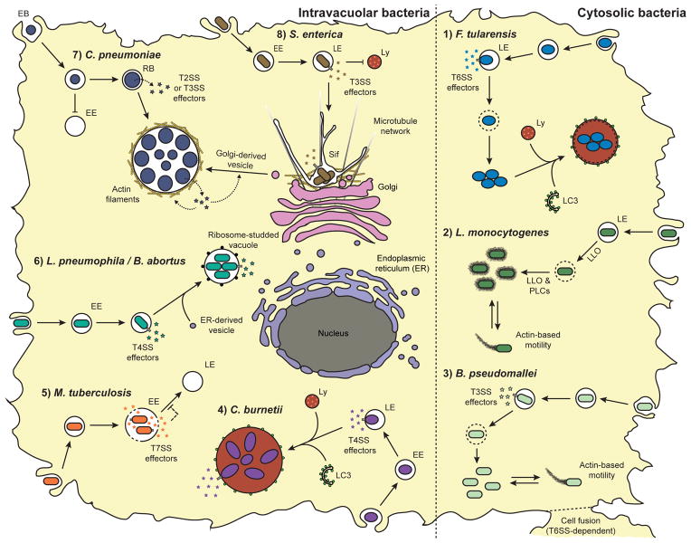
Intracellular bacteria are often clinically relevant pathogens that infect virtually every cell type found in host organisms. However, myeloid cells, especially macrophages, constitute the primary cells targeted by most species of intracellular bacteria. Paradoxically, macrophages possess an extensive antimicrobial arsenal and are efficient at killing microbes. In addition to their ability to detect and signal the presence of pathogens, macrophages sequester and digest microorganisms using the phagolysosomal and autophagy pathways or, ultimately, eliminate themselves through the induction of programmed cell death. Consequently, intracellular bacteria influence numerous host processes and deploy sophisticated strategies to replicate within these host cells. Although most intracellular bacteria have a unique intracellular life cycle, these pathogens are broadly categorized into intravacuolar and cytosolic bacteria. Following phagocytosis, intravacuolar bacteria reside in the host endomembrane system and, to some extent, are protected from the host cytosolic innate immune defenses. However, the intravacuolar lifestyle requires the generation and maintenance of unique specialized bacteria-containing vacuoles and involves a complex network of host-pathogen interactions. Conversely, cytosolic bacteria escape the phagolysosomal pathway and thrive in the nutrient-rich cytosol despite the presence of host cell-autonomous defenses. The understanding of host-pathogen interactions involved in the pathogenesis of intracellular bacteria will continue to provide mechanistic insights into basic cellular processes and may lead to the discovery of novel therapeutics targeting infectious and inflammatory diseases.


Figures

Figure 1
Figure 1
Lifestyles of intracellular bacterial pathogens. (1) F. tularensis escapes a late endosome (LE)-like vacuole in a T6SS-dependent manner. Following replication in the cytosol, F. tularensis may retranslocate to a membrane-bound compartment resembling an autolysosome. (2) L. monocytogenes escapes the phagolysosomal pathway using the T2SS (Sec) effectors LLO and PLCs. L. monocytogenes replicates rapidly in the cytosol and hijacks the host actin polymerization machinery to move within and between cells. (3) B. pseudomallei escapes into the cytosol in a T3SS-dependent manner. B. pseudomallei performs actin-based motility and promotes host cell fusion. (4) C. burnetii is adapted to the phagolysosomal pathway and resides in a spacious phagolysosomal-like compartment. The Dot/Icm system (T4SS) is required for recruiting the autophagosomal marker LC3 and for vacuole biogenesis. (5) M. tuberculosis arrests phagosome maturation at the early endosome (EE) stage in a T7SS-dependent manner. (6) L. pneumophila and B. abortus segregate from the endocytic route at the EE stage, recruit endoplasmic reticulum (ER)-derived vesicles, and form ribosome-studded specialized vacuoles in a T4SS-dependent manner. (7) C. pneumoniae segregates from the endocytic route and form a unique inclusion vacuole by recruiting Golgi-derived vesicles. C. pneumoniae effectors promote Golgi fragmentation and generate actin filaments around the inclusion. Chlamydia is found in two different forms: the non-replicating infectious elementary body (EB) and the intracytoplasmic replicative reticulate body (RB). T2SS and T3SS effectors are thought to be involved in the intracellular life cycle of Chlamydia. (8) S. enterica replicates in a late endosome (LE)-like compartment that excludes lysosomal degradation enzymes. The S. enterica containing vacuole migrates to the microtubule-organizing centre (MTOC) and forms Salmonella-induced filaments (Sif) along microtubules in a T3SS-dependent manner.

References

    1. Fauci AS, Morens DM. The perpetual challenge of infectious diseases. N Engl J Med. 2012;366:454–461. - PubMed
    1. Price JV, Vance RE. The macrophage paradox. Immunity. 2014;41:685–693. - PubMed
    1. Dumler JS, Choi KS, Garcia-Garcia JC, Barat NS, Scorpio DG, Garyu JW, Grab DJ, Bakken JS. Human granulocytic anaplasmosis and Anaplasma phagocytophilum. Emerg Infect Dis. 2005;11:1828–1834. - PMC - PubMed
    1. Franco MP, Mulder M, Gilman RH, Smits HL. Human brucellosis. Lancet Infect Dis. 2007;7:775–786. - PubMed
    1. Piggott JA, Hochholzer L. Human melioidosis. A histopathologic study of acute and chronic melioidosis. Arch Pathol. 1970;90:101–111. - PubMed

Publication types

Cite

AltStyle によって変換されたページ (->オリジナル) /