This site needs JavaScript to work properly. Please enable it to take advantage of the complete set of features!
Skip to main page content
U.S. flag

An official website of the United States government

Dot gov

The .gov means it’s official.
Federal government websites often end in .gov or .mil. Before sharing sensitive information, make sure you’re on a federal government site.

Https

The site is secure.
The https:// ensures that you are connecting to the official website and that any information you provide is encrypted and transmitted securely.

NIH NLM Logo
Log in
Access keys NCBI Homepage MyNCBI Homepage Main Content Main Navigation
. 2016 May 31;113(22):6277-82.
doi: 10.1073/pnas.1600488113. Epub 2016 May 18.

Broadly neutralizing epitopes in the Plasmodium vivax vaccine candidate Duffy Binding Protein

Affiliations

Broadly neutralizing epitopes in the Plasmodium vivax vaccine candidate Duffy Binding Protein

Edwin Chen et al. Proc Natl Acad Sci U S A. .

Abstract

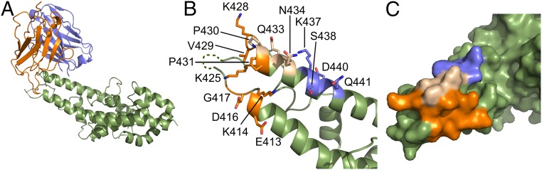
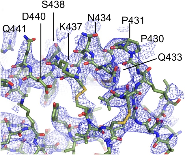
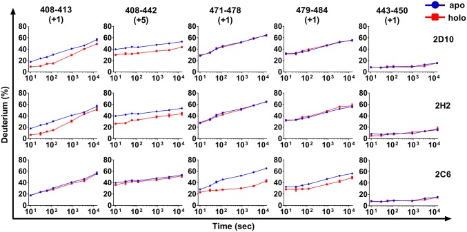
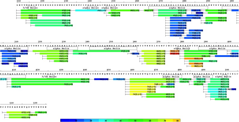
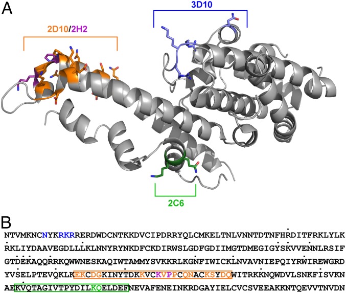
Plasmodium vivax Duffy Binding Protein (PvDBP) is the most promising vaccine candidate for P. vivax malaria. The polymorphic nature of PvDBP induces strain-specific immune responses, however, and the epitopes of broadly neutralizing antibodies are unknown. These features hamper the rational design of potent DBP-based vaccines and necessitate the identification of globally conserved epitopes. Using X-ray crystallography, small-angle X-ray scattering, hydrogen-deuterium exchange mass spectrometry, and mutational mapping, we have defined epitopes for three inhibitory mAbs (mAbs 2D10, 2H2, and 2C6) and one noninhibitory mAb (3D10) that engage DBP. These studies expand the currently known inhibitory epitope repertoire by establishing protective motifs in subdomain three outside the receptor-binding and dimerization residues of DBP, and introduce globally conserved protective targets. All of the epitopes are highly conserved among DBP alleles. The identification of broadly conserved epitopes of inhibitory antibodies provides critical motifs that should be retained in the next generation of potent vaccines for P. vivax malaria.

Keywords: Duffy Binding Protein; Plasmodium vivax; broadly neutralizing; epitopes; malaria.


Conflict of interest statement

The authors declare no conflict of interest.

Figures

Fig. 1.
Fig. 1.
Crystal structure of the DBP-II/2D10scFv complex. (A) Overall structure of the DBP-II/2D10scFv complex shown in ribbon representation. The DBP-II domain is colored in green. The scFv heavy chain (VH) is in blue, and the light chain (VL) is in orange. (B) Ribbon representation of DBP-II mapping the 2D10 epitopes. Residues contacted by the scFv are shown in stick form. Residues contacted by the heavy chain are colored blue, residues contacted by the light chain are in orange, and residues contacted by both are in beige. Residues not contacted by mAb are in green. Regions of disorder are shown as a dotted line. (C) Surface representation of the DBP-II. Color scheme is as in B.
Fig. S1.
Fig. S1.
2Fo-Fc electron density contoured at 1σ around the epitope clearly identifies contact sites in DBP. The mAb is removed for clarity, and only DBP and associated electron density are shown. In this orientation, clear electron density for residues 433–441 that compose part of the epitope for 2D10 is observed.
Fig. 2.
Fig. 2.
mAbs 2D10 and 2H2 share an epitope that is distinct from that of 2C6. (A) Plot of scattering intensity (I) against scattering momentum (Q) and statistical fit of theoretical scatter from the DBP-II/2D10scFv crystal structure (red line) with the experimental SAXS profile of the DBP-II/2D10Fab complex (red line) with a χ2 value of 2.3. (B) SAXS pairwise comparison of 2D10, 2H2, and 2C6. (C) Plot of scattering intensity (I) against scattering momentum (Q) and statistical fit of theoretical scatter from the DBP-II/2D10scFv crystal structure (red line) with the experimental SAXS profile of the DBP-II/2H2Fab complex (red line), with a χ2 value of 1.9. (D) Plot of scattering intensity (I) against scattering momentum (Q) and statistical fit of theoretical scatter from the DBP-II/2D10scFv crystal structure (red line) with the experimental SAXS profile of the DBP-II/2C6Fab complex (red line), with a χ2 value of 5.1.
Fig. S2.
Fig. S2.
Heavy and light chain alignments for mAbs 2D10, 2H2, and 2C6. Gray indicates similar residues; black indicates identical residues.
Fig. 3.
Fig. 3.
Comparison of the kinetics of HDX for five regions of DBP in the presence of various mAbs (holo state, depicted in red) and in the absence of the mAb (apo state, depicted in blue). Each region (column) is represented by a peptic peptide and its charge state, as measured by mass spectrometry. Each row represents a state bound with a mAb; the antibody is listed on the right. Those regions showing reduced rates or extents of exchange for the holo state (red) are considered to contain the epitopes. Those regions showing no difference are examples of region that do not contain the epitopes, and can be viewed as controls.
Fig. S3.
Fig. S3.
Sequence coverage of peptic digestion with an optimized quenching/reducing condition and HDX heat map for apo PvDBP. Each bar represents a peptide analyzed for deuterium content by mass spectrometry. Average deuterium uptake level and SD for the duplicate analysis across all exchange time points are listed on each bar. Bars are also color-coded, with warmer colors indicating higher deuterium uptake levels. Stable secondary structural features identified by crystallography (PDB ID code 3RRC) are represented by boxes underneath the corresponding segments.
Fig. 4.
Fig. 4.
Determination of mAb epitopes by surface mutant libraries and ELISA. mAbs 2D10, 2H2, 3D10, and 2C6 were tested for binding against a panel of Sal-1 DBP surface mutants as described in Experimental Procedures. (A) Sal-1 DBP wild type. (B) Mutant 6 (K414A, P430A, P431A, N434A, K437A, Q441A). (C) Mutant 9 (N434R, K437R). (D) Mutant 1 (N218S, R221G, K222S, R223G). (E) Mutant 12 (K479A, Q480A). (F) Mutant 8 (K428A, P430A). All error bars are ±1 SD, calculated from three replicate experiments.
Fig. S4.
Fig. S4.
Alignment of mutant sequences. An alignment of the sequences of all mutants tested in this study and wild type Sal-1 DBP, with mutated residues highlighted in black and the sub domains of DBP (SD1, red; SD2, blue; and SD3, green) outlined above the alignment.
Fig. S5.
Fig. S5.
Binding of monoclonal antibodies to a panel of Sal-1 DBP mutants. mAbs 2D10, 2H2, 3D10, and 2C6 were tested for binding against a panel of Sal-1 DBP mutants and BSA as described in Experimental Procedures. (A) BSA. (B) Mutant 2. (C) Mutant 3. (D) Mutant 4. (E) Mutant 5. (F) Mutant 7. (G) Mutant 10. (H) Mutant 11. (I) Mutant 13. (J) Mutant 14. (K) Mutant 15. (L) Mutant 16.
Fig. 5.
Fig. 5.
Competition ELISA demonstrates that 2D10 and 2H2 share the same epitope. mAbs 2D10, 2H2, 3D10, and 2C6 were tested at different molar ratios to 2D10 (×ばつ, ×ばつ, and ×ばつ) for the ability to compete with 2D10 for binding to DBP. All error bars are ±1 SD, calculated from three replicate experiments.
Fig. 6.
Fig. 6.
Epitopes of 2D10, 2H2, 2C6, and 3D10 mapped on PvDBP reveal that the epitopes are broadly conserved. (A) Epitopes are mapped on the surface of PvDBP with 2D10 in orange, 2H2 specific residues in purple, 3D10 in blue, and 2C6 in green. (B) Sequence of the Sal-1 DBP-II region with identified polymorphic sites indicated by dots above the individual residues. Structurally, mutationally, and HDX-MS (boxes)-identified epitopes are highlighted for 2D10 in orange, 2H2 in purple, for 2C6 in green, and for 3D10 in blue.

References

    1. Carlton JM, Sina BJ, Adams JH. Why is Plasmodium vivax a neglected tropical disease? PLoS Negl Trop Dis. 2011;5(6):e1160. - PMC - PubMed
    1. Tjitra E, et al. Multidrug-resistant Plasmodium vivax associated with severe and fatal malaria: A prospective study in Papua, Indonesia. PLoS Med. 2008;5(6):e128. - PMC - PubMed
    1. Genton B, et al. Plasmodium vivax and mixed infections are associated with severe malaria in children: A prospective cohort study from Papua New Guinea. PLoS Med. 2008;5(6):e127. - PMC - PubMed
    1. Guerra CA, et al. The international limits and population at risk of Plasmodium vivax transmission in 2009. PLoS Negl Trop Dis. 2010;4(8):e774. - PMC - PubMed
    1. Price RN, et al. Vivax malaria: Neglected and not benign. Am J Trop Med Hyg. 2007;77(6) Suppl:79–87. - PMC - PubMed

Publication types

MeSH terms

Associated data

Cite

AltStyle によって変換されたページ (->オリジナル) /