This site needs JavaScript to work properly. Please enable it to take advantage of the complete set of features!
Skip to main page content
U.S. flag

An official website of the United States government

Dot gov

The .gov means it’s official.
Federal government websites often end in .gov or .mil. Before sharing sensitive information, make sure you’re on a federal government site.

Https

The site is secure.
The https:// ensures that you are connecting to the official website and that any information you provide is encrypted and transmitted securely.

NIH NLM Logo
Log in
Access keys NCBI Homepage MyNCBI Homepage Main Content Main Navigation
. 2016 Mar 9:7:290.
doi: 10.3389/fmicb.2016.00290. eCollection 2016.

Virulence of Cryptococcus sp. Biofilms In Vitro and In Vivo using Galleria mellonella as an Alternative Model

Affiliations

Virulence of Cryptococcus sp. Biofilms In Vitro and In Vivo using Galleria mellonella as an Alternative Model

Tatiane Benaducci et al. Front Microbiol. .

Abstract

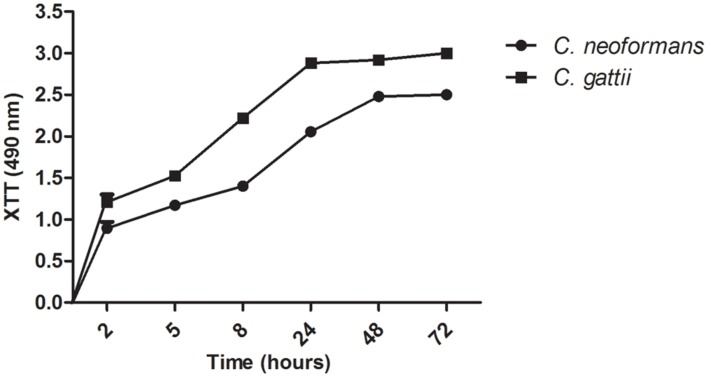
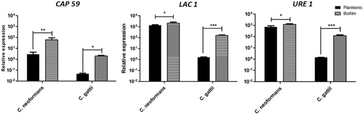
Cryptococcus neoformans and C. gattii are fungal pathogens that are most commonly found in infections of the central nervous system, which cause life-threatening meningoencephalitis and can grow as a biofilm. Biofilms are structures conferring protection and resistance of microorganism to the antifungal drugs. This study compared the virulence of planktonic and biofilm cells of C. neoformans and C. gattii in Galleria mellonella model, as well as, the quantification of gene transcripts LAC1, URE1, and CAP59 by real time PCR. All three of the genes showed significantly increased expressions in the biofilm conditions for two species of Cryptococcus, when compared to planktonic cells. C. neoformans and C. gattii cells in the biofilm forms were more virulent than the planktonic cells in G. mellonella. This suggests that the biofilm conditions may contribute to the virulence profile. Our results contribute to a better understanding of the agents of cryptococcosis in the host-yeast aspects of the interaction.

Keywords: Cryptococcus sp.; Galleria mellonella; biofilms; virulence.


Figures

FIGURE 1
FIGURE 1
Scanning electron microscopy (SEM) showing yeast communities adhered to surfaces of glass coverslips indicating the formation of mature biofilms. (A) Biofilm of Cryptococcus neoformans ATCC 90112 at ×ばつ magnification. (B) Biofilm of C. neoformans ATCC 90112 at ×ばつ magnification. (C) Biofilm of C. gattii 56990 at ×ばつ magnification. (D) Biofilm of C. gattii 56990 at ×ばつ magnification. The blue arrows indicate the self-produced matrix.
FIGURE 2
FIGURE 2
Confocal laser microscopy showing metabolically active cells in mature biofilms. Staining with FUN1 fluorochrome shows the cytoplasm of metabolically active cells dyed orange–yellow. Staining with concavalin associated with Alexa Fluor 488 (CAAF) shows the presence of mannose in the cell wall of Cryptococcus sp. Orthogonal images show the thickness and architecture of the biofilms. (A) Mature biofilm of C. neoformans ATCC 90112. (B) Thickness of mature biofilm of C. neoformans ATCC 90112. (C) Mature biofilm of C. gattii ATCC 56990. (D) Thickness of mature biofilm of C. gattii ATCC 56990.
FIGURE 3
FIGURE 3
Kinetics of biofilm formation by Cryptococcus sp. in microdilution plates as determined by the XTT reduction assay.
FIGURE 4
FIGURE 4
Expression of the CAP59, laccase (LAC1), and urease (URE1) genes normalized by GAPDH in biofilms and planktonic cells of C. neoformans and C. gattii. Graphs are plotted in power of 10 to better visualization the results. *p < 0.5, **p < 0.01, and ***p < 0.001.
FIGURE 5
FIGURE 5
Comparison of virulence from planktonic and biofilm cells of C. neoformans ATCC 90112 and C. gattii ATCC 56990 using Galleria mellonella as the infection model. The differences were statistically significant (p < 0.05). Graphs represent the average of three repetitions.

References

    1. Albuquerque P. C., Fonseca F. L., Dutra F. F., Bozza M. T., Frases S., Casadevall A., et al. (2014). Cryptococcus neoformans glucuronoxylomannan fractions of different molecular masses are functionally distinct. Future Microbiol. 9 147–161. 10.2217/fmb.13.163 - DOI - PMC - PubMed
    1. Albuquerque P. C., Rodrigues M. L. (2012). Research trends on pathogenic Cryptococcus species in the last 20 years: a global analysis with focus on Brazil. Future Microbiol. 7 319–329. 10.2217/fmb.11.162 - DOI - PubMed
    1. Alspaugh J. A., Perfect J. R., Heitman J. (1998). Signal transduction pathways regulating differentiation and pathogenicity of Cryptococcus neoformans. Fungal Genet. Biol. 25 1–14. 10.1006/fgbi.1998.1079 - DOI - PubMed
    1. Bach M. C., Tally P. W., Godofsky E. W. (1997). Use of cerebrospinal fluid shunts in patients having acquired immunodeficiency syndrome with cryptococcal meningitis and uncontrollable intracranial hypertension. Neurosurgery 41 1280–1282. 10.1097/00006123-199712000-00008 - DOI - PubMed
    1. Banerjee U., Gupta K., Venugopal P. (1997). A case of prosthetic valve endocarditis caused by Cryptococcus neoformans var. neoformans. J. Med. Vet. Mycol. 35 139–141. 10.1080/02681219780001031 - DOI - PubMed

LinkOut - more resources

Cite

AltStyle によって変換されたページ (->オリジナル) /