This site needs JavaScript to work properly. Please enable it to take advantage of the complete set of features!
Skip to main page content
U.S. flag

An official website of the United States government

Dot gov

The .gov means it’s official.
Federal government websites often end in .gov or .mil. Before sharing sensitive information, make sure you’re on a federal government site.

Https

The site is secure.
The https:// ensures that you are connecting to the official website and that any information you provide is encrypted and transmitted securely.

NIH NLM Logo
Log in
Access keys NCBI Homepage MyNCBI Homepage Main Content Main Navigation
. 2015 Dec 23:8:657.
doi: 10.1186/s13071-015-1255-x.

New insights into the evolution of the Trypanosoma cruzi clade provided by a new trypanosome species tightly linked to Neotropical Pteronotus bats and related to an Australian lineage of trypanosomes

Affiliations

New insights into the evolution of the Trypanosoma cruzi clade provided by a new trypanosome species tightly linked to Neotropical Pteronotus bats and related to an Australian lineage of trypanosomes

Luciana Lima et al. Parasit Vectors. .

Abstract

Background: Bat trypanosomes are implicated in the evolution of the T. cruzi clade, which harbours most African, European and American trypanosomes from bats and other trypanosomes from African, Australian and American terrestrial mammals, including T. cruzi and T. rangeli, the agents of the American human trypanosomiasis. The diversity of bat trypanosomes globally is still poorly understood, and the common ancestor, geographical origin, and evolution of species within the T. cruzi clade remain largely unresolved.

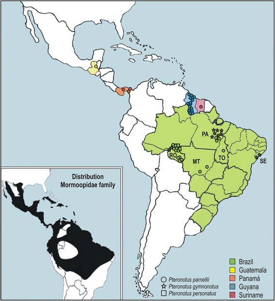
Methods: Trypanosome sequences were obtained from cultured parasites and from museum archived liver/blood samples of bats captured from Guatemala (Central America) to the Brazilian Atlantic Coast. Phylogenies were inferred using Small Subunit (SSU) rRNA, glycosomal glyceraldehyde phosphate dehydrogenase (gGAPDH), and Spliced Leader (SL) RNA genes.

Results: Here, we described Trypanosoma wauwau n. sp. from Pteronotus bats (Mormoopidae) placed in the T. cruzi clade, then supporting the bat-seeding hypothesis whereby the common ancestor of this clade likely was a bat trypanosome. T. wauwau was sister to the clade T. spp-Neobats from phyllostomid bats forming an assemblage of trypanosome species exclusively of Noctilionoidea Neotropical bats, which was sister to an Australian clade of trypanosomes from indigenous marsupials and rodents, which possibly evolved from a bat trypanosome. T. wauwau was found in 26.5% of the Pteronotus bats examined, and phylogeographical analysis evidenced the wide geographical range of this species. To date, this species was not detected in other bats, including those that were sympatric or shared shelters with Pteronotus. T. wauwau did not develop within mammalian cells, and was not infective to Balb/c mice or to triatomine vectors of T. cruzi and T. rangeli.

Conclusions: Trypanosoma wauwau n. sp. was linked to Pteronotus bats. The positioning of the clade T. wauwau/T.spp-Neobats as the most basal Neotropical bat trypanosomes and closely related to an Australian lineage of trypanosomes provides additional evidence that the T. cruzi clade trypanosomes likely evolved from bats, and were dispersed in bats within and between continents from ancient to unexpectedly recent times.


Figures

Fig. 1
Fig. 1
Geographical origin of Trypanosoma wauwau isolates obtained from hemocultures and archive blood/tissue samples from Pteronotus bats captured in Central and South America
Fig. 2
Fig. 2
Barcoding and phylogeographical analysis of new trypanosomes from Neotropical bats. Phylogenetic analysis of V7V8 SSU rRNA sequences of trypanosomes from cultures and bat blood/tissue samples from Pteronotus and Phyllostomidae bats from Central and South America. The numbers on the nodes are bootstrap values (P/ML) derived from 500 replicates
Fig. 3
Fig. 3
Positioning of T. wauwau in the phylogenetic tree of Trypanosoma. Phylogenetic tree (ML) inferred using whole SSU rRNA sequences from the trypanosomes of Noctilionoidea Neotropical bats: T. wauwau, to date found exclusively in Pteronotus bats clustered with the trypanosomes of the clade T. spp. Neobats (Panamanian, Colombian and Brazilian Phyllostomidae bats) close to the clade of Australian bats. The analyses included species of all major clades of Trypanosoma and trypanosomatids of other genera as outgroups (1.728 characters, –Ln = -8870.359849). The numbers at the nodes correspond respectively to P, ML (500 replicates) and BI support values. Codes within parenthesis are GenBank accession numbers
Fig. 4
Fig. 4
Phylogenetic relationships between T. wauwau and the other T. cruzi clade trypanosomes. ML phylogenetic analysis based on the concatenated sequences of V7V8 SSU rRNA and gGAPDH genes (1.691 characters, –Ln = 8457.739334) from seven isolates (genotypes Pt1 and Pt2) of T. wauwau, two isolates of T. spp. Neobats, other 24 bat trypanosomes, and 13 trypanosomes from other mammals. Species of the clade T. lewisi were used as outgroup. The numbers at the nodes correspond respectively to P, ML (500 replicates) and BI support values
Fig. 5
Fig. 5
The SL RNA primary and secondary structures of T. wauwau and its closest trypanosomes. a, Aligned sequences of the SL RNA transcripts from T. wauwau (Pt1 and Pt2 genotypes), and other T. cruzi clade trypanosomes. b, Network of SL RNA transcript sequences and almost identical secondary structures shared by Neotropical T. wauwau and Australian T. sp H25 (differences are indicated by arrows in the T. wauwau SL structure), and highly similar to that of T. livingstonei from African bats. Numbers in nodes correspond to bootstrap values estimated by 500 replicates using the same parameters optimized for network inferences
Fig. 6
Fig. 6
Pteronotus parnellii and developmental forms of T. wauwau: Light microscopy of Giemsa-stained forms: A, trypomastigotes in bat blood smear. B, flagellates co-cultivated with Hi-5 insect cells: Supernatants of early cultures showing small and rounded division forms (a, b, c), multiple fission forms united by the posterior extremity exhibiting various nuclei, kinetoplasts and flagella (a, c), rosettes of epimastigotes attached by the flagella (b), epimastigotes largely varying in shape and size (b, d-f), log-phase regular epimastigote that multiply by binary fission (d), small trypomastigotes with terminal kinetoplast of stationary cultures (g). C, Epimastigotes (a, b) and trypomastigotes (b) with noticeable undulant membrane, and small trypomastigotes (a) in the supernatant of LLC-MK2 mammalian cells at 37 °C. Trypomastigotes are indicated by black stars. Nucleus (N); Kinetoplast (K); Flagellum (F), Undulant Membrane (UM). Scale bars: 10 μm
Fig. 7
Fig. 7
Morphology of T. wauwau developmental forms in culture assessed by SEM. Small dividing forms and rounded flagellates common in early cultures (a, b),rosettes of epimastigotes attached by the flagella (c), large multiple fission form showing the anterior ends and the flagella of many parasites that remained united by the posterior extremity (d), epimastigotes of variable size and shape likely originated from multiple and irregular division forms (e, f, h), log-phase regular epimastigotes and binary division (g), small flagellate resembling the trypomastigotes of stationary-phase cultures (i)
Fig. 8
Fig. 8
Ultrastructural features of T. wauwau revealed by TEM microscopy. Cultured epimastigotes: transversal section showing three nuclei with large and condensed nucleolus and a single kinetoplast (a); acidocalcisomes (b), flagellum with a conspicuous paraxial structure (b’, c), highly compacted disk-shaped kinetoplast (b, c), enlarged mitochondria filled with many cristae (c), structure resembling a short cytostome-cytopharynx complex (d). Nucleus (N), Kinetoplast (K), Flagellum (F), Acidocalcisomes (Ac), Mitochondria (M), Cytostome (Cy), Paraxial structure (PR)

References

    1. Hamilton PB, Teixeira MMG, Stevens JR. The evolution of Trypanosoma cruzi: the ‘bat seeding’ hypothesis. Trends Parasitol. 2012;28:136–141. doi: 10.1016/j.pt.201201006. - DOI - PubMed
    1. Hamilton PB, Cruickshank C, Stevens JR, Teixeira MMG, Mathews F. Parasites reveal movement of bats between the New and Old Worlds. Mol Phylogenet Evol. 2012;63:521–526. doi: 10.1016/j.ympev.201201007. - DOI - PMC - PubMed
    1. Lima L, Maia da Silva F, Neves L, Attias M, Takata CS, Campaner M, et al. Evolutionary insights from bat trypanosomes: morphological, developmental and phylogenetic evidence of a new species, Trypanosoma (Schizotrypanum) erneyi sp. nov. in African bats closely related to Trypanosoma(Schizotrypanum) cruzi and allied species. Protist. 2012;163:856–872. doi: 10.1016/j.protis.201112003. - DOI - PubMed
    1. Lima L, Espinosa-Álvarez O, Hamilton PB, Neves L, Takata CSA, Campaner M, et al. Trypanosoma livingstonei: a new species from African bats supports the bat seeding hypothesis for the Trypanosoma cruzi clade. Parasit Vectors. 2013;6(1):221. doi: 10.1186/1756-3305年6月22日1. - DOI - PMC - PubMed
    1. Lima L, Espinosa-Álvarez O, Ortiz P, Trejo-Varon JA, Carranza JC, Pinto CM, et al. Genetic diversity of Trypanosoma cruzi in bats, and multilocus phylogenetic and phylogeographical analyses supporting Tcbat as an independent DTU (discrete typing unit) Acta Trop. 2015;151:166–177. doi: 10.1016/j.actatropica.2015年07月01日5. - DOI - PubMed

Publication types

MeSH terms

Substances

LinkOut - more resources

Cite

AltStyle によって変換されたページ (->オリジナル) /