This site needs JavaScript to work properly. Please enable it to take advantage of the complete set of features!
Skip to main page content
U.S. flag

An official website of the United States government

Dot gov

The .gov means it’s official.
Federal government websites often end in .gov or .mil. Before sharing sensitive information, make sure you’re on a federal government site.

Https

The site is secure.
The https:// ensures that you are connecting to the official website and that any information you provide is encrypted and transmitted securely.

NIH NLM Logo
Log in
Access keys NCBI Homepage MyNCBI Homepage Main Content Main Navigation
doi: 10.1038/srep17609.

Environmental marine pathogen isolation using mesocosm culture of sharpsnout seabream: striking genomic and morphological features of novel Endozoicomonas sp

Affiliations

Environmental marine pathogen isolation using mesocosm culture of sharpsnout seabream: striking genomic and morphological features of novel Endozoicomonas sp

Pantelis Katharios et al. Sci Rep. .

Abstract

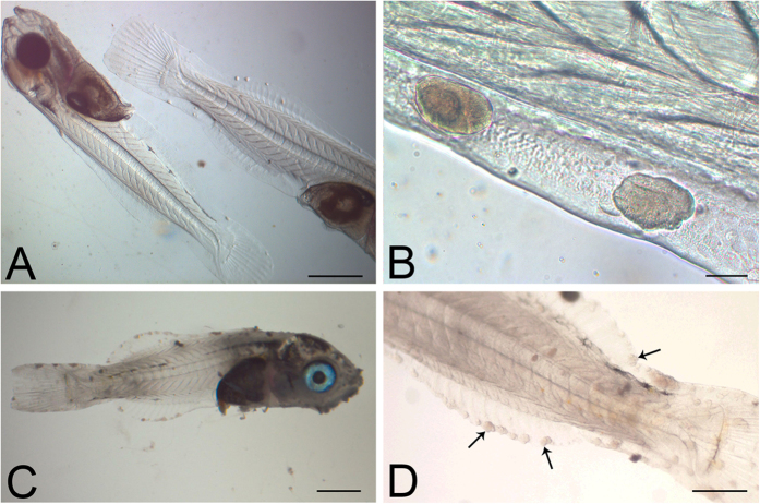
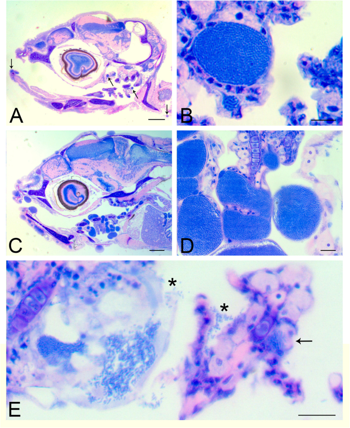
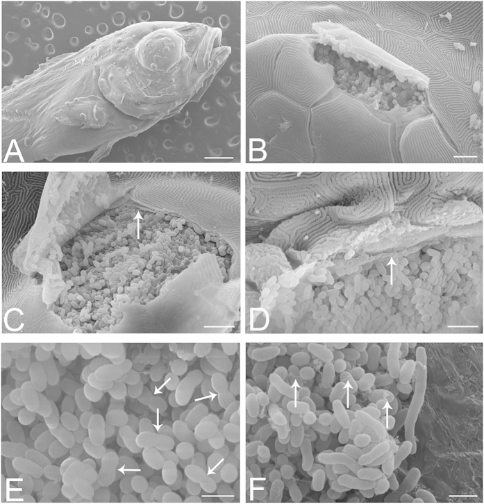
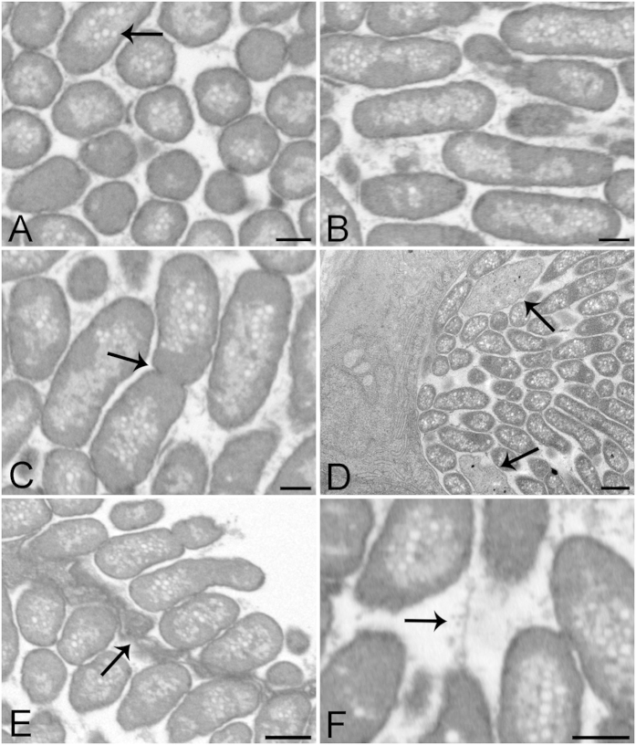
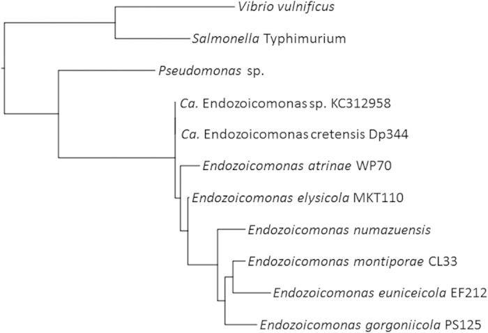
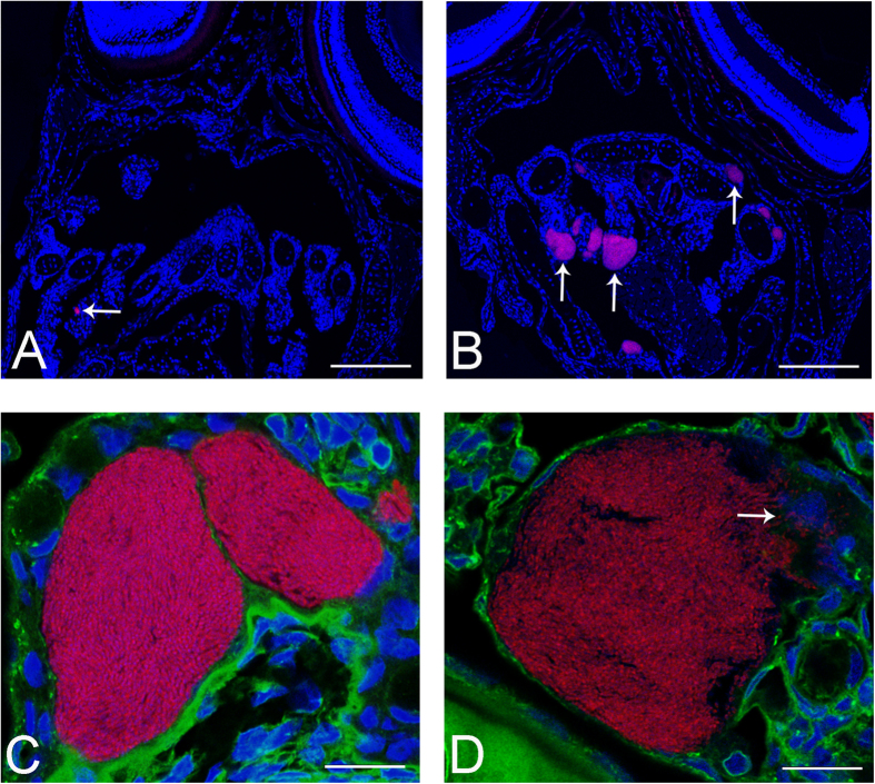
Aquaculture is a burgeoning industry, requiring diversification into new farmed species, which are often at risk from infectious disease. We used a mesocosm technique to investigate the susceptibility of sharpsnout seabream (Diplodus puntazzo) larvae to potential environmental pathogens in seawater compared to control borehole water. Fish exposed to seawater succumbed to epitheliocystis from 21 days post hatching, causing mortality in a quarter of the hosts. The pathogen responsible was not chlamydial, as is often found in epitheliocystis, but a novel species of the γ-proteobacterial genus Endozoicomonas. Detailed characterisation of this pathogen within the infectious lesions using high resolution fluorescent and electron microscopy showed densely packed rod shaped bacteria. A draft genome sequence of this uncultured bacterium was obtained from preserved material. Comparison with the genome of the Endozoicomonas elysicola type strain shows that the genome of Ca. Endozoicomonas cretensis is undergoing decay through loss of functional genes and insertion sequence expansion, often indicative of adaptation to a new niche or restriction to an alternative lifestyle. These results demonstrate the advantage of mesocosm studies for investigating the effect of environmental bacteria on susceptible hosts and provide an important insight into the genome dynamics of a novel fish pathogen.


Figures

Figure 1
Figure 1. Bright field images of representative whole larvae.
(A,B) Larvae 21 dph showing epitheliocystis infection, the cysts most readily apparent in the periphery of the fins. (C,D) Larvae 28 dph, heavily infected with epitheliocystis affecting fins and skin. Several cysts are indicated with arrows. Images were taken immediately after larval euthanasia. Scale bars represent: (A,C) 1 mm, (B) 50 μm, (D) 500 μm.
Figure 2
Figure 2. Histopathology of representative infected larvae.
(A) Section of the head of a 21 dph larva with cysts (arrows) in gills and skin. (B) A typical large cyst in the gills of a 21 dph infected larva with particulate appearance, and a small one below it, are visible. (C) Section of the head of an infected larva 28 dph, with extensive and multiple cysts in the epithelium of the skin, mouth and gills. (D) Higher magnification of the gill lamellae from (C) showing bacterial-sized particles within cysts. (E) An empty inclusion from a 21 dph larva with freed bacteria (*) attaching to the next filament. There is also a newly formed small inclusion (arrow) at the base of the lamella. Scale bars represent: (A,C) 200 μm, (B) 10 μm, (D,E) 20 μm.
Figure 3
Figure 3. SEM of infected larvae at 21 dph.
(A) Surface view of larva, showing cysts on skin and fins. (B) A ruptured cyst beneath the skin appears to be densely packed with bacteria. (C) Ruptured cyst revealing the bacteria to be separated from the epithelial cells by a membrane (arrow) with a fibrous-like appearance, and with multiple fenestrations or perforations. Should the cyst microenvironment be tightly regulated by the enveloping cells, then presumably these would need to form tight junctional contacts with each other and with the cyst itself. (D) Bacteria appear to be in intimate contact with the surrounding membrane (arrow), which appears to partially fold around the bacteria, creating shallow bays for them. (E) Within a cyst, bacteria appear to be dividing (arrows). (F) Another view shows elongated bacterial forms and filaments (arrows). Scale bars represent: (A) 500 μm, (B) 10 μm, (C,D) 5 μm, (E,F) 2 μm.
Figure 4
Figure 4. Representative FIB-SEM and TEM images of epitheliocysts from larvae 24 dph and 20 dph respectively.
(A,B) FIB-SEM images from a cyst 3D volume at cross and longitudinal planes showing bacterial size, orientation, shape and compactness. Bacteria are surrounded by a densely stained single membrane containing a dense homogeneous layer. The centres of the bacteria appear lighter with many pale vacuoles (arrow). (C) FIB-SEM image of dividing bacterium (arrow). Granular material is visible between the bacteria. (D) TEM image of a transverse section of 20 dph larva showing the edge of a cyst with bacteria cut lengthwise and in cross-section. Two amorphous bodies are also present (arrows). (E) FIB-SEM of two neighbouring cysts separated by a cyst boundary (arrow). This tissue presents involuted membranes, partially enveloping the bacteria. (F) FIB-SEM image showing a network of thin filaments between the bacteria. Scale bars represent: (AC,F) 500 nm, (D,E) 1 μm.
Figure 5
Figure 5. Phylogenetic analysis of the novel epitheliocystis agent within the γ-proteobacteria.
Representative 16S rRNA gene sequence Dp344 amplified from 21 dph larva, compared against all Endozoicomonas species, contextualised with other γ-proteobacteria. Sequences aligned with muscle, phylogeny by PhyML. Bootstraps within the Endozoicomonas genus are all over 85%. A further nine sequenced clones showed 1-2 mismatches relative to Dp344 over the length of the 16S rRNA gene (data not shown).
Figure 6
Figure 6. FISH of larvae using probes based on Endozoicomonas 16S rRNA gene sequences.
All sections are hybridised with Endo-0474-Cy3 (red) for bacteria and are counterstained with DAPI. (A) Section of larva 20 dph with one bacteria-containing cyst visible (arrow). (B) Section of larva 24 dph with several cysts in the gills visible (arrows). Sections from 28 dph contained many more labelled cysts (data not shown). (C,D) High resolution confocal images (deconvolved) of Endozoicomonas epitheliocysts from larvae 24 dph additionally labelled with concanavalin A-Alexa488, showing large, granular bacteria-containing cysts lying within the gill epithelium, displacing the nuclei of surrounding cells. Arrow (D) indicates several macrophages attacking the bacteria where the cyst is ruptured. Scale bars represent: (A,B) 100 μm, (C,D) 10 μm.

References

    1. Divinach P. & Kentouri M. Hatchery techniques for specific diversification in Mediterranean finfish larviculture. Cahiers Options Mediterraneennes 47, 75–87 (2000).
    1. Papandroulakis N., Mylonas C. C., Maingot E. & Divanach P. First results of greater amberjack (Seriola dumerili) larval rearing in mesocosm. Aquaculture 250, 155–161, 10.1016/j.aquaculture.2005年02月03日6 (2005). - DOI
    1. Papandroulakis N., Kentouri M., Maingot E. & Divanach P. Mesocosm: A Reliable Technology for Larval Rearing of Diplodus puntazzo and Diplodus sargus sargus. Aquaculture International 12, 345–355, 10.1023/B:AQUI.0000042134.21211.ab (2004). - DOI
    1. Bergh Ø. The dual myths of the healthy wild fish and the unhealthy farmed fish. Dis Aquat Org 75, 159–164 (2007). - PubMed
    1. Segner H. et al. Health of farmed fish: its relation to fish welfare and its utility as welfare indicator. Fish Physiol Biochem 38, 85–105, 10.1007/s10695-011-9517-9 (2011). - DOI - PubMed

Publication types

Substances

LinkOut - more resources

Cite

AltStyle によって変換されたページ (->オリジナル) /