This site needs JavaScript to work properly. Please enable it to take advantage of the complete set of features!
Skip to main page content
U.S. flag

An official website of the United States government

Dot gov

The .gov means it’s official.
Federal government websites often end in .gov or .mil. Before sharing sensitive information, make sure you’re on a federal government site.

Https

The site is secure.
The https:// ensures that you are connecting to the official website and that any information you provide is encrypted and transmitted securely.

NIH NLM Logo
Log in
Access keys NCBI Homepage MyNCBI Homepage Main Content Main Navigation
. 2015 Oct 21;7(11):2996-3008.
doi: 10.1093/gbe/evv202.

Evolutionary and Phylogenetic Analysis of the Hepaciviruses and Pegiviruses

Affiliations

Evolutionary and Phylogenetic Analysis of the Hepaciviruses and Pegiviruses

Julien Thézé et al. Genome Biol Evol. .

Abstract

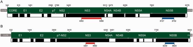
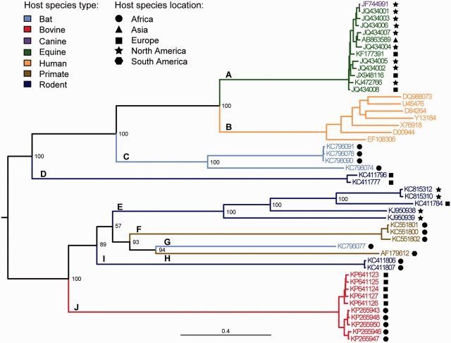
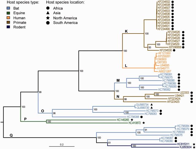
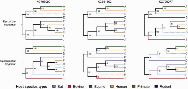
The known genetic diversity of the hepaciviruses and pegiviruses has increased greatly in recent years through the discovery of viruses related to hepatitis C virus and human pegivirus in bats, bovines, equines, primates, and rodents. Analysis of these new species is important for research into animal models of hepatitis C virus infection and into the zoonotic origins of human viruses. Here, we provide the first systematic phylogenetic and evolutionary analysis of these two genera at the whole-genome level. Phylogenies confirmed that hepatitis C virus is most closely related to viruses from horses whereas human pegiviruses clustered with viruses from African primates. Within each genus, several well-supported lineages were identified and viral diversity was structured by both host species and location of sampling. Recombination analyses provided evidence of interspecific recombination in hepaciviruses, but none in the pegiviruses. Putative mosaic genome structures were identified in NS5B gene region and were supported by multiple tests. The identification of interspecific recombination in the hepaciviruses represents an important evolutionary event that could be clarified by future sampling of novel viruses. We also identified parallel amino acid changes shared by distantly related lineages that infect similar types of host. Notable parallel changes were clustered in the NS3 and NS4B genes and provide a useful starting point for experimental studies of the evolution of Hepacivirus host-virus interactions.

Keywords: cross-species transmission; hepatitis C virus; host range; human pegivirus; parallel molecular evolution; recombination.


Figures

F<sc>ig</sc>. 1.—
Fig. 1.—
Genome structures (green) of the hepacivirus and pegivirus reference genomes. (A) HCV strain H77 and (B) simian pegivirus strain NC_001837. Positions of gene boundaries are marked above each structure. Black and white bars below each structure indicate the regions included in the genome-wide alignment (black) and regions that were unalignable (white). The locations of the NS3 and NS5B subgenomic regions that were analyzed separately are indicated in red and blue, respectively.
F<sc>ig</sc>. 2.—
Fig. 2.—
Phylogeny of the Hepaciviruses. Estimated from the genome-wide amino acid alignment using ML inference. Statistical support for phylogenetic nodes was assessed using a bootstrap approach (100 replicates). Tip and branch labels are colored by host species type while the sampling location of nonhuman hosts is denoted by the adjacent symbol. Letters indicate the different Hepacivirus lineages discussed in the main text.
F<sc>ig</sc>. 3.—
Fig. 3.—
Phylogeny of the Pegiviruses. Estimated from the genome-wide amino acid alignment using ML inference. Statistical support for phylogenetic nodes was assessed using a bootstrap approach (100 replicates). Tip and branch labels are colored by host species type while the sampling location of nonhuman hosts is denoted by the adjacent symbol. Letters indicate the different Pegivirus lineages discussed in the main text.
F<sc>ig</sc>. 4.—
Fig. 4.—
Tree topologies of recombinant hepacivirus lineages. Trees were estimated from the putative recombinant fragment (bottom row) and from remainder of the genome (top row) using ML. In each case the tree topology was fixed for all sequences except the putative recombinant sequence in question (shown along top, and highlighted in bold and with dashed branch line in each tree). Tip and branch labels are colored by host species type.

References

    1. Anthony SJ, et al. 2013. A strategy to estimate unknown viral diversity in mammals. MBio 4:e00598–13. - PMC - PubMed
    1. Baechlein C, et al. 2015. Identification of a novel hepacivirus in domestic cattle from Germany. J Virol. 89:7007–7015. - PMC - PubMed
    1. Bissig K-D, et al. 2010. Human liver chimeric mice provide a model for hepatitis B and C virus infection and treatment. J Clin Invest. 120:924–930. - PMC - PubMed
    1. Boni MF, de Jong MD, van Doorn HR, Holmes EC. 2010. Guidelines for identifying homologous recombination events in influenza A virus. PLoS One 5:e10434. - PMC - PubMed
    1. Boni MF, Posada D, Feldman MW. 2007. An exact nonparametric method for inferring mosaic structure in sequence triplets. Genetics 176:1035–1047. - PMC - PubMed

Publication types

MeSH terms

LinkOut - more resources

Cite

AltStyle によって変換されたページ (->オリジナル) /