This site needs JavaScript to work properly. Please enable it to take advantage of the complete set of features!
Skip to main page content
U.S. flag

An official website of the United States government

Dot gov

The .gov means it’s official.
Federal government websites often end in .gov or .mil. Before sharing sensitive information, make sure you’re on a federal government site.

Https

The site is secure.
The https:// ensures that you are connecting to the official website and that any information you provide is encrypted and transmitted securely.

NIH NLM Logo
Log in
Access keys NCBI Homepage MyNCBI Homepage Main Content Main Navigation
doi: 10.1038/srep11717.

Pathogenic diversity amongst serotype C VGIII and VGIV Cryptococcus gattii isolates

Affiliations

Pathogenic diversity amongst serotype C VGIII and VGIV Cryptococcus gattii isolates

Jéssica Rodrigues et al. Sci Rep. .

Abstract

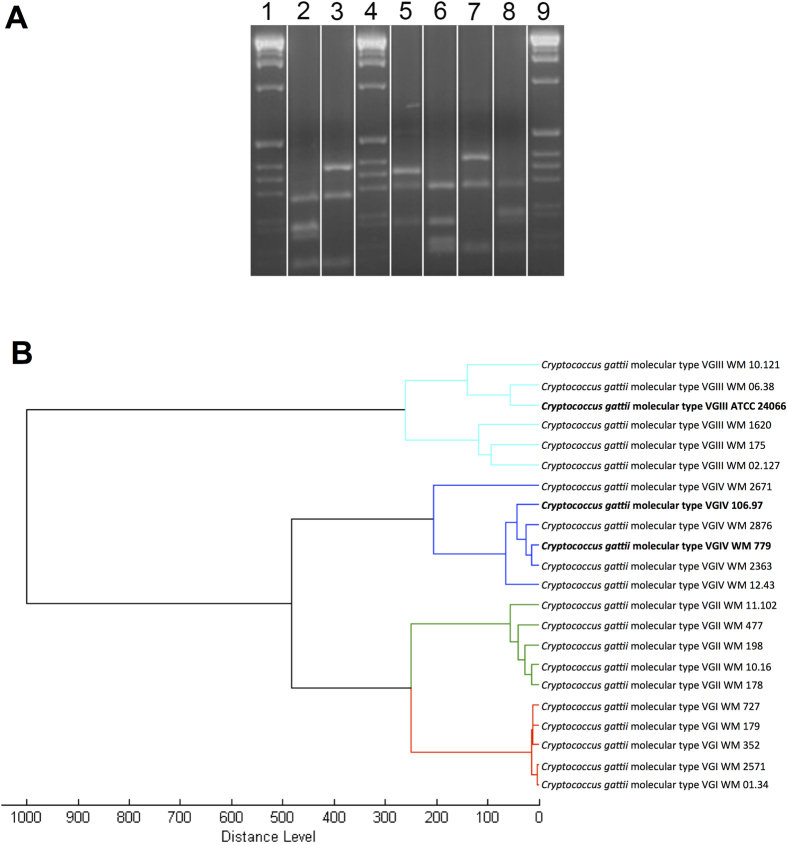
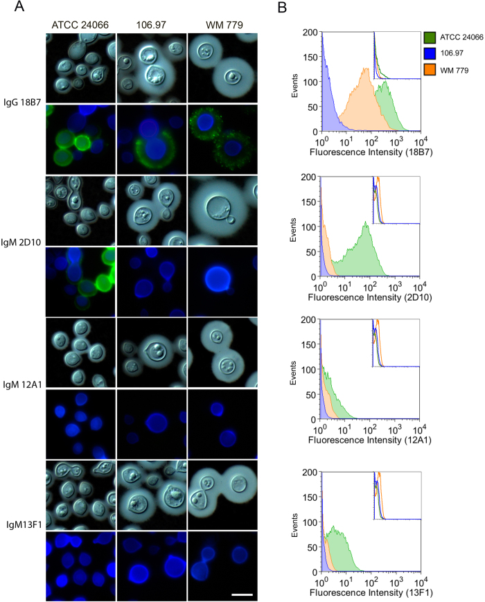
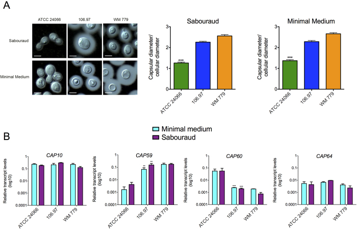
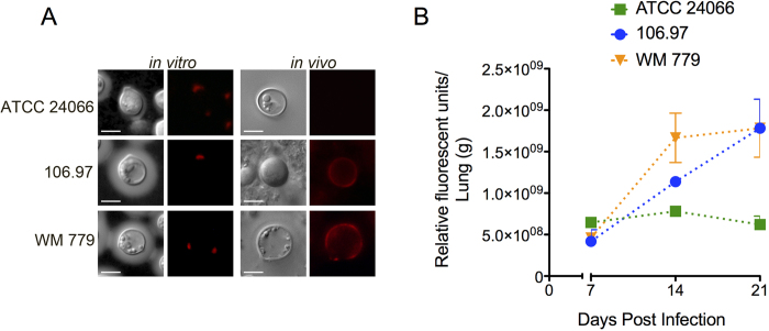
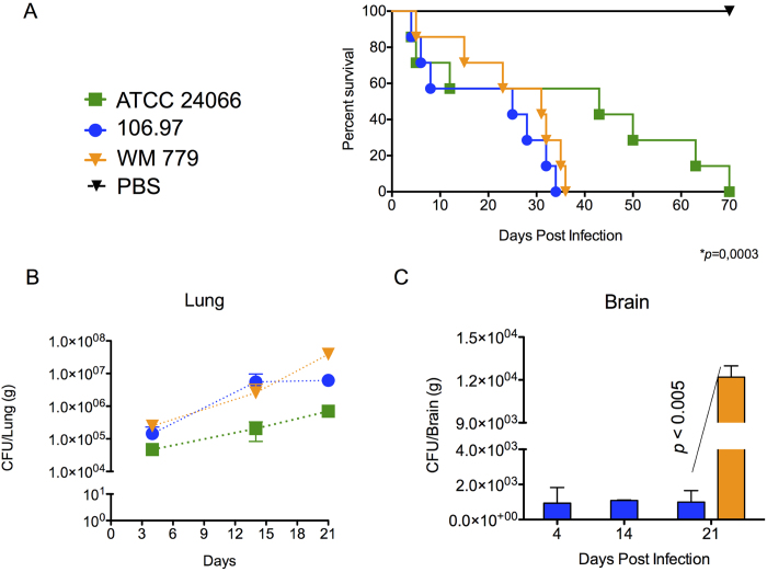
Cryptococcus gattii is one of the causative agents of human cryptococcosis. Highly virulent strains of serotype B C. gattii have been studied in detail, but little information is available on the pathogenic properties of serotype C isolates. In this study, we analyzed pathogenic determinants in three serotype C C. gattii isolates (106.97, ATCC 24066 and WM 779). Isolate ATCC 24066 (molecular type VGIII) differed from isolates WM 779 and 106.97 (both VGIV) in capsule dimensions, expression of CAP genes, chitooligomer distribution, and induction of host chitinase activity. Isolate WM 779 was more efficient than the others in producing pigments and all three isolates had distinct patterns of reactivity with antibodies to glucuronoxylomannan. This great phenotypic diversity reflected in differential pathogenicity. VGIV isolates WM 779 and 106.97 were similar in their ability to cause lethality and produced higher pulmonary fungal burden in a murine model of cryptococcosis, while isolate ATCC 24066 (VGIII) was unable to reach the brain and caused reduced lethality in intranasally infected mice. These results demonstrate a high diversity in the pathogenic potential of isolates of C. gattii belonging to the molecular types VGIII and VGIV.


Figures

Figure 1
Figure 1. Molecular type and MALDI-TOF analyses of the C. gattii isolates.
A. Molecular type analysis. Determination of the major molecular types via electrophoretic separation of URA5 gene restriction patterns after double digestion with HhaI and Sau96I obtained from isolates 106.97 (VGIV (2)), ATCC 24066 (VGIII (3)), WM179 (VGI reference strain (5)), WM178 (VGII reference strain (6)), WM175 (VGIII reference strain (7)) and WM 779 (VGIV reference strain (8)), 1, 4, 9 = 1 Kb extension ladder (Invitrogen, USA). The gel showed in this panel is representative of different experiments performed under the same experimental conditions. Cropping lines are indicated in white and the unedited gel is available as Supplemental Figure 1. B. MSP dendrograms grouping mass spectra of C. gattii isolates according to their major molecular type. Isolates used in this analysis other than the three currently studied (bold) were obtained from the in-house MALDI_Biotyper BDAL MSP library at Westmead Hospital, Westmead Millennium Culture Collection at Sydney University – Sydney Medical School.
Figure 2
Figure 2. General phenotypic analysis of C. gattii.
A. Growth curves of each isolate in Sabouraud broth or in minimal medium at both 30 oC and 37 oC. B. Urease activity from the three serotype C isolates and from S. cerevisiae control strain RSY113. C. Visual analysis of pigmentation after fungal growth in solid media supplemented with L-DOPA in different temperatures. D. Quantitative analysis of pigmentation demonstrated that isolate WM 779 was significantly more efficient in producing pigments than isolates 106.97 and ATCC 24066.
Figure 3
Figure 3. Reactivity of surface GXM with different mAbs.
A. Microscopic analysis of fungal cells after sequential staining with GXM mAbs (green), calcofluor White (blue) and counter-staining with India ink. Each mAb used for GXM detection is indicated on the left. Scale bar: 5 μm. B. Flow cytometry analysis of the reactivity of GXM-binding mAbs with the serotype C C. gattii isolates. Insets denotes the fluorescence of fungal cells in systems where primary antibodies were not used.
Figure 4
Figure 4. Glycosyl composition of polysaccharide extracts from the C. gattii isolates analyzed in this study.
Monosaccharides were obtained after methanolysis of polysaccharide extracts for further GC-MS analysis, as detailed in the Methods section.
Figure 5
Figure 5. Capsular dimensions and expression of CAP genes in C. gattii.
A. India ink counter-staining (left panels) and corresponding capsular dimensions (right panels) of C. gattii after growth in Sabouraud or minimal media. Capsular dimensions of the ATCC 24066 isolate were significantly smaller than those observed for 106.97 and WM 779 isolates (***p = <0.0005). Scale bar: 5 μm. B. Expression of CAP genes under the conditions used for determination of capsular dimensions. CAP59 and CAP60 had their expression significantly altered in the ATCC 24066 isolate (***p = <0.0001; **p = <0.005).
Figure 6
Figure 6. Analysis of C. gattii chitooligomers and host-derived chitinase activity.
A. In vitro and in vivo detection of chitooligomers using TRITC-WGA. The typical polarized pattern of chitooligomer detection was observed in vitro. In vivo analysis revealed annular patterns of oligomer detection in isolates 106.97 and WM 779 and negative staining for the ATCC 24066 isolate. Scale bar, 5 μm. B. Pulmonary chitinase activity after infection with C. gattii. From day 14 to 21 post infection, chitinase activity was significantly higher when mice was infected with isolates 106.97 or WM 779, in comparison to the ATCC 24066 isolate (P < 0.05).
Figure 7
Figure 7. Mortality curves
(A) and fungal burden (B,C) after infection of mice with serotype C isolates of C. gattii. A. Mice infected with the ATCC 24066 isolate lived longer than those infected with isolates 106.97 and WM 779 (P = 0.0003). B. Pulmonary burden after infection with isolate ATCC 24066 was always smaller (P<0.05) in comparison with systems where animals were inoculated with isolates 106.97 or WM 779. C. Brain colonization after infection of mice with isolates 106.96 and WM 779. Isolate WM 779 took longer to infect the brain of mice, but fungal burden 21 days post-infection was significantly higher for this isolate. Negative results were obtained when fungal colonization was analyzed after infection with isolate ATCC 24066. Isolate 106.97, blue bars; isolate WM 779, orange bars.

References

    1. Park B. J. et al. Estimation of the current global burden of cryptococcal meningitis among persons living with HIV/AIDS. AIDS 23, 525–30 (2009). - PubMed
    1. Chayakulkeeree M. & Perfect J. R. Cryptococcosis. Infect Dis Clin North Am 20, 507–44, v-vi (2006). - PubMed
    1. Ngamskulrungroj P. et al. Genetic diversity of the Cryptococcus species complex suggests that Cryptococcus gattii deserves to have varieties. PLoS One 4, e5862 (2009). - PMC - PubMed
    1. Huston S. M. & Mody C. H. Cryptococcosis: an emerging respiratory mycosis. Clin Chest Med 30, 253–64, vi (2009). - PubMed
    1. Lin X. Cryptococcus neoformans: morphogenesis, infection, and evolution. Infect Genet Evol 9, 401–16 (2009). - PubMed

Publication types

MeSH terms

LinkOut - more resources

Cite

AltStyle によって変換されたページ (->オリジナル) /