This site needs JavaScript to work properly. Please enable it to take advantage of the complete set of features!
Skip to main page content
U.S. flag

An official website of the United States government

Dot gov

The .gov means it’s official.
Federal government websites often end in .gov or .mil. Before sharing sensitive information, make sure you’re on a federal government site.

Https

The site is secure.
The https:// ensures that you are connecting to the official website and that any information you provide is encrypted and transmitted securely.

NIH NLM Logo
Log in
Access keys NCBI Homepage MyNCBI Homepage Main Content Main Navigation
. 2015 Aug;185(8):2105-17.
doi: 10.1016/j.ajpath.2015年04月01日7. Epub 2015 Jun 13.

Activation of autophagy and nucleotide-binding domain leucine-rich repeat-containing-like receptor family, pyrin domain-containing 3 inflammasome during Leishmania infantum-associated glomerulonephritis

Affiliations

Activation of autophagy and nucleotide-binding domain leucine-rich repeat-containing-like receptor family, pyrin domain-containing 3 inflammasome during Leishmania infantum-associated glomerulonephritis

Kevin J Esch et al. Am J Pathol. 2015 Aug.

Abstract

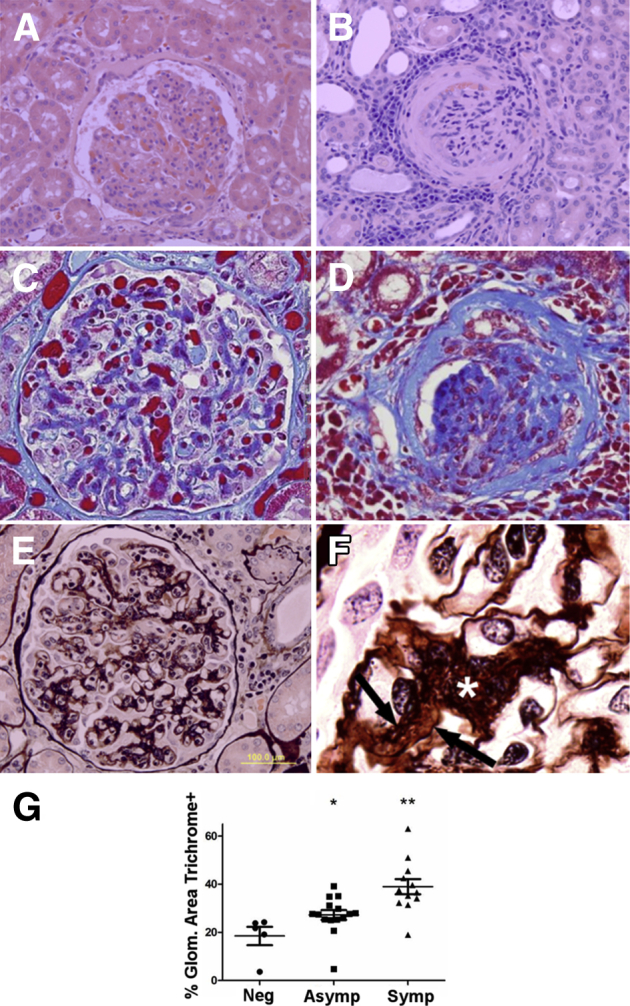
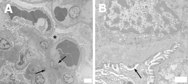
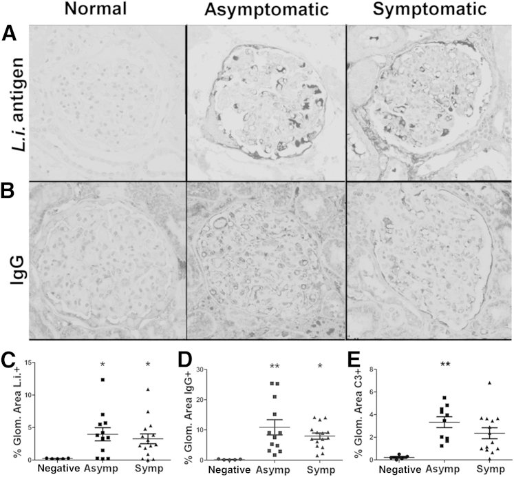
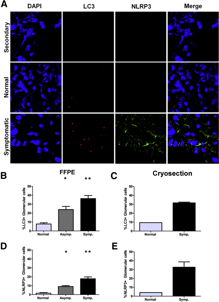
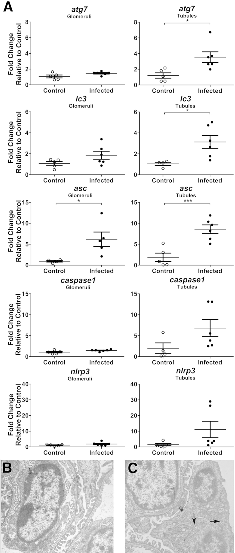
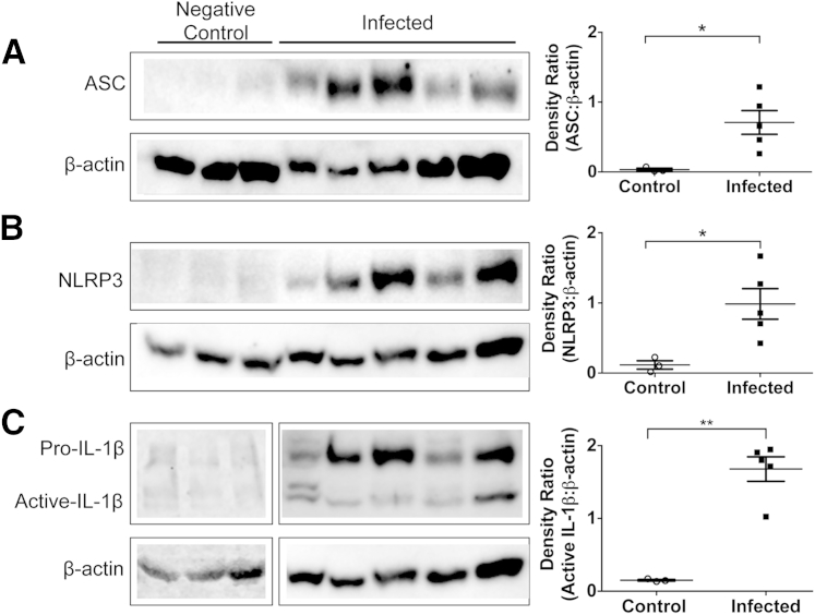
Chronic kidney disease is a major contributor to human and companion animal morbidity and mortality. Renal complications are sequelae of canine and human visceral leishmaniasis (VL). Despite the high incidence of infection-mediated glomerulonephritis, little is known about pathogenesis of VL-associated renal disease. Leishmania infantum-infected dogs are a naturally occurring model of VL-associated glomerulonephritis. Membranoproliferative glomerulonephritis type I [24 of 25 (96%)], with interstitial lymphoplasmacytic nephritis [23 of 25 (92%)], and glomerular and interstitial fibrosis [12 of 25 (48%)] were predominant lesions. An ultrastructural evaluation of glomeruli from animals with VL identified mesangial cell proliferation and interposition. Immunohistochemistry demonstrated significant Leishmania antigen, IgG, and C3b deposition in VL dog glomeruli. Asymptomatic and symptomatic dogs had increased glomerular nucleotide-binding domain leucine-rich repeat-containing-like receptor family, pyrin domain containing 3 and autophagosome-associated microtubule-associated protein 1 light chain 3 associated with glomerular lesion severity. Transcriptional analyses from symptomatic dogs confirmed induction of autophagy and inflammasome genes within glomeruli and tubules. On the basis of temporal VL staging, glomerulonephritis was initiated by IgG and complement deposition. This deposition preceded presence of nucleotide-binding domain leucine-rich repeat-containing-like receptor family, pyrin domain containing 3-associated inflammasomes and increased light chain 3 puncta indicative of autophagosomes in glomeruli from dogs with clinical VL and renal failure. These findings indicate potential roles for inflammasome complexes in glomerular damage during VL and autophagy in ensuing cellular responses.


Figures

Figure 1
Figure 1
Visceral leishmaniasis (VL)–associated glomerular lesions are present in asymptomatic dogs, with glomerular fibrosis in symptomatic VL animals. A: Asymptomatic VL. Glomeruli have diffuse mesangial and endocapillary hypercellularity with glomerular tuft hypersegmentation and segmental thickening of Bowman capsule. Hematoxylin and eosin was used. B: Symptomatic VL. Multiple renal glomeruli are sclerotic or contain segmental synechiae, with prominent lymphoplasmacytic and histiocytic interstitial nephritis and fibrosis of Bowman capsule and glomerular tuft. Hematoxylin and eosin was used. C and D: Trichrome staining identifies progressively increased collagen (blue) within the mesangium of asymptomatic dogs (C) and throughout the glomerular tuft and Bowman capsule with synechiae and glomerulosclerosis in symptomatic dogs (D). Masson's trichrome was used. E: Symptomatic dog glomeruli have robust mesangial expansion, mesangial cell hypercellularity, and interposition. Glomerular basement membranes often are split, with double contours. Periodic acid–Schiff–methenamine silver was used. F: Mesangial cell interposition and mesangium expansion (asterisk), with double contours because of segmental splitting of the glomerular basement membranes (arrows). Periodic acid–Schiff–methenamine silver was used. G: Glomerular area positive for collagen is significantly increased with progression of VL. Percentage glomerular (Glom.) area collagen positive in normal controls, asymptomatic (Asymp) VL, and symptomatic (Symp) VL, gated via color-threshold analysis in ImageJ version 1.48. SEM analysis was conducted via one-way analysis of variance with Tukey post test. P < 0.05, ∗∗P < 0.01. Original magnifications: ×ばつ400 (AE); ×ばつ1000 (F). Neg, negative.
Figure 2
Figure 2
Ultrastructural lesions of visceral leishmaniasis (VL)–associated glomerulonephritis are type I membranoproliferative glomerulonephritis. Renal cortical sections harvested from symptomatic VL dogs. Renal tissue was analyzed via transmission electron microscopy. A: Glomerular section with mesangial cell hypercellularity and capillary interposition (asterisk). Prominent mesangial and subendothelial deposits (arrows). B: Prominent subendothelial deposits within glomerular capillary, segmental thickening of glomerular basement membrane. Deposits associated with mild to moderate blunting and fusion of podocyte foot processes (arrow) with microvillous transformation. Representative of glomerular lesions from five VL dogs. Original magnifications: ×ばつ4320 (A); ×ばつ27,600 (B).
Figure 3
Figure 3
Visceral leishmaniasis (VL)–associated membranoproliferative glomerulonephritis result of Leishmania infantum antigen, IgG, and C3 deposition. Formalin-fixed, paraffin-embedded renal cortical sections from L. infantum–infected dogs or negative controls evaluated for endocapillary and mesangial deposits of L. infantum antigen (A and C), IgG (B and D), and complement protein C3 (E). A:Leishmania infantum antigen immunohistochemistry with hematoxylin counterstain. B: IgG immunohistochemistry with hematoxylin counterstain. C: Percentage glomerular (Glom.) area positive for L. infantum antigen in control, asymptomatic (Asymp), and symptomatic (Symp) VL, gated via threshold analysis in ImageJ. D: Percentage glomerular area positive for IgG in control, asymptomatic, and symptomatic VL, gated via threshold analysis in ImageJ. E: Percentage glomerular area positive for C3 in control, asymptomatic, and symptomatic VL, gated via threshold analysis in ImageJ. CE: Statistical analysis was performed via one-way analysis of variance with Tukey post test. Data are given as means ± SEM (CE). P < 0.05, ∗∗P < 0.01. Original magnification, ×ばつ400 (A and B).
Figure 4
Figure 4
Visceral leishmaniasis (VL)–associated membranoproliferative glomerulonephritis associated with elevated vacuolar-associated light chain 3 (LC3) autophagy protein and nucleotide-binding domain leucine-rich repeat–containing-like receptor family, pyrin domain containing 3 (NLRP3) inflammasomes. Frozen and/or formalin-fixed, paraffin-embedded renal cortical sections labeled with immunofluorescence antibodies, imaged, and counted via confocal microscopy evaluated presence of LC3-positive autophagy-associated vacuoles and presence of NLRP3-positive inflammasomes. A: Immunofluorescence of renal glomeruli with antibody control sample (top panels), Leishmania infantum–negative dogs (middle panels), and dogs with symptomatic VL (bottom panels). Cells stained with DAPI and labeled for NLRP3 and LC3. B and C: Percentage LC3-positive cells from total nucleated glomerular cells in L. infantum–negative, asymptomatic (Asymp.), and symptomatic (Symp.) dogs. D and E: Percentage NLRP3-positive cells from total glomerular cells of L. infantum–negative, asymptomatic, and symptomatic dogs. Data were analyzed via one-way analysis of variance with Tukey post test for significance. P < 0.05, ∗∗P < 0.01.
Figure 5
Figure 5
Glomeruli and tubules from visceral leishmaniasis–infected dogs express higher levels of autophagy and inflammasome-associated genes. A: Laser-capture microdissected glomeruli and tubules analyzed for RNA transcription by RT-PCR. Analysis of Atg7, Lc3, Asc, caspase1, and NLRP3 relative to average CT value of healthy control cells via SYBR Green (Life Technologies, Grand Island, NY). GAPDH was used as endogenous control reference gene. B and C: Apoptosis-associated speck-like protein containing a caspase activation and recruitment domain (Asc)−/− mice infected 4 weeks with 106 metacyclic Leishmania infantum promastigotes. B: Both wild-type and Asc−/− glomeruli develop mild podocyte foot process blunting and fusion compared with uninfected controls. Transmission electron microscopy (TEM) was used. C:Asc−/− mice have increased multivesicular bodies (arrows) within hypertrophied podocytes. TEM was used. Representative of ultrastructural glomerular lesions from three infected Asc−/− mice and three WT infected mice. Data are given as means ± SEM (A). n = 5 per group (A). P < 0.05, ∗∗∗P < 0.001. Original magnification, ×ばつ13,000 (B and C).
Figure 6
Figure 6
Inflammasome proteins are increased in Leishmania infantum–infected dogs compared with uninfected controls, although intracellular IL-1β is decreased. Kidney protein lysates or peripheral blood mononuclear cells (5 μg) were electrophoresed and transferred to polyvinylidene difluoride (PVDF) for protein-target identification. PDVF was probed for apoptosis-associated speck-like protein containing a caspase activation and recruitment domain (ASC; A), nucleotide-binding domain leucine-rich repeat–containing-like receptor family, pyrin domain containing 3 (NLRP3; B), and IL-1β (C); membranes were stripped and reprobed for β-actin as loading control (A and B). Bands were digitally imaged, and photon densitometry was calculated for band semiquantification as a ratio to β-actin. Statistical analysis was performed with t-test with Welsh correction. Data are given as means ± SEM (AC). n = 3 (control); n = 5 (infected). P < 0.05, ∗∗P < 0.01.

References

    1. Benderitter T., Casanova P., Nashkidachvili L., Quilici M. Glomerulonephritis in dogs with canine leishmaniasis. Ann Trop Med Parasitol. 1988;82:335–341. - PubMed
    1. Costa F.A., Goto H., Saldanha L.C., Silva S.M., Sinhorini I.L., Silva T.C., Guerra J.L. Histopathologic patterns of nephropathy in naturally acquired canine visceral leishmaniasis. Vet Pathol. 2003;40:677–684. - PubMed
    1. Dutra M., Martinelli R., de Carvalho E.M., Rodrigues L.E., Brito E., Rocha H. Renal involvement in visceral leishmaniasis. Am J Kidney Dis. 1985;6:22–27. - PubMed
    1. Liborio A.B., Rocha N.A., Oliveira M.J., Franco L.F., Aguiar G.B., Pimentel R.S., Abreu K.L., Silva G.B., Jr., Daher E.F. Acute kidney injury in children with visceral leishmaniasis. Pediatr Infect Dis J. 2012;31:451–454. - PubMed
    1. Sayari M., Avizeh R., Barati F. Microscopic evaluation of renal changes in experimental canine visceral leishmaniosis after chemo- and immunotherapy. Pak J Biol Sci. 2008;11:1630–1633. - PubMed

Publication types

MeSH terms

Cite

AltStyle によって変換されたページ (->オリジナル) /