This site needs JavaScript to work properly. Please enable it to take advantage of the complete set of features!
Skip to main page content
U.S. flag

An official website of the United States government

Dot gov

The .gov means it’s official.
Federal government websites often end in .gov or .mil. Before sharing sensitive information, make sure you’re on a federal government site.

Https

The site is secure.
The https:// ensures that you are connecting to the official website and that any information you provide is encrypted and transmitted securely.

NIH NLM Logo
Log in
Access keys NCBI Homepage MyNCBI Homepage Main Content Main Navigation
. 2015 Apr 8;10(4):e0123407.
doi: 10.1371/journal.pone.0123407. eCollection 2015.

Peripheral opioid antagonist enhances the effect of anti-tumor drug by blocking a cell growth-suppressive pathway in vivo

Affiliations

Peripheral opioid antagonist enhances the effect of anti-tumor drug by blocking a cell growth-suppressive pathway in vivo

Masami Suzuki et al. PLoS One. .

Abstract

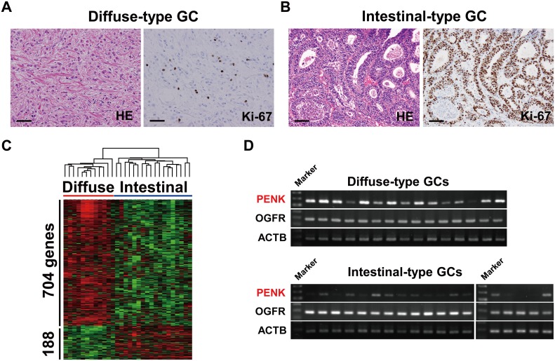
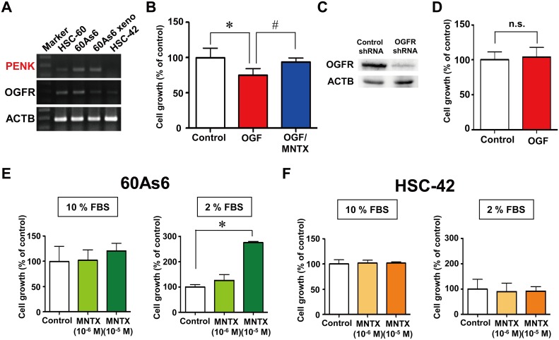
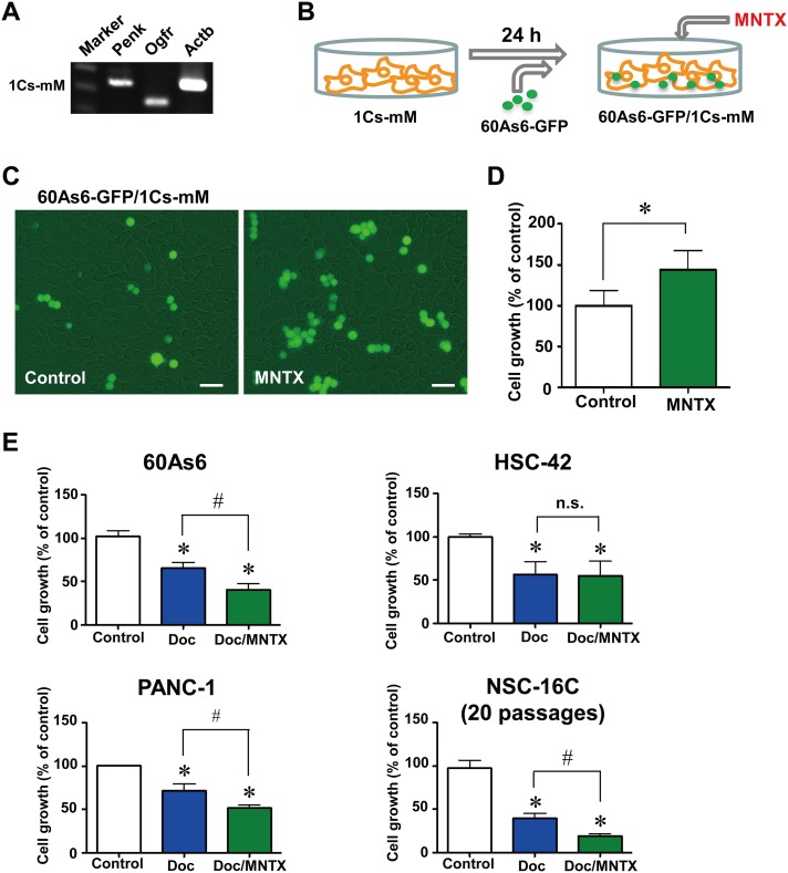
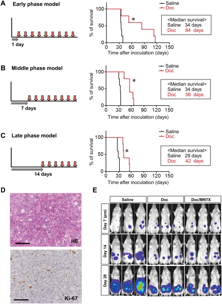
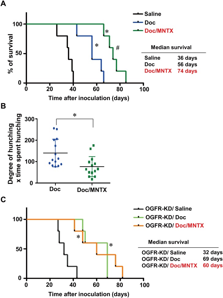
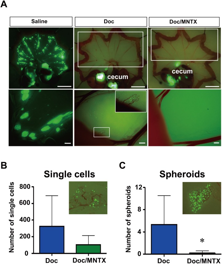
The dormancy of tumor cells is a major problem in chemotherapy, since it limits the therapeutic efficacy of anti-tumor drugs that only target dividing cells. One potential way to overcome chemo-resistance is to "wake up" these dormant cells. Here we show that the opioid antagonist methylnaltrexone (MNTX) enhances the effect of docetaxel (Doc) by blocking a cell growth-suppressive pathway. We found that PENK, which encodes opioid growth factor (OGF) and suppresses cell growth, is predominantly expressed in diffuse-type gastric cancers (GCs). The blockade of OGF signaling by MNTX releases cells from their arrest and boosts the effect of Doc. In comparison with the use of Doc alone, the combined use of Doc and MNTX significantly prolongs survival, alleviates abdominal pain, and diminishes Doc-resistant spheroids on the peritoneal membrane in model mice. These results suggest that blockade of the pathways that suppress cell growth may enhance the effects of anti-tumor drugs.


Conflict of interest statement

Competing Interests: The authors have declared that no competing interests exist.

Figures

Fig 1
Fig 1. PENK encoding opioid growth factor (OGF) is preferentially expressed in diffuse-type gastric cancers (GCs).
Representative histological image (hematoxylin-eosin, HE) and Ki-67 immunostaining of diffuse-type GC (A) and intestinal-type GC (B). Scale bar, 50 μm. C, supervised clustering analysis of 892 specifically expressed genes in 12 diffuse-type and 18 intestinal-type GCs. By the Wilcoxon u-test (p<0.05) and a 2-fold change, 188 genes were selected as specific genes for 18 intestinal-type GCs, and 704 genes were selected as specific genes for 12 diffuse-type GCs. The results of a two-dimensional hierarchical clustering analysis of the 892 selected genes are shown. D, RT-PCR analyses of OGF signaling molecules, PENK and its receptor OGFR, in diffuse-type and intestinal-type GCs.
Fig 2
Fig 2. Blockade of OGF signaling by methylnaltrexone (MNTX) increased the growth of a diffuse-type GC cell line, but not an intestinal-type GC cell line, under low nutrient conditions.
A, RT-PCR analyses of PENK and OGFR in diffuse-type GC cell lines, HSC-60 cells and highly metastatic 60As6 cells, 60As6 xenograft (60As6 xeno) and the intestinal-type GC cell line HSC-42. B, growth of 60As6 cells treated with OGF (10-4 M), methylnaltrexone (MNTX, 10-6 M), or a combination of these compounds for 72 h (mean ± SD, n = 3–6 per group, *p<0.05, control vs. OGF, #p <0.05, OGF vs. OGF/MNTX). C, western blot analysis of OGFR protein in 60As6 cells with a stable transfectant of OGFR shRNA or control shRNA. D, growth of the stable transfectant of OGFR shRNA of 60As6 cells in the presence or absence of OGF (10-4 M) for 72 h. Non-targeting control shRNA was used as a control (mean ± SD, n = 3 each). Growth of 60As6 cells (E) and HSC-42 cells (F) treated with MNTX (10-6 and 10-5 M) or a vehicle for 72 h under normal nutrient (10% FBS) and low nutrient (2% FBS) conditions. (mean ± SD, n = 4 each, *p<0.05, vs. control).
Fig 3
Fig 3. Blockade of OGF signaling by MNTX increased the growth of diffuse-type GC cells co-cultured with mesothelial cells.
A, RT-PCR analyses of Penk and Ogfr, in mouse mesothelial cells (1Cs-mM). B, schematic illustration of the system for co-culture of 60As6-GFP and 1Cs-mM. C, Growth of 60As6-GFP cells co-cultured with 1Cs-mM cells in the presence or absence of MNTX (10-5 M) for 72 h. Scale bar, 20 μm. D, the growth of 60As6-GFP cells was calculated (mean ± SD, n = 3 each, *p<0.05). E, growth of the diffuse-type GC cell line 60As6 cells, the intestinal-type GC cell line HSC-42 cells, the pancreatic cancer cell line PANC-1 cells, and primary cultured GC cells derived from the ascites of a patient NSC-16C cells treated with Doc (10-9 M) or a vehicle for 48 h, and subsequently treated with Doc, Doc/MNTX (10-6 M) or a vehicle for 48 h. Cells were counted with a hemacytometer (mean ± SD, n = 4 each, *p<0.05, vs. control, #p<0.05, Doc vs. Doc/MNTX).
Fig 4
Fig 4. Mouse models of intraperitoneal low-dose Doc therapy corresponding to 3 different phases (early, middle, and late) in the progression of peritoneal dissemination.
Survival curves for the early phase (A), middle phase (B), and late phase (C) of a peritoneal metastasis model. Administration of Doc (0.5 mg/kg) was started 1 day (A), 7 days (B) or 14 days (C) after the inoculation of 60As6-Luc cells, and was continued until the endpoint criteria were reached (n = 5, *p<0.05, vs. saline). D, representative histological image (hematoxylin-eosin, HE) and Ki-67 immunostaining of 60As6 xenograft. Scale bar, 50 μm. E, detection of the progression of peritoneal dissemination in real-time using an in vivo photon-counting analysis of mice treated with saline, Doc or Doc/MNTX (0.3 mg/kg). Beginning on day 7 after inoculation, the mice were divided into 4 groups based on photon counts. The mice were then treated intraperitoneally with the above reagents twice weekly until the endpoint criteria were met (middle phase).
Fig 5
Fig 5. Combined use of Doc and MNTX significantly prolonged survival and alleviated abdominal pain in model mice.
A, survival curves of middle-phase peritoneal metastasis model mice treated with saline, Doc, or Doc/MNTX (0.3 mg/kg) (n = 5, *p<0.05, vs. saline, #p<0.05, Doc vs. Doc/MNTX). Drug administration was started 7 days after the inoculation of 60As6-Luc cells. Mice were treated with Doc or a combination of Doc and MNTX 2 times a week until the endpoint criteria were met. B, visceral pain-related behavior of peritoneal metastasis model mice. Visceral pain-related behavior was assessed in terms of the degree of hunching and the time spent hunching before each drug treatment at 35 days after the inoculation of 60As6-Luc cells (mean ± SD, n = 14 each, *p<0.05). C, survival curves of peritoneal metastasis mice of OGFR-shRNA transfected 60As6 cells (OGFR-KD) treated with saline, Doc, or Doc/MNTX (0.3 mg/kg) (n = 5, *p<0.05, vs. saline, no significance in Doc vs. Doc/MNTX).
Fig 6
Fig 6. Combined use of Doc and MNTX significantly suppressed peritoneal metastasis.
A, representative image of 60As6-GFP cells attached to the mesentery of saline-, Doc- and Doc/MNTX-treated mice. Drug administration started from 7 days to 35 days (saline) and 49 days (Doc or Doc/MNTX) after the inoculation of 60As6-GFP cells. Mice were treated with saline, Doc, or Doc/MNTX 2 times a week. Scale bar, upper: 10 μm, lower: 1 μm. Numbers of single cells (B) and spheroids (C) on the mesentery of Doc- and Doc/MNTX-treated mice 49 days after the inoculation of 60As6-GFP cells. Drug administration started from 7 days to 49 days after inoculation. Mice were treated with saline twice a week. (mean ± SD, n = 6 each, *p<0.05).

References

    1. Aguirre-Ghiso JA. Models, mechanisms and clinical evidence for cancer dormancy. Nat Rev Cancer. 2007;7: 834–846. - PMC - PubMed
    1. Ishii H, Iwatsuki M, Ieta K, Ohta D, Haraguchi N, Mimori K, et al. Cancer stem cells and chemoradiation resistance. Cancer Sci. 2008;99: 1871–1877. 10.1111/j.1349-7006.2008.00914.x - DOI - PMC - PubMed
    1. Ito K, Bernardi R, Morotti A, Matsuoka S, Saqlio G, Ikeda Y, et al. PML targeting eradicates quiescent leukaemia-initiating cells. Nature. 2008;453: 1072–1078. 10.1038/nature07016 - DOI - PMC - PubMed
    1. Nagini S. Carcinoma of the stomach: a review of epidemiology, pathogenesis, molecular genetics and chemoprevention. World J Gastroentest Oncol. 2012;4: 156–169. 10.4251/wjgo.v4.i7.156 - DOI - PMC - PubMed
    1. Lauren P. The two histological main types of gastric carcinoma: diffuse and so-called intestinal-type carcinoma. An attempt at a histo-clinical classification. Acta Pathol. Microbiol. Scand. 1965;64: 31–49. - PubMed

Publication types

MeSH terms

Cite

AltStyle によって変換されたページ (->オリジナル) /