This site needs JavaScript to work properly. Please enable it to take advantage of the complete set of features!
Skip to main page content
U.S. flag

An official website of the United States government

Dot gov

The .gov means it’s official.
Federal government websites often end in .gov or .mil. Before sharing sensitive information, make sure you’re on a federal government site.

Https

The site is secure.
The https:// ensures that you are connecting to the official website and that any information you provide is encrypted and transmitted securely.

NIH NLM Logo
Log in
Access keys NCBI Homepage MyNCBI Homepage Main Content Main Navigation
Clinical Trial
. 2015 Apr 15;194(8):3883-9.
doi: 10.4049/jimmunol.1402202. Epub 2015 Mar 13.

Perturbed T cell IL-7 receptor signaling in chronic Chagas disease

Affiliations
Clinical Trial

Perturbed T cell IL-7 receptor signaling in chronic Chagas disease

M Cecilia Albareda et al. J Immunol. .

Abstract

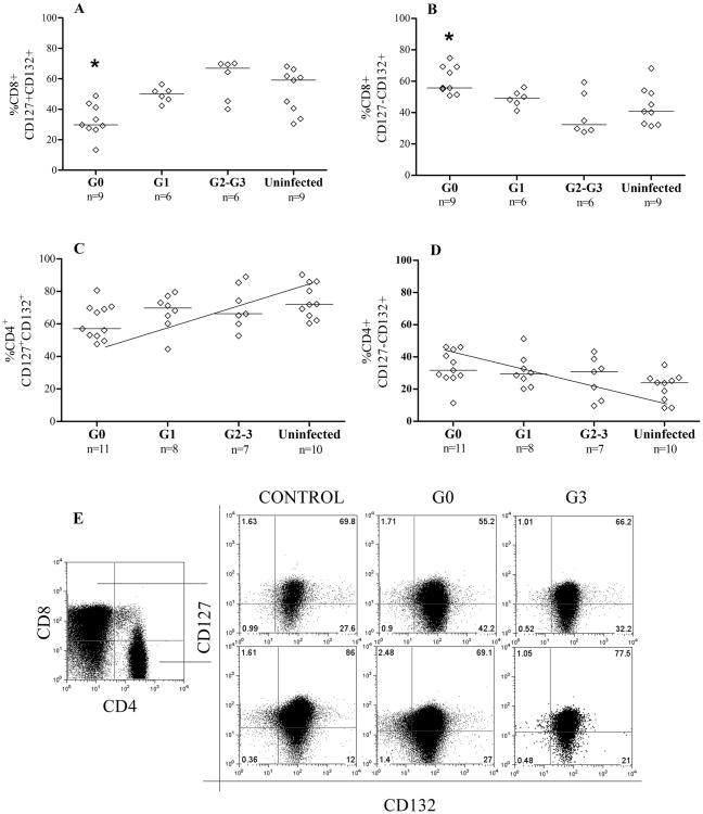
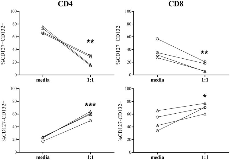
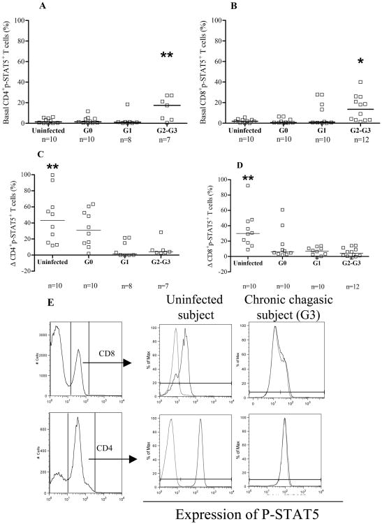
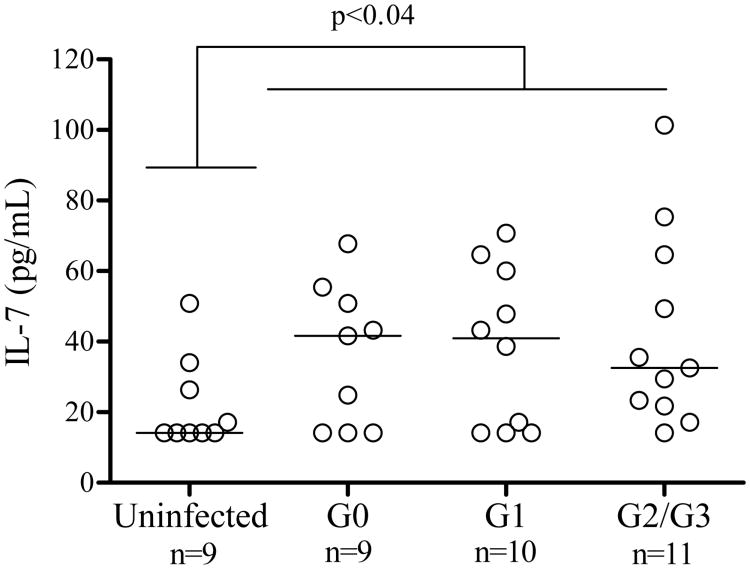
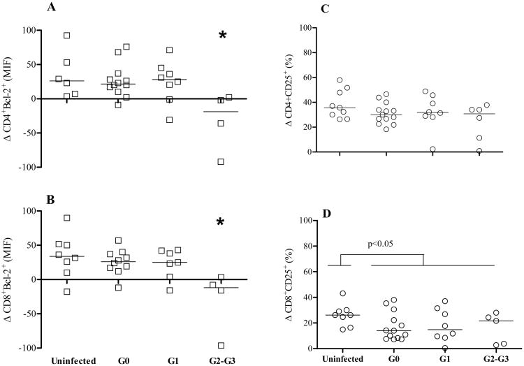
We have previously demonstrated that immune responses in subjects with chronic Trypanosoma cruzi infection display features common to other persistent infections with signs of T cell exhaustion. Alterations in cytokine receptor signal transduction have emerged as one of the cell-intrinsic mechanisms of T cell exhaustion. In this study, we performed an analysis of the expression of IL-7R components (CD127 and CD132) on CD4(+) and CD8(+) T cells and evaluated IL-7-dependent signaling events in patients at different clinical stages of chronic chagasic heart disease. Subjects with no signs of cardiac disease showed a decrease in CD127(+)CD132(+) cells and a reciprocal gain of CD127(-)CD132(+) in CD8(+) and CD4(+) T cells compared with either patients exhibiting heart enlargement or uninfected controls. T. cruzi infection, in vitro, was able to stimulate the downregulation of CD127 and the upregulation of CD132 on T cells. IL-7-induced phosphorylation of STAT5 as well as Bcl-2 and CD25 expression were lower in T. cruzi-infected subjects compared with uninfected controls. The serum levels of IL-7 were also increased in chronic chagasic patients. The present study highlights perturbed IL-7/IL-7R T cell signaling through STAT5 as a potential mechanism of T cell exhaustion in chronic T. cruzi infection.


Figures

Figure 1
Figure 1
Cell-surface expression of IL-7 receptor components in subjects with chronic T. cruzi infection. PBMC were stained for CD8, CD4, CD127 and CD132 and analyzed by flow cytometry. Lymphocytes were gated by side scatter vs forward scatter light. Each point represents the expression of CD127 and CD132 on the total CD8+ and CD4+ T cell compartments in one patient. Median values are indicated by the horizontal lines. Statistically significantly differences (*p < 0.05) in the frequencies of CD8+CD127+CD132+ (A) and CD8+CD127-CD132+ (B) in G0 patients vs uninfected or G2-G3 clinical groups. [C-D], Positive and negative trend in the percentages of CD127+CD132+ and CD127-CD132+ CD4+ T cells, respectively, as clinical stage becomes more severe. [E] Representative dot plots of the expression of CD127 and CD132 in CD4+ and CD8+ T cells from an uninfected control, an asymptomatic patient (G0) and a severe cardiomyopathy patient (G3). The numbers in each quadrant represents the percentage of expression of each of the 4 possible subsets based on the expression of CD127 and CD132 IL-7 receptor components.
Figure 2
Figure 2
Modulation of the IL-7R components after in vitro infection of T cells with T. cruzi. PBMCs from two chronic Chagas disease subjects and two uninfected controls were cocultured with T. cruzi in a 1:1 ratio for 48hs, stained for CD8, CD4, CD127 and CD132 and analyzed by flow cytometry. Lymphocytes were gated by side scatter vs forward scatter light. Each point represents the expression of CD127 and CD132 on total CD8+ (right panel) and CD4+ (left panel) T cells from media and T. cruzi stimulated wells in each subject. しろさんかく Uninfected controls; しろまる Chagas disease patient
Figure 3
Figure 3
STAT-5 activation in CD4+ and CD8+ T cells in chronic Chagas disease. PBMC were stimulated with 100 ng/ml IL-7 and evaluated for p-STAT5+ induction in CD4+ and CD8+ T cells by flow cytometry. Lymphocytes were gated in side scatter vs forward sacatter light. [A-B] Each point represents the basal levels of CD4+p-STAT5+ and CD8+p-STAT5+ T cells, respectively. Differences among groups were evaluated by ANOVA followed by Bonferroni. Median values are indicated by the horizontal lines. [C-D] Each point represents the induced expression of p-STAT5 for individual subjects, calculated as the difference in the percentage of CD4+p-STAT5+ or CD8+p-STAT5+ T cells, respectively, between IL-7-stimulated and unstimulated samples. Differences among groups were evaluated by ANOVA followed by Bonferroni. Median values are indicated by the horizontal lines. *p<0.05, **p<0.01 [E] Representative CD4+/CD8+ histogram plots of PBMC from an uninfected control and a T. cruzi-infected subject with severe cardiomyopathy. Grey lines indicate the basal expression of p-STAT5 and the black lines the expression of p-STAT5 after IL-7 stimulation.
Figure 4
Figure 4
Serum IL-7 concentration in chronic Chagas disease patients. Serum levels were measured in 30 chronically T. cruzi-infected subjects with different clinical stages of the disease and 9 uninfected controls using an IL-7 a capture ELISA assay. Each point represents the IL7 serum levels in each subject. Median values are indicated by the horizontal lines.
Figure 5
Figure 5
Impaired IL-7 functional responses in CD4+ and CD8+ T cells from chronic Chagas disease patients. PBMC from healthy donors (n=9) and chronically T. cruzi-infected subjects (G0 n=13; G1 n=8; G2-G3 n=6) were cultured for 2 days in complete medium in the presence or absence of 10ng/ml of IL-7. Each point represents the differences in Bcl-2 MFI (Δ Blc-2 MFI) [A-B] and in the percentage of CD25+ T cells (Δ% CD25) [C-D] between IL7-stimulated and unstimulated samples in CD4+ or CD8 T cell compartments. [A-B] (*) Analysis of variance (ANOVA) followed by Bonferroni test for multiple comparisons. G2-G3 p < 0.05 compared to G0, G1 and uninfected subjects. [D] Mann-Whitney U-test between T. cruzi-infected and uninfected subjects, p < 0.05 compared to T. cruzi-infected subjects.

References

    1. World Health Organization. Research Priorities for Chagas disease, Human African Trypanosomiasis and Leishmaniasis. Tech Rep Ser. 2012:1–100. - PubMed
    1. Schmunis GA, Yadon ZE. Chagas disease: a Latin American health problem becoming a world health problem. Acta Trop. 2010;115:14–21. - PubMed
    1. Albareda MC, Laucella SA, Alvarez MG, Armenti AH, Bertochi G, Tarleton RL, Postan M. Trypanosoma cruzi modulates the profile of memory CD8+ T cells in chronic Chagas' disease patients. Int Immunol. 2006;18:465–471. - PubMed
    1. Albareda MC, Olivera GC, Laucella SA, Alvarez MG, Fernandez ER, Lococo B, Viotti R, Tarleton RL, Postan M. Chronic human infection with Trypanosoma cruzi drives CD4+ T cells to immune senescence. J Immunol. 2009;183:4103–4108. - PMC - PubMed
    1. Albareda MC, Olivera GC, De Rissio AM, Postan M. Short Report: Assessment of CD8 + T Cell Differentiation in Trypanosoma cruzi -Infected Children. Am J Trop Med Hyg. 2010;82:861–864. - PMC - PubMed

Publication types

MeSH terms

Substances

Cite

AltStyle によって変換されたページ (->オリジナル) /