This site needs JavaScript to work properly. Please enable it to take advantage of the complete set of features!
Skip to main page content
U.S. flag

An official website of the United States government

Dot gov

The .gov means it’s official.
Federal government websites often end in .gov or .mil. Before sharing sensitive information, make sure you’re on a federal government site.

Https

The site is secure.
The https:// ensures that you are connecting to the official website and that any information you provide is encrypted and transmitted securely.

NIH NLM Logo
Log in
Access keys NCBI Homepage MyNCBI Homepage Main Content Main Navigation
. 2015 Jan 6:8:1.
doi: 10.1186/s13071-014-0608-1.

Sm16, a major component of Schistosoma mansoni cercarial excretory/secretory products, prevents macrophage classical activation and delays antigen processing

Affiliations

Sm16, a major component of Schistosoma mansoni cercarial excretory/secretory products, prevents macrophage classical activation and delays antigen processing

David E Sanin et al. Parasit Vectors. .

Abstract

Background: Schistosoma mansoni cercariae penetrate the skin by releasing excretory/secretory (E/S) products known as 0-3hRP, which are associated with immune modulation through Toll like receptor (TLR) signalling. Furthermore, these secretions contain Sm16, which when given to cells as a recombinant protein inhibits human monocyte derived cytokine responses to TLR4 and TLR3 ligands. Nonetheless, the extent and mechanism(s) of these inhibitory effects remain largely uncharacterized.

Methods: Murine bone marrow derived macrophages were exposed to different fractions of 0-3hRP, obtained via ultracentrifugation, or recombinant Sm16. These cells were exposed to the parasite molecules in combination with different TLR ligands, or Interferon gamma, and tested for the production of the cytokines IL-10 and IL-12p40, and their ability to process antigen.

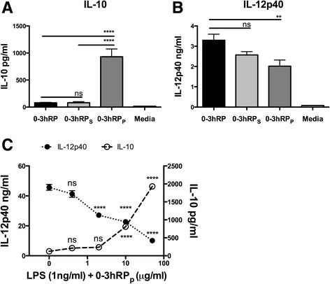
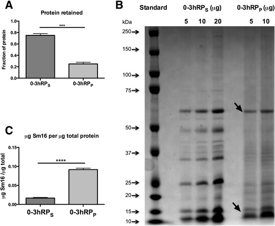
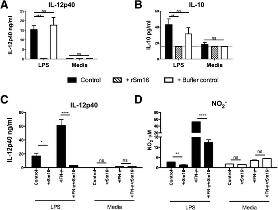
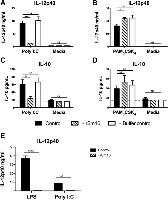
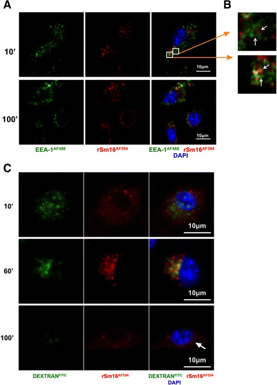
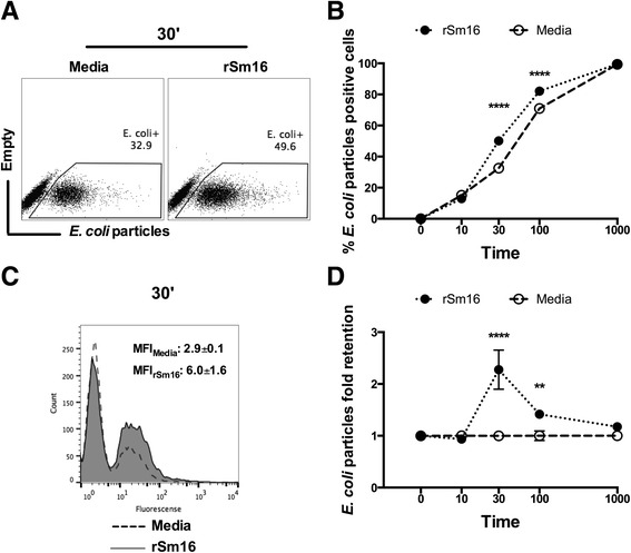
Results: The immunomodulatory function of 0-3hRP is enriched predominantly in the pellet fraction, which contains a greater proportion of Sm16, also corroborating the ability of recombinant Sm16 to inhibit macrophage activation in response to TLR ligands. We further demonstrate that Sm16 blocks classical activation of macrophages to LPS or IFN-γ stimulation in vitro, and that inhibition of macrophage classical activation is independent of TLR2 recognition. Finally we show that Sm16 shares the altered intracellular processing observed for 0-3hRP, and is able to delay antigen processing by macrophages.

Conclusions: Collectively, our findings show that Sm16 is a major component of S. mansoni cercarial E/S products, and is partly responsible for its immune-regulatory properties. Moreover, we propose that the mechanism employed by Sm16 to exert its inhibitory function is likely to be linked with alteration of endosomal trafficking and is not dependent on particular TLR receptors. Finally, we suggest that accumulation of Sm16 in the skin after percutaneous infection with S. mansoni cercariae could contribute to limiting dermal inflammation.


Figures

Figure 1
Figure 1
The pelleted fraction of cercarial E/S products (0-3hRP P ) induces abundant IL-10 from macrophages and blocks LPS driven IL-12p40. The presence of (A) IL-10 and (B) IL-12p40 in culture supernatants from BMMΦs exposed to 0-3hRP (50 μg/ml), 0-3hRPS (50 μg/ml), 0-3hRPP (25 μg/ml), or left un-stimulated (Media). (C) IL-12p40 (closed circles, left axis) and IL-10 (open circles, right axis) in culture supernatants of BMMΦs exposed to LPS (1 ng/ml) and increasing doses of 0-3hRPP. Bars, or symbols, are mean values ± SEM of 6 technical replicates and are representative of three independent experiments. ANOVA and Tukey’s or Dunnett’s test were performed to examine statistically significant differences between selected means (** = p < 0.01; **** = p < 0.0001; ns = p > 0.05).
Figure 2
Figure 2
Sm16 is enriched in pellet fraction of cercarial E/S products. Three preparations of 0-3hRP were fractionated by ultracentrifugation and the pellet fractions restored to the original volume of each preparation. (A) Protein content of each fraction expressed as a percentage of the total protein present in the original preparation. Bars = mean + SEM; statistical significance was tested using two tailed t-test (*** = p < 0.001). (B) SDS PAGE gel of 0-3hRP fractions (5, 10 and 20 μg) stained for protein. Black arrows highlight bands identified by mass spectrometry. (C) Equivalent volumes of 0-3hRPS (78 μg) and 0-3hRPP (10 μg) based on the original preparation, were processed for Western blot analysis alongside rSm16 (1 μg) probed using rabbit anti-rSm16 antibody, estimated as relative concentration of Sm16 / μg 0-3hRP. Bars are mean + SEM, two tailedt-test show statistically significant differences (**** = p < 0.0001).
Figure 3
Figure 3
Recombinant Sm16 blocks activation of BMMΦs in response to LPS and IFN-γ. The presence of (A) IL-12p40 and (B) IL-10 in culture supernatants from BMMΦs exposed to LPS (1 ng/ml) (black bars), or Media, in the presence of rSm16 (10 μg/ml) (hatched bars), or an equivalent volume of protein buffer (open bars). (C) IL-12p40 and (D) nitric oxide (NO2 ) in culture supernatants from BMMΦs exposed to LPS (1 ng/ml) plus IFN-γ (25U/ml) in the presence, or absence of rSm16 (10 μg/ml). Bars = means + SEM of 3 technical replicates. Dotted line represents minimum level of cytokine detection by ELISA. Statistically significant differences tested by ANOVA and Bonferroni’s or Sidak’s test between selected means (* = p < 0.05; ** = p < 0.01; *** = p < 0.001; **** = p < 0.0001; ns = p > 0.05). Results are representative of three independent experiments.
Figure 4
Figure 4
Recombinant Sm16 blocks cytokine production in BMMΦs exposed to TLR4 and TLR3, but not TLR2 ligands. (A-B) IL-12p40 and (C-D) IL-10 in culture supernatants from BMMΦs exposed to (A&C) Poly I:C (25 μg/ml), (B & D) Pam3CSK4 (5 μg/ml), or Media, plus rSm16 (10 μg/ml) (hatched bars) or an equivalent volume of protein buffer (open bars). Stimulus only controls (black bars) are also given. (E) BMMΦs from TLR2−/− mice exposed to LPS (1 ng/ml), or Poly I:C (25 μg/ml) (solid bars), plus rSm16 (10 μg/ml) (hatched bars), and supernatants tested for IL-12p40 by ELISA. Means + SEM of 3 technical replicates are presented. Dotted line represents minimum level of cytokine detection. ANOVA and Bonferroni’s or Sidak’s test were performed to examine statistically significant differences between the means (* = p < 0.05; ** = p < 0.01; *** = p < 0.001; **** = p < 0.0001; ns = p > 0.05). Results are representative of three independent experiments.
Figure 5
Figure 5
Uptake of fluorescently labelled rSm16 by BMMΦs. Representative confocal images of (A) BMMΦs exposed to labelled rSm16AF594 (red) stained with DAPI (blue) within EEA-1+ endosomes (green) 10 min and 100 min after ligand stimulation. (B) Insert showing EEA-1+ endosomes containing rSm16AF594 (2 μm x 2 μm) (C) BMMΦs exposed for 100 min to rSm16AF594 (red) and DEXTRANFITC (green) washed and imaged after 10, 60 and 100 min. (63x objective, scale bar = 10 μm; acquired using a Zeiss LSM 710 invert microscope).
Figure 6
Figure 6
Recombinant Sm16 delays antigen processing by BMMΦs. BMMΦs were exposed for 10, 30, 100 or 1000 min to AlexaFluor®488 conjugated E. coli BioParticles® (100 particles per cell) in the absence (Media, open circles), or presence of rSm16 (10 μg/ml) (closed circles). (A) Representative flow plots of BMMΦs 30 min after exposure to E. coli particles. (B) Mean percentages ± SEM of BMMΦs containing labelled E. coli BioParticles at each time point, and (C) representative overlaid histograms including the MFI ± SD of each group. (D) Mean ± SEM fold-retention of labelled E. coli BioParticles within rSm16-treated BMMφs, relative to the respective MFI of BMMΦs treated with Media control, arbitrarily set to 1. Statistically significant differences between the means of rSm16 treated and Media controls at each time point were examined by ANOVA and Sidak’s test (** = p < 0.01; **** = p < 0.0001).

References

    1. Curwen RS, Ashton PD, Sundaralingam S, Wilson RA. Identification of novel proteases and immunomodulators in the secretions of schistosome cercariae that facilitate host entry. Mol Cell Proteomics. 2006;5:835–844. doi: 10.1074/mcp.M500313-MCP200. - DOI - PubMed
    1. Wilson RA. Virulence factors of schistosomes. Microbes Infect. 2012;14:1442–1450. doi: 10.1016/j.micinf.201209001. - DOI - PubMed
    1. Paveley RA, Aynsley SA, Cook PC, Turner JD, Mountford AP. Fluorescent imaging of antigen released by a skin-invading helminth reveals differential uptake and activation profiles by antigen presenting cells. PLoS Negl Trop Dis. 2009;3:e528. doi: 10.1371/journal.pntd.0000528. - DOI - PMC - PubMed
    1. McKerrow JH, Salter J. Invasion of skin by Schistosoma cercariae. Trends Parasitol. 2002;18:193–195. doi: 10.1016/S1471-4922(02)02309-7. - DOI - PubMed
    1. Salter JP, Lim KC, Hansell E, Hsieh I, McKerrow JH. Schistosome invasion of human skin and degradation of dermal elastin are mediated by a single serine protease. J Biol Chem. 2000;275:38667–38673. doi: 10.1074/jbc.M006997200. - DOI - PubMed

Publication types

Cite

AltStyle によって変換されたページ (->オリジナル) /