This site needs JavaScript to work properly. Please enable it to take advantage of the complete set of features!
Skip to main page content
U.S. flag

An official website of the United States government

Dot gov

The .gov means it’s official.
Federal government websites often end in .gov or .mil. Before sharing sensitive information, make sure you’re on a federal government site.

Https

The site is secure.
The https:// ensures that you are connecting to the official website and that any information you provide is encrypted and transmitted securely.

NIH NLM Logo
Log in
Access keys NCBI Homepage MyNCBI Homepage Main Content Main Navigation
. 2014 Dec 9:5:5691.
doi: 10.1038/ncomms6691.

Critical role of lysine 134 methylation on histone H2AX for γ-H2AX production and DNA repair

Affiliations

Critical role of lysine 134 methylation on histone H2AX for γ-H2AX production and DNA repair

Kenbun Sone et al. Nat Commun. .

Abstract

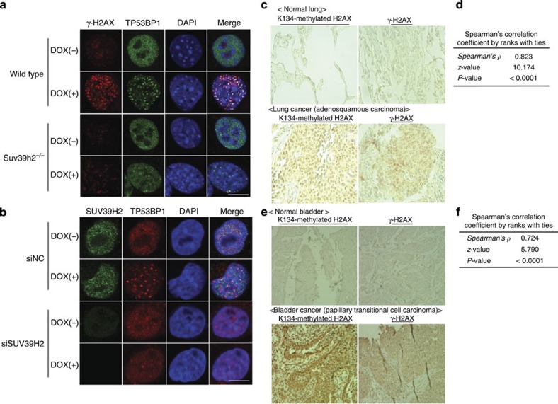
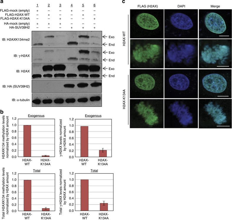
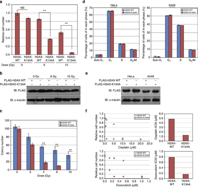
The presence of phosphorylated histone H2AX (γ-H2AX) is associated with the local activation of DNA-damage repair pathways. Although γ-H2AX deregulation in cancer has previously been reported, the molecular mechanism involved and its relationship with other histone modifications remain largely unknown. Here we find that the histone methyltransferase SUV39H2 methylates histone H2AX on lysine 134. When H2AX was mutated to abolish K134 methylation, the level of γ-H2AX became significantly reduced. We also found lower γ-H2AX activity following the introduction of double-strand breaks in Suv39h2 knockout cells or on SUV39H2 knockdown. Tissue microarray analyses of clinical lung and bladder tissues also revealed a positive correlation between H2AX K134 methylation and γ-H2AX levels. Furthermore, introduction of K134-substituted histone H2AX enhanced radio- and chemosensitivity of cancer cells. Overall, our results suggest that H2AX methylation plays a role in the regulation of γ-H2AX abundance in cancer.


Conflict of interest statement

Y. Nakamura is a stock holder and a scientific advisor of Oncotherapy Science, and also has research grants from Oncotherapy Science. All other authors declare no competing financial interests.

Figures

Figure 1
Figure 1. SUV39H2 is overexpressed in human lung cancer.
(a) In vitro methyltransferase analysis of SUV39H2. Recombinant histone H2AX and 3H-SAM were incubated in the presence or absence of recombinant SUV39H2, and the reaction products were analysed by SDS–PAGE followed by fluorography (upper panel) and stained for total protein(lower panel). (b) SUV39H2 mRNA levels in 14 lung cancer cases (NSCLC: 9 cases; SCLC: 5 cases) and 16 normal tissues. (c) Quantitative real-time PCR analysis was performed in 14 lung cancer samples and 16 normal tissues (the brain, breast, colon, oesophagus, eye, heart, liver, pancreas, rectum, spleen, stomach, kidney, bladder, testis, placenta and lung) and the result is shown by box-whisker plot. For statistical analysis, Kruskal–Wallis (*P<0.05) and Student’s t-test (**P<0.05) were performed. (d) Representative cases for positive SUV39H2 expression in lung cancer tissues and normal adult tissues. ADC, adenocarcinoma; SCC, squamous cell carcinoma. Original magnification, ×ばつ 100 (lung ×ばつ 200). (e) Expression levels of SUV39H2 in 65 normal lung samples, 19 large cell lung carcinoma samples, 45 lung adenocarcinoma samples and 27 squamous cell lung carcinoma samples. SUV39H2 is overexpressed in all three types of lung cancer. Expression profile is derived from Oncomine database.
Figure 2
Figure 2. SUV39H2 methylates lysine 134 on histone H2AX both in vitro and in vivo.
(a) The MS/MS spectrum corresponding to the dimethylated histone H2AX peptide (residues 128–142). A 28-Da increase indicates dimethylated Lys 134. Score and Expect show Mascot Ion Score and Expectation value in Mascot Database search results are shown, respectively. (b) Validation of K134 methylation on histone H2AX. Recombinant histone H2AX-WT or H2AX-K134A proteins and 3H-SAM were incubated in the presence of recombinant SUV39H2, and the reaction products were analysed by SDS–PAGE followed by fluorography. The membrane was stained with Ponceau S (lower panel). (c) Amino acid sequence alignment of human histone H2A family. Lysine 134 is located in the unique sequence portion of H2AX. (d) Amino acid sequence alignment of H2AX unique sequence portion. (e) Determination of the titre and specificity of the anti-dimethylated K134 H2AX antibody analysed by enzyme-linked immunosorbent assay. (f) Validation of the anti-dimethylated K134 H2AX antibody. Recombinant H2AX-WT or H2AX-K134A proteins and SAM were incubated in the presence or absence of recombinant SUV39H2, and the reaction products were analysed by WB analysis. The intensity of each H2AX K134 dimethylation signal was normalized by the corresponding H2AX. SAHH, S-adenosyl-L-homocysteine hydrolase. Results are the mean±s.d. (n=3). (g) Immunocytochemical analysis of HeLa cells. Cells were stained with an anti-FLAG antibody (green), an anti-H2AX K134me2 antibody (red) and 4′,6-diamidino-2-phenylindole dihydrochloride (DAPI (blue)). Non-transfected HeLa cells were used as negative control. Scale bars, 10 μm. (h) 293T cells were co-transfected with a FLAG-H2AX-WT or a FLAG-H2AX-K134A, and an empty vector (HA-Mock) or HA-SUV39H2. The samples were immunoblotted with anti-dimethylated K134 H2AX and anti-FLAG antibodies following IP with anti-FLAG. Results are the mean±s.d. (n=3).
Figure 3
Figure 3. SUV39H2 is critical for γ-H2AX production.
(a) Effects of SUV39H2 knockdown on the γ-H2AX levels. RERF-LC-AI cells were transfected with two control siRNAs (siNC and siEGFP) and SUV39H2 siRNAs (#1 and #2), and were treated with 1 μM of doxorubicin 48 h after transfection with siRNAs. Cells were harvested 2 h after treatment with doxorubicin, followed by SDS–PAGE. Western blotting was performed using anti-SUV39H2, anti-SUV39H1, anti-dimethylated K134 H2AX, anti-γ-H2AX, anti-H2AX, anti-phospho-ATM, anti-ATM, anti-ATR, anti-phosph-Chk2 and anti-α-Tubulin (internal control) antibodies. (b) The intensity of γ-H2AX levels was normalized by α-Tubulin and averaged. Results are the mean±s.d. (n=3). P-values were calculated using Student’s t-test (*P<0.05). (c) RERF-LC-AI cells were transfected with two control siRNAs (siNC and siEGFP) and SUV39H2 siRNAs (#1 and #2), and were treated with 1 μM of doxorubicin 48 h after transfection with siRNAs. Cells were harvested 2 h after treatment with doxorubicin and IP was conducted using an anti-H2AX antibody. Input and immunoprecipitated samples were immunoblotted with anti-SUV39H2, anti-α-Tubulin, anti-dimethylated K134 H2AX and anti-H2AX antibodies. (d) RERF-LC-AI and HeLa cells were treated with 1 μM of doxorubicin for 2 h before lysis. The samples were immunoblotted with anti-SUV39H2, anti-H2AXK134me2 and anti-α-Tubulin antibodies. The intensity of SUV39H2 and H2AK134me2 levels was normalized by α-Tubulin expression levels. Results are the mean±s.d. (n=3). (e) Immunocytochemical analysis of RERF-LC-AI cells. Cells were transfected with control siRNA and SUV39H2 siRNA, and were treated with 1 μM of doxorubicin 48 h after transfection with siRNAs for 1 h. Cells were stained with an anti-SUV39H2 antibody (green), an anti-γ-H2AX antibody (red) and 4′,6-diamidino-2-phenylindole dihydrochloride (DAPI (blue)) 2 h after treatment with doxorubicin. Scale bar, 10 μm. (f) γ-H2AX intensity examined in e was quantified with the Image J software and averaged. Results are the means±s.d. of ten independent cells. P-values were calculated using Student’s t-test (***P<0.001).
Figure 4
Figure 4. H2AXK134 methylation by SUV39H2 plays an important role in γ-H2AX production.
(a) FLAG-H2AX-WT or a FLAG-H2AX-K134A were co-expressed with HA-SUV39H2 and treated with 1 μM of doxorubicin 48 h post transfection. The samples were immunoblotted with anti-H2AXK134me2, anti-γ-H2AX, anti-FLAG, anti-phospho ATM and ATR antibodies after immunoprecipitating with anti-FLAG M2 agarose. Input protein levels of phospho-ATM, ATR, α-Tubulin and HA are also shown. (b) Quantification of WB results shown in a. γ-H2AX, phospho ATM and ATR levels after doxorubicin treatment normalized by FLAG or α-Tubulin expression levels and averaged. Results are the mean±s.d. (n=3). (c) In vitro kinase and pull-down assay. Biotin-tagged unmodified H2AX peptides or biotin-tagged K134 dimethylated H2AX peptides were mixed with enzyme source (whole-cell lysates of HeLa cells). Peptides were precipitated and immunoblotted with an anti-γ-H2AX antibody. (d) The intensity of γ-H2AX signal was normalized by peptide amount and averaged. Results are the means±s.d. (n=3). (e) Comparison of ATR and phospho-ATM amounts bound to unmodified-H2AX peptides and K134-dimethylated H2AX peptides. Biotin-tagged peptides were mixed with 5 μl of enzyme source (whole-cell lysates of HeLa cells). Peptides were precipitated and immunoblotted with anti-ATR and anti-phospho-ATM antibodies. Results are the means±s.d. (n=3). (f) The schematic drawing of strategy for in vitro kinase assays. (g) Biotin-tagged unmodified H2AX peptides, biotin-tagged K134-dimethylated H2AX peptides and biotin-tagged K134R H2AX peptides were mixed with enzyme source (immunoprecipitated ATM) in the presence or absence of KU-55933. The samples were immunoblotted with an anti-γ-H2AX antibody.
Figure 5
Figure 5. H2AXK134 methylation and γ-H2AX levels are correlated in clinical tissues.
(a) Immunocytochemical analysis of MEF-WT and MEF-Suv39h2−/−. Cells were treated with 1 μM of doxorubicin for 1 h and stained with an anti-γ-H2AX antibody (red), an anti-TP53BP1 antibody (green) and 4′,6-diamidino-2-phenylindole dihydrochloride (DAPI; blue). Scale bar, 10 μm. (b) Immunocytochemical analysis of RERF-LC-AI cells after knockdown of SUV39H2. Cells were transfected with siNC and siSUV39H2. After 48 h, siRNA-transfected cells were treated with 1 μM of doxorubicin for 1 h and stained with an anti-SUV39H2 antibody (red), an anti-TP53BP1 antibody (green) and DAPI (blue). Scale bar, 10 μm. (c) Immunohistochemical stainings of K134-methylated H2AX and γ-H2AX in clinical lung tissues. Typically stained normal and tumour tissues are shown. Detailed clinical information and data are described in Supplementary Table 2. (d) Correlation of staining between K134-methylated H2AX and γ-H2AX was statistically calculated using Spearman’s correlation coefficient by ranks with ties. (e) H2AXK134 methylation and γ-H2AX were co-expressed in clinical bladder tissues. Immunohistochemical stainings of K134-methylated H2AX and γ-H2AX in clinical bladder tissues. Typically stained normal and tumour tissues are shown. Detailed clinical information and data are described in Supplementary Table 3. (f) Correlation of staining between K134-methylated H2AX and γ-H2AX in clinical bladder tissues was statistically calculated using Spearman’s correlation coefficient by ranks with ties.
Figure 6
Figure 6. Establishment of the dominant-negative system using HeLa cells transfected with FLAG-H2AX-WT and FLAG-H2AX-K134A.
(a) HeLa cells were transfected with a FLAG-H2AX-WT, FLAG-H2AX-K134A or FLAG with HA-SUV39H2 or an HA-control. After 48 h, cells were treated with 1 μM of doxorubicin for 1 h and samples immunoblotted with anti-H2K134me2, anti-γ-H2AX, anti-H2AX, anti-HA (SUV39H2) and anti-α-Tubulin antibodies. (b) Quantification of exogenous and endogenous K134-dimethylated or γ-H2AX amount. The intensity of K134-dimethylated H2AX and γ-H2AX levels was normalized against H2AX. Results are the mean±s.d. (n=3). Total amount shows the sum of endogenous and exogenous expression levels. Results are the mean±s.d. (n=3). (c) HeLa cells were transfected with FLAG-H2AX-WT or FLAG-H2AX-K134A, and treated with 7.5 (μg ml−1) of aphidicolin to synchronize the cell cycle 48 h after transfection. Next, the cells were stained with an anti-FLAG antibody ( green) and 4′,6-diamidino-2-phenylindole dihydrochloride (DAPI; blue) 13 h after release from G1 arrest. Scale bars, 10 μm.
Figure 7
Figure 7. SUV39H2-dependent H2AXK134 methylation regulates radiosensitivity and chemosensitivity of cancer cells.
(a) HeLa cells were transfected with a FLAG-H2AX-WT or a FLAG-H2AX-K134A expression vector and HA-SUV39H2, then irradiated with 6 or 10 Gy of ionizing radiation 24 and 72 h post transfection. Cell viability was measured 24 h after the second irradiation. Results are the mean±s.d. (n=3). P-values were calculated using Student’s t-test (**P<0.01). n.s., not significant. (b) Expression of FLAG-H2AX-WT and FLAG-H2AX-K134A in HeLa cells from a. Samples were immunoblotted with anti-FLAG and anti-α-Tubulin antibodies. (c) Clonogenicity assays of HeLa cells transfected with H2AX-WT and H2AX-K134A. Cells were irradiated with 0, 2, 4, 6 and 10 Gy of ionizing radiation 24 h post transfection. Subsequently, the cells were cultured in EMEM with 0.8 mg ml−1 of Geneticin/G-418 for 15 days. Results are the means±s.d. (n=3). P-values=Student’s t-test (**P<0.01). (d) Detailed cell cycle kinetics in HeLa cells 48 h after transfection with FLAG-H2AX and FLAG-K134A. (e) Expression of FLAG-H2AX-WT and FLAG-H2AX-K134A in HeLa and A549 cells from d. (f) HeLa cells overexpressing FLAG-H2AX-WT or FLAG-H2AX-K134A with HA-SUV39H2 were treated with cisplatin or doxorubicin 24 h post transfection. Cell viability was measured 48 h after the drug treatment.

References

    1. Moore J. D. & Krebs J. E. Histone modifications and DNA double-strand break repair. Biochem. Cell Biol. 82, 446–452 (2004). - PubMed
    1. Lowndes N. F. & Toh G. W. DNA repair: the importance of phosphorylating histone H2AX. Curr. Biol. 15, R99–R102 (2005). - PubMed
    1. Pinto D. M. & Flaus A. Structure and function of histone H2AX. Subcell. Biochem. 50, 55–78 (2010). - PubMed
    1. Bartova E., Krejci J., Harnicarova A., Galiova G. & Kozubek S. Histone modifications and nuclear architecture: a review. J. Histochem. Biochem. 56, 711–721 (2008). - PMC - PubMed
    1. Bonner W. M. et al. GammaH2AX and cancer. Nat. Rev. Cancer 8, 957–967 (2008). - PMC - PubMed

MeSH terms

Cite

AltStyle によって変換されたページ (->オリジナル) /