This site needs JavaScript to work properly. Please enable it to take advantage of the complete set of features!
Skip to main page content
U.S. flag

An official website of the United States government

Dot gov

The .gov means it’s official.
Federal government websites often end in .gov or .mil. Before sharing sensitive information, make sure you’re on a federal government site.

Https

The site is secure.
The https:// ensures that you are connecting to the official website and that any information you provide is encrypted and transmitted securely.

NIH NLM Logo
Log in
Access keys NCBI Homepage MyNCBI Homepage Main Content Main Navigation
Comparative Study
. 2014 Nov 14;9(11):e112410.
doi: 10.1371/journal.pone.0112410. eCollection 2014.

NK cell activity differs between patients with localized and diffuse cutaneous leishmaniasis infected with Leishmania mexicana: a comparative study of TLRs and cytokines

Affiliations
Comparative Study

NK cell activity differs between patients with localized and diffuse cutaneous leishmaniasis infected with Leishmania mexicana: a comparative study of TLRs and cytokines

Isabel Cristina Cañeda-Guzmán et al. PLoS One. .

Abstract

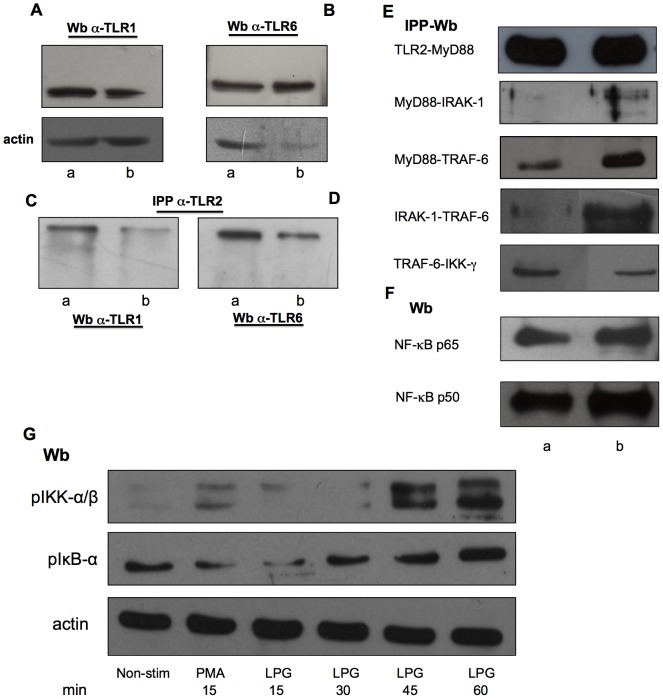
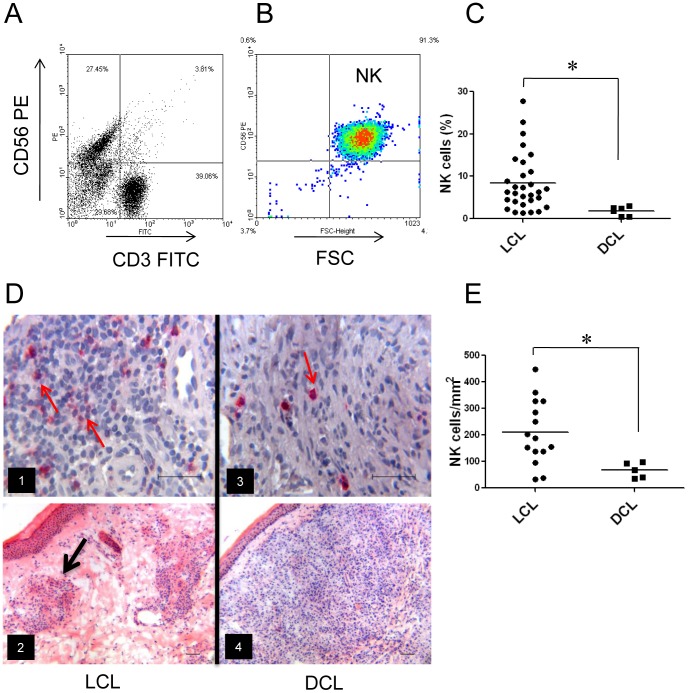
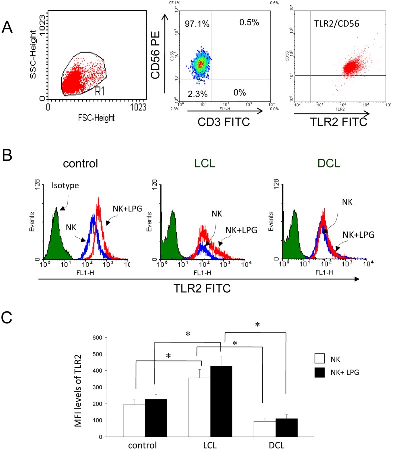
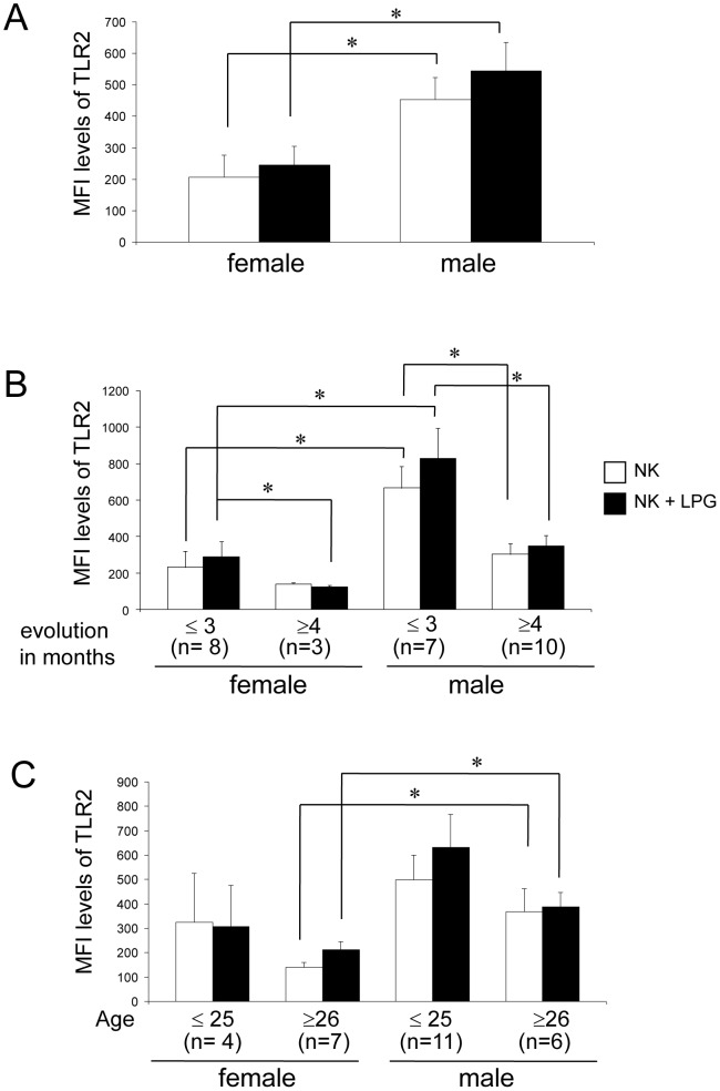
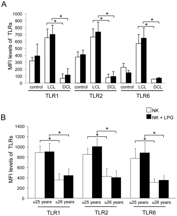
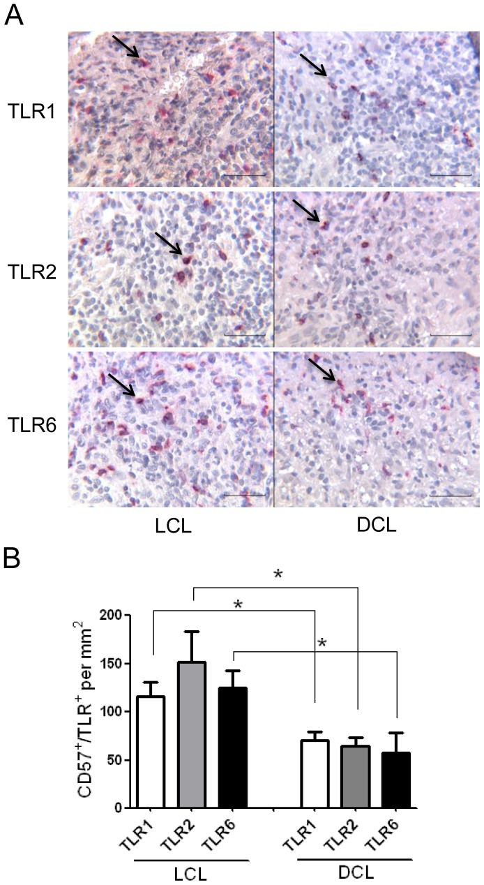
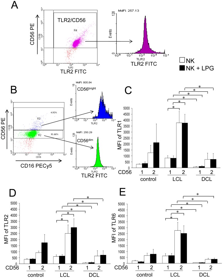
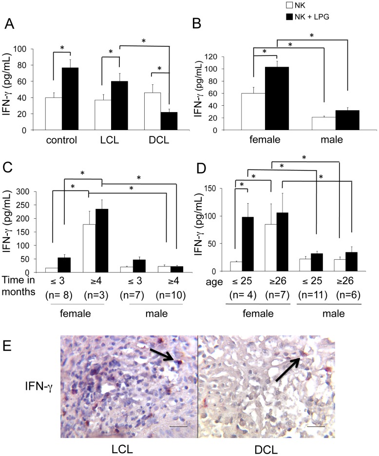
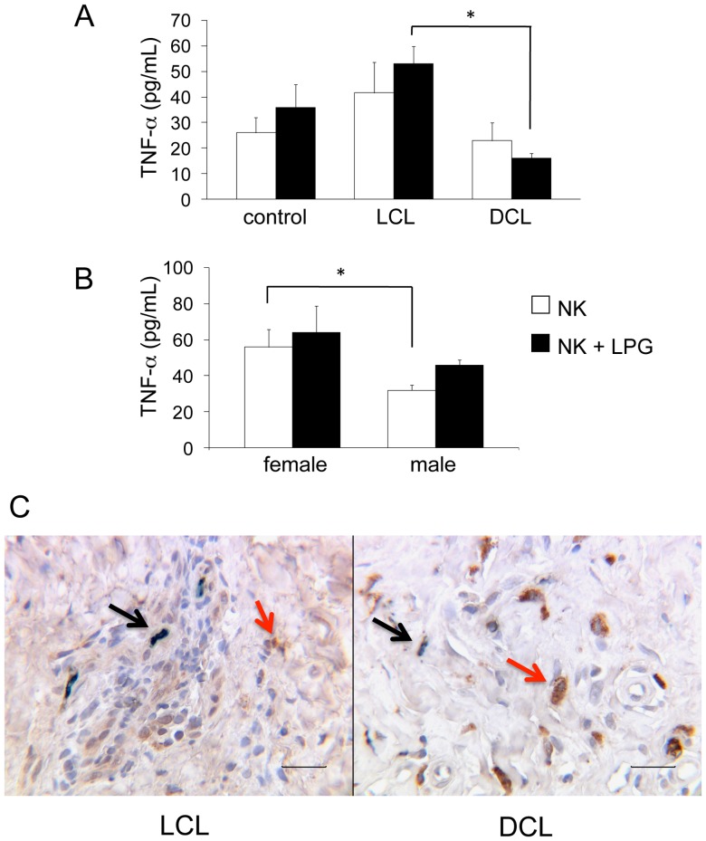
Leishmania mexicana causes localized (LCL) or diffuse cutaneous leishmaniasis (DCL). The cause of dissemination in DCL remains unknown, yet NK cells possibly play a role in activating leishmanicidal mechanisms during innate and adaptive immune responses. We had previously shown that Leishmania lipophosphoglycan (LPG) is a ligand for TLR2, activating human NK cells. We have now analyzed NK cells in LCL and DCL patients. NK numbers and effector mechanisms differed drastically between both groups of patients: DCL patients showed reduced NK cell numbers; diminished IFN-γ and TNF-α production; and lower TLR2, TLR1, and TLR6 expression as compared to LCL patients. The altered protein expression found in NK cells of DCL patients correlated with their down-regulation of IFN-γ gene expression in LPG-stimulated and non-stimulated cells as compared to LCL patients. NK cell response was further analyzed according to gender, age, and disease evolution in LCL patients showing that female patients produced higher IFN-γ levels throughout the disease progression, whereas TLR2 expression diminished in both genders with prolonged disease evolution and age. We furthermore show the activation pathway of LPG binding to TLR2 and demonstrated that TLR2 forms immunocomplexes with TLR1 and TLR6. In addition to the reduced NK cell numbers in peripheral blood, DCL patients also showed reduced NK cell numbers in the lesions. They were randomly scattered within the lesions, showing diminished cytokine production, which contrasts with those of LCL lesions, where NK cells produced IFN-γ and TNF-α and were found within organized granulomas. We conclude that in DCL patients the reduced NK-cell numbers and their diminished activity, evidenced by low TLR expression and low cytokine production, are possibly involved in the severity of the disease. Our results provide new information on the contribution of NK cells in Leishmania infections of the human host.


Conflict of interest statement

Competing Interests: The authors have declared that no competing interests exist.

Figures

Figure 1
Figure 1. LPG promotes binding of TLR signaling proteins and NF-κB nuclear translocation.
(A and B) Western blot shows TLR1 and TLR6 in NK cells. (C and D) Immunoprecipitations were performed with anti-TLR2 in non-stimulated (lane a) and LPG-stimulated NK cells after 1 h of co-incubation (lane b). (C) Immunoprecipitates were subjected to Western blotting and probed with anti-TLR1 and (D) anti-TLR6 antibodies, respectively. A representative immunoblot from four different experiments is shown. (E) NK cell lysates in non-stimulated and LPG-stimulated NK cells were immunoprecipitated with anti-TLR2, anti-MyD88, anti-IRAK-1 and anti-TRAF-6 antibodies and Western blotted with anti-MyD88, anti-IRAK-1, anti-TRAF-6 and anti-IKK-γ antibodies. (F) Nuclear translocation of p50 and p65 NF-κB isoforms were analyzed in non-stimulated and LPG-stimulated NK cells. (G) Phosphorilation of pIKK-α/β and pIκB-α were analyzed in non-stimulated NK cells (Non-stim) or in cells stimulated with PMA (15 min) or LPG (15, 30, 45 and 60 min). A representative immunoblot from two different experiments is show.
Figure 2
Figure 2. NK cells in patients infected with Leishmania mexicana.
(A) Representative flow cytometry plots of peripheral blood from patients before separation of NK cells from PBMC. (B) NK cells after purification (CD56+/CD3-). (C) Percentage of NK cells from patients with cutaneous leishmaniasis in peripheral blood [LCL (n = 30) and DCL (n = 6)]. Each dot represents a patient and horizontal line represents the mean of each group. (D) Immunostaining of NK cells in lesions shown NK cells. (D1) NK cells in LCL patients stained in red (CD57+) some of which are marked with red arrows. (D3) NK cells in lesions of DCL patients. (D2) H&E staining of lesions from LCL patients showing granuloma (black arrow). (D4) H&E staining of lesion from DCL patient. (E) Number of NK cells per mm2 in lesions. Each dot represents a patient and the horizontal line represents the mean. Images are representative of LCL patients (n = 15) and DCL patients (n = 5). *p≤0.05 was considered significant. Scale bar = 50 μm.
Figure 3
Figure 3. Analysis of TLR2 expression on NK cells.
(A) Representative flow CD56+/CD3- expression in membranes of NK cells; right image: NK cells were gated and analyzed for TLR2 (TLR2+/CD56+) expression. (B) Representative histograms showing the MFI levels of TLR2 expression on NK cells from control and patients (LCL and DCL). (C) Quantitation of TLR2 expression on NK cells. しろいしかく Non-stimulated cells, ▪ LPG-stimulated cells. Data are representative of healthy controls (n = 21), LCL patients (n = 28) and DCL patients (n = 6). These results are the mean ±SEM. *p≤0.05 was considered significant.
Figure 4
Figure 4. TLR2 expression in NK cells in LCL patients (n = 28).
Analysis according to: (A) gender, (B) disease evolution (≤3 or ≥4 months) or (C) age (≤25 or ≥26 years). しろいしかく Non-stimulated NK cells, ▪ LPG-stimulated NK cells. Cell surface expression is indicated by MFI. These results are the mean ± SEM. *p≤0.05 was considered significant.
Figure 5
Figure 5. TLR1, TLR2 and TLR6 expression on NK cells from controls and patients (LCL and DCL).
(A) Analysis according to disease form. (B) TLR2, TLR1 and TLR6 expression according to age (≤25 or ≥26 years) in LCL patients. しろいしかく Non-stimulated NK cells, ▪ LPG-stimulated NK cells. Cell surface expression is indicated by MFI. These results are the mean ±SEM. *Significant differences were observed between LCL and DCL patients for all TLRs in LPG-stimulated and non-stimulated cells.
Figure 6
Figure 6. TLR1, TLR2 and TLR6 expression on NK cells in lesions of 3 LCL and 3 DCL patients.
(A) Double immunohistochemistry staining of TLR expression (TLR1, TLR2 and TLR6) on NK cells (CD57). (B) Number of NK cells expressing TLRs per mm2. Results are the mean ±SEM. Scale bar = 50 μm. Black arrows show double positive cells.
Figure 7
Figure 7. TLR1, TLR2 and TLR6 expression on NK-cell subsets (CD56bright and CD56dim) of patients and controls.
(A) Representative flow cytometry dot plot (TLR2 vs. CD56) and histogram of TLR2 expression in NK cells. (B) This gate was subsequently analyzed for TLR2 expression on NK-cell subsets. Dot plot (CD16+/CD56+) and histogram for CD56bright (blue box) and CD56dim (green box) NK cells. (C) TLR1 expression. (D) TLR2 expression. (E) TLR6 expression. しろいしかく Non-stimulated NK cells; ▪ LPG-stimulated NK cells. Lane 1: CD56dim; lane 2: CD56bright NK cells. Cell surface expression is indicated by MFI. Results are the mean ±SEM. *Significant differences were observed between NK cell subsets of LCL and DCL patients for all 3 TLRs in LPG-stimulated and non-stimulated cells. LCL patients (n = 9), DCL patients (n = 4) and controls (n = 4).
Figure 8
Figure 8. IFN-γ production by NK cells.
(A) IFN-γ production of peripheral blood NK cells [control subjects (n = 21), LCL patients (n = 28) and DCL patients (n = 6)]. Analysis in LCL patients [female (n = 11) and male (n = 17)] according to: (B) gender; (C) disease duration; (D) age. (E) Double immunohistochemical labelling (CD57+/IFN-γ+) in lesions of patients (LCL and DCL) showed redish-brown staining generated by the combination of a red AP substrate used for NK cells and DAB Black used for IFN-γ staining. しろいしかく Non-stimulated NK cells. ▪ LPG-stimulated NK cells. Mean ±SEM is shown. *p≤0.05 was considered significant. Scale bar = 50 μm. Black arrows show double positive cells.
Figure 9
Figure 9. TNF-α production by NK cells.
(A) TNF-α production in peripheral blood [control subjects (n = 21), LCL patients (n = 28) and DCL patients (n = 6)]. (B) Analysis of TNF-α production of LCL patients according to gender [female (n = 11) and male (n = 17)]. (C) Double immunohistochemistry (CD57+/TNF-α+) staining of lesions of LCL and DCL patients showed dark green staining induced by the combination of a green substrate (Stay Green/AP) used for NK cells and DAB (brown) used for TNF-α staining. しろいしかく Non-stimulated NK cells, ▪ LPG-stimulated NK cells. Mean ±SEM is shown. *p≤0.05 was considered significant. Scale bar = 50 μm. Double positive cells CD57+/TNF-α+ (black arrows) and single positive cells CD57-/TNF-α+ (red arrows).

References

    1. Salaiza-Suazo N, Volkow P, Pérez-Tamayo R, Moll H, Gillitzer R, et al. (1999) Treatment of two patients with diffuse cutaneous leishmaniasis caused by Leishmania mexicana modifies the immunohistological profile but not the disease outcome. Trop Med Int Health 4: 801–811. - PubMed
    1. MEXICO World Health Organization website. Available: www.who/int/leishmaniasis/resources/MEXICO.pdf. Accessed December 5, 2013.
    1. Becker I, Salaiza N, Aguirre M, Delgado J, Carrillo-Carrasco N, et al. (2003) Leishmania lipophosphoglycan (LPG) activates NK cells through toll-like receptor-2. Mol Biochem Parasitol 130: 65–74. - PubMed
    1. de Veer MJ, Curtis JM, Baldwin TM, DiDonato JA, Sexton A, et al. (2003) MyD88 is essential for clearance of Leishmania major: possible role for lipophosphoglycan and Toll-like receptor 2 signaling. Eur J Immunol 33: 2822–2831. - PubMed
    1. Kamhawi S (2006) Phlebotomine sand flies and Leishmania parasites: friends or foes? Trends Parasitol 22: 439–445. - PubMed

Publication types

MeSH terms

LinkOut - more resources

Cite

AltStyle によって変換されたページ (->オリジナル) /