This site needs JavaScript to work properly. Please enable it to take advantage of the complete set of features!
Skip to main page content
U.S. flag

An official website of the United States government

Dot gov

The .gov means it’s official.
Federal government websites often end in .gov or .mil. Before sharing sensitive information, make sure you’re on a federal government site.

Https

The site is secure.
The https:// ensures that you are connecting to the official website and that any information you provide is encrypted and transmitted securely.

NIH NLM Logo
Log in
Access keys NCBI Homepage MyNCBI Homepage Main Content Main Navigation
. 2014;181(13-14):1715-1723.
doi: 10.1007/s00604-014-1198-5. Epub 2014 Feb 18.

Multiplex isothermal solid-phase recombinase polymerase amplification for the specific and fast DNA-based detection of three bacterial pathogens

Affiliations

Multiplex isothermal solid-phase recombinase polymerase amplification for the specific and fast DNA-based detection of three bacterial pathogens

Sebastian Kersting et al. Mikrochim Acta. 2014.

Abstract

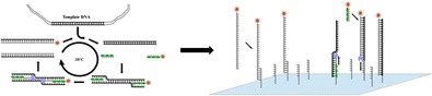
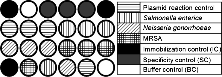
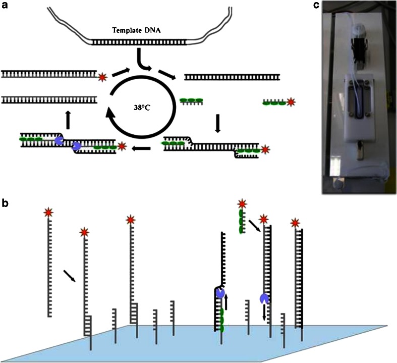
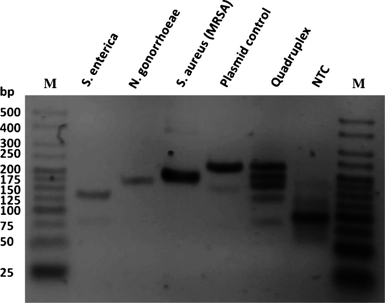
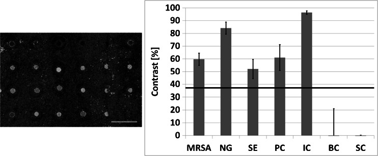
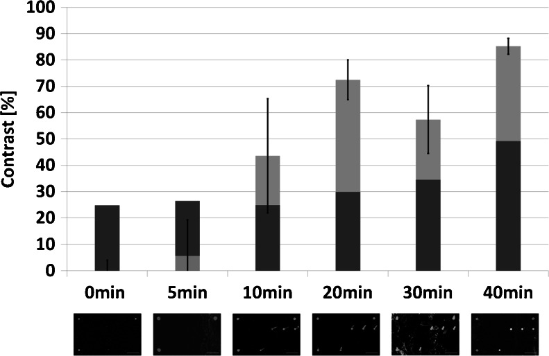
We report on the development of an on-chip RPA (recombinase polymerase amplification) with simultaneous multiplex isothermal amplification and detection on a solid surface. The isothermal RPA was applied to amplify specific target sequences from the pathogens Neisseria gonorrhoeae, Salmonella enterica and methicillin-resistant Staphylococcus aureus (MRSA) using genomic DNA. Additionally, a positive plasmid control was established as an internal control. The four targets were amplified simultaneously in a quadruplex reaction. The amplicon is labeled during on-chip RPA by reverse oligonucleotide primers coupled to a fluorophore. Both amplification and spatially resolved signal generation take place on immobilized forward primers bount to expoxy-silanized glass surfaces in a pump-driven hybridization chamber. The combination of microarray technology and sensitive isothermal nucleic acid amplification at 38 °C allows for a multiparameter analysis on a rather small area. The on-chip RPA was characterized in terms of reaction time, sensitivity and inhibitory conditions. A successful enzymatic reaction is completed in <20 min and results in detection limits of 10 colony-forming units for methicillin-resistant Staphylococcus aureus and Salmonella enterica and 100 colony-forming units for Neisseria gonorrhoeae. The results show this method to be useful with respect to point-of-care testing and to enable simplified and miniaturized nucleic acid-based diagnostics. FigureThe combination of multiplex isothermal nucleic acid amplification with RPA and spatially-resolved signal generation on specific immobilized oligonucleotides.

Keywords: DNA sensor; Isothermal amplification; Microchip; Point-of-care; RPA.


Figures

Figure
Figure
The combination of multiplex isothermal nucleic acid amplification with RPA and spatially-resolved signal generation on specific immobilized oligonucleotides
Fig. 1
Fig. 1
Array layout for on-chip RPA experiments. Each subarray consists of 24 spots. In total nine of these subarrays were spotted on each slide. All subarrays contained target specific oligonucleotides for on-chip amplification and hybridization and different types of controls: Specificity control (SC), buffer control (BC) and immobilization control (IC)
Fig. 2
Fig. 2
Principle of labeling RPA, on-chip RPA and photograph of one programmable hybridization chamber. a Schematic reaction mechanism of the labeling RPA in solution: two oligonucleotide primers form a complex with a recombinase (green ovals) and are directed to homologous sequences on the target sequence were they are able to invade the DNA double strand. The polymerase (blue) elongates the strand leading to duplication. RPA runs continuously at 38 °C. A labeling of the amplification product is achieved by using a reverse primer coupled with a Cy5 reporter dye (red). b Two mechanisms for signal generation during the on-chip RPA. (i) Single-stranded and labeled amplicons from asymmetric RPA in solution above the biochip can hybridize specifically to immobilized forward primers. (ii) The forward primer attached to the surface serves as starting point for solid-phase RPA c Photograph of one chamber in the programmable hybridization station used for on-chip RPA experiments. The chamber is temperature adjustable and features an individually programmable pump-driven mixing system (not in the picture) with a reaction volume of 45 μL. Slides placed in the chamber are sealed with a silicone o-ring
Fig. 3
Fig. 3
Agarose gel electrophoresis (3 %) with all singleplex and multiplex RPA reactions after clean-up, using 250 pg of genomic or plasmid DNA. Amplified products can be identified by size: Salmonella enterica (133 bp) Neisseria gonnorhoeae (165 bp), methicillin-resistant Staphylococcus aureus (MRSA) (193 bp) and plasmid control (227 bp). A successful quadruplex RPA is indicated by the four distinct bands visible, while in the no-template control (NTC) no specific band is present. M = Molecular weight marker HyperLadder V (Bioline)
Fig. 4
Fig. 4
Quadruplex on-chip RPA experiment for the detection of methicilin-resistant Staphylococcus aureus (MRSA), Neisseria gonorrhoeae (NG), Salmonella enterica (SE) and a plasmid (amplification control) (PC) in parallel. Laser scanner images of one subarray (left side) showing clearly visible signals at positions where amplification-specific sequences are immobilized (compare with Fig. 1). Quantitative analysis (right side) exhibit signals well above the dynamic limit of detection (LOD) (horizontal line), while no signals above LOD occur in non-specific or buffer control positions. IC: immobilization control; BC: buffer control; SC: specificity control
Fig. 5
Fig. 5
Determination of the reaction time for on-chip RPA experiments showing a signal after 20 min of isothermal amplification for the specific detection of Salmonella enterica. After 10 min also target specific signals are visible but are not constant across the whole slide resulting in a high standard derivation. Dark grey: Independently calculated dynamic LOD for every assay. Light grey: Signal for Salmonella enterica-specific positions on slides

References

    1. Mullis K, Faloona F, Scharf S, Saiki R, Horn G, Erlich H. Specific enzymatic amplification of DNA in vitro: the polymerase chain reaction. Cold Spring Harb Symp Quant Biol. 1986;51(Pt 1):263–273. doi: 10.1101/SQB.1986.051.01.032. - DOI - PubMed
    1. Yang S, Rothman RE. PCR-based diagnostics for infectious diseases: uses, limitations, and future applications in acute-care settings. Lancet Infect Dis. 2004;4:337–348. doi: 10.1016/S1473-3099(04)01044-8. - DOI - PMC - PubMed
    1. Zhang Y, Ozdemir P. Microfluidic DNA amplification—a review. Anal Chim Acta. 2009;638:115–125. doi: 10.1016/j.aca.2009年02月03日8. - DOI - PubMed
    1. Gill P, Ghaemi A. Nucleic acid isothermal amplification technologies: a review. Nucleosides Nucleotides Nucleic Acids. 2008;27:224–243. doi: 10.1080/15257770701845204. - DOI - PubMed
    1. Notomi T. Loop-mediated isothermal amplification of DNA. Nucleic Acids Res. 2000;28:63e. doi: 10.1093/nar/28.12.e63. - DOI - PMC - PubMed
Cite

AltStyle によって変換されたページ (->オリジナル) /