This site needs JavaScript to work properly. Please enable it to take advantage of the complete set of features!
Skip to main page content
U.S. flag

An official website of the United States government

Dot gov

The .gov means it’s official.
Federal government websites often end in .gov or .mil. Before sharing sensitive information, make sure you’re on a federal government site.

Https

The site is secure.
The https:// ensures that you are connecting to the official website and that any information you provide is encrypted and transmitted securely.

NIH NLM Logo
Log in
Access keys NCBI Homepage MyNCBI Homepage Main Content Main Navigation
Review
. 2014 Jul 3;8(7):e2918.
doi: 10.1371/journal.pntd.0002918. eCollection 2014 Jul.

Merits and pitfalls of currently used diagnostic tools in mycetoma

Affiliations
Review

Merits and pitfalls of currently used diagnostic tools in mycetoma

Wendy W J van de Sande et al. PLoS Negl Trop Dis. .

Abstract

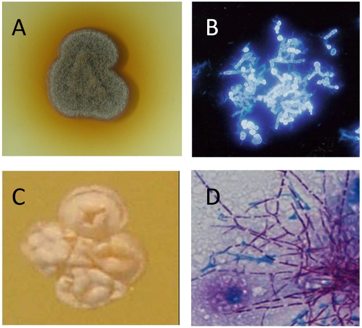
Treatment of mycetoma depends on the causative organism and since many organisms, both actinomycetes (actinomycetoma) and fungi (eumycetoma), are capable of producing mycetoma, an accurate diagnosis is crucial. Currently, multiple diagnostic tools are used to determine the extent of infections and to identify the causative agents of mycetoma. These include various imaging, cytological, histopathological, serological, and culture techniques; phenotypic characterisation; and molecular diagnostics. In this review, we summarize these techniques and identify their merits and pitfalls in the identification of the causative agents of mycetoma and the extent of the disease. We also emphasize the fact that there is no ideal diagnostic tool available to identify the causative agents and that future research should focus on the development of new and reliable diagnostic tools.


Conflict of interest statement

The authors have declared that no competing interests exist.

Figures

Figure 1
Figure 1. Different grains obtained from eumycetoma and actinomycetoma lesions.
In this figure different grain types are shown. A: An eumycetoma surgical excision with numerous black grains, indicative for M. mycetomatis. B: An actinomycetoma surgical biopsy with numerous yellow grains, indicative for S. somaliensis. C: Grains of Madurella mycetomatis fixed in formalin. D: Histological slide of a Madurella mycetomatis grain inside subcutaneous tissue. The grain is clearly seen as a round brown structure (arrow) (×ばつ100). E: Histological slide of a S. somaliensis grain inside subcutaneous tissue (arrow) (×ばつ400).
Figure 2
Figure 2. Imaging techniques used in mycetoma.
A: A radiograph of the foot, showing soft tissue shadow (arrow), and multiple large cavities (c) in line with eumycetoma. B: A typical sonogram of scrotal eumycetoma. In this sonogram multiple cavities with thick walls and multiple hyper-reflective echoes (arrow) are seen, which are in line with grains. C: An MRI of the foot, showing massive soft tissue and bone destruction. In this MRI, grains appear as conglomerates of small (2–5 mm) round hyperintense lesions (arrow).
Figure 3
Figure 3. Obtaining grains via Fine Needle Aspiration.
Figure 4
Figure 4. Flow diagram of histological identification of causative agents of mycetoma, based on references , , .
Figure 5
Figure 5. Identifying mycetoma causative agents by culture.
A: Madurella mycetomatis grown on sabouraud agar. B: Microscopic appearance of Madurella mycetomatis stained with calcofluor white. C: N. brasiliensis colony. D: Microscopic appearance of N. brasiliensis.

References

    1. Ahmed AO, van Leeuwen W, Fahal A, van de Sande WWJ, Verbrugh H, et al. (2004) Mycetoma caused by Madurella mycetomatis: a neglected infectious burden. Lancet Infect Dis 4: 566–574. - PubMed
    1. Ahmed A, van de Sande WWJ, Fahal A, Bakker-Woudenberg IA, Verbrugh H, et al. (2007) Management of mycetoma: major challenge in tropical mycoses with limited international recognition. Curr Opin Infect Dis 20: 146–151. - PubMed
    1. Mhmoud NA, Ahmed SA, Fahal AH, de Hoog GS, Gerrits van den Ende AH, et al. (2012) Pleurostomophora ochracea, a novel agent of human eumycetoma with yellow grains. J Clin Microbiol 50: 2987–2994. - PMC - PubMed
    1. Van de Sande WWJ (2013) Global burden of human mycetoma: a systematic review and meta-analysis. PLoS Negl Trop Dis 7: e2550. - PMC - PubMed
    1. de Hoog GS, van Diepeningen AD, Mahgoub el S, van de Sande WW (2012) New species of Madurella, causative agents of black-grain mycetoma. J Clin Microbiol 50: 988–994. - PMC - PubMed

Publication types

Cite

AltStyle によって変換されたページ (->オリジナル) /