This site needs JavaScript to work properly. Please enable it to take advantage of the complete set of features!
Skip to main page content
U.S. flag

An official website of the United States government

Dot gov

The .gov means it’s official.
Federal government websites often end in .gov or .mil. Before sharing sensitive information, make sure you’re on a federal government site.

Https

The site is secure.
The https:// ensures that you are connecting to the official website and that any information you provide is encrypted and transmitted securely.

NIH NLM Logo
Log in
Access keys NCBI Homepage MyNCBI Homepage Main Content Main Navigation
Review
. 2014 Apr 29;6(5):1929-73.
doi: 10.3390/v6051929.

Hantavirus reservoirs: current status with an emphasis on data from Brazil

Affiliations
Review

Hantavirus reservoirs: current status with an emphasis on data from Brazil

Renata Carvalho de Oliveira et al. Viruses. .

Abstract

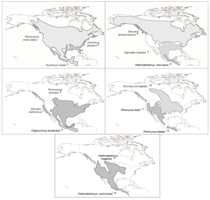
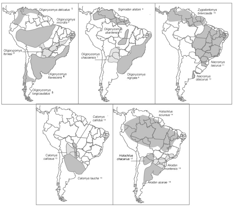
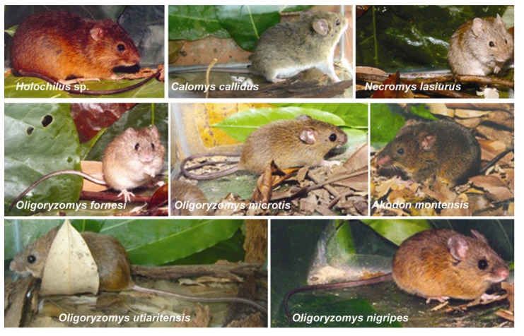
Since the recognition of hantavirus as the agent responsible for haemorrhagic fever in Eurasia in the 1970s and, 20 years later, the descovery of hantavirus pulmonary syndrome in the Americas, the genus Hantavirus has been continually described throughout the World in a variety of wild animals. The diversity of wild animals infected with hantaviruses has only recently come into focus as a result of expanded wildlife studies. The known reservoirs are more than 80, belonging to 51 species of rodents, 7 bats (order Chiroptera) and 20 shrews and moles (order Soricomorpha). More than 80 genetically related viruses have been classified within Hantavirus genus; 25 recognized as human pathogens responsible for a large spectrum of diseases in the Old and New World. In Brazil, where the diversity of mammals and especially rodents is considered one of the largest in the world, 9 hantavirus genotypes have been identified in 12 rodent species belonging to the genus Akodon, Calomys, Holochilus, Oligoryzomys, Oxymycterus, Necromys and Rattus. Considering the increasing number of animals that have been implicated as reservoirs of different hantaviruses, the understanding of this diversity is important for evaluating the risk of distinct hantavirus species as human pathogens.


Figures

Figure 1
Figure 1
Distribution of the North American hantavirus primary reservoirs. Number after species names refer to the hantavirus genotype or specie (*) associate: (1) Sin Nombre virus* and Monongahela virus; (2) Bayou virus*; (3) Catacamas virus and Playa de Oro virus; (4) Prospect Hill virus; (5) Black Creek virus* and Muleshoe virus*; (6) Rio Segundo virus*; (7) Isla Vista virus; (8) Blue River virus and New York virus*; (9) Choclo virus; (10) Bloodland Lake virus; (11) Limestone canyon virus; (12) Montano virus; (13) El Moro Canyon virus* and Huitzilac virus; (14) Carrizal virus, see Supplementary Table 2.
Figure 2
Figure 2
Distribution of the main South American hantavirus reservoirs. Number after species names refer to the hantavirus genotype or specie (*) associate: (1) Maporal virus; (2) Rio Mamore virus*; (3) Anajatuba virus; (4) Central Plata virus and Lechiguanas virus; (5) Andes virus* andOran virus; (6) Castelo dos Sonhos virus; (7) Bermejo virus; (8) Juquitiba virus and Itapua virus; (9) Caño Delgadito virus*; (10) Calabazo virus; (11) Araraquara virus; (12) Maciel virus; (13) Laguna Negra virus*; (14) Rio Mearin virus; (15) Ape Aime-Itapua virus and Jabora virus; (16) Pergamino virus; (17) Alto Paraguay virus, see Supplementary Table 2.
Figure 3
Figure 3
(a) Akodon montensis. Image courtesy of Cibele Bonvicino, Ph.D. (b) Electron micrograph. Morphology of Jabora hantavirus particule obtained from lung sample of Akodon montensis. Scale bar: 100 nm. Image courtesy of Debora Barreto, Ph.D. and Monika Barth, Ph.D.
Figure 4
Figure 4
Rodents reservoirs of the hantaviruses described in Brazil. Image courtesy of Cibele Bonvicino, Ph.D.

References

    1. Lee H.W., Lee P.W., Johnson K.M. Isolation of the etiologic agent of Korean hemorrhagic fever. J. Infect. Dis. 1978;137:298–308. doi: 10.1093/infdis/137.3.298. - DOI - PubMed
    1. Lee P.W., Gajdusek D.C., Gibbs C.J., Xu Z.Y. Aetiological relation between Korean haemorrhagic fever with renal syndrome in People’s Republic of China. Lancet. 1980;1:819–820. - PubMed
    1. Brummer-Korvenkontio M., Vaheri A., Hovi T., von Bonsdorff C.H., Vuorimies J., Manni T., Penttinen K., Oker-Blom N., Lähdevirta J. Nephropathia epidemica: Detection of antigen in bank voles and serologic diagnosis of human infection. J. Infect. Dis. 1980;141:131–134. doi: 10.1093/infdis/141.2.131. - DOI - PubMed
    1. Lee P.W., Goldgaber D., Gibbs C.J., Gajdusek D.C., Yanagihara R., Svedmyr A., Hlaca D., Vesenjak-Hirjan J., Gligic A. Other serotypes of hemorrhagic fever with renal syndrome viruses in Europe. Lancet. 1982;2:1405–1406. - PubMed
    1. Avsic-Zupanc T., Xiao S.Y., Stojanovic R., Gligic A., van der Groen G., LeDuc J.W. Characterization of Dobrava virus: A Hantavirus from Slovenia, Yugoslavia. J. Med. Virol. 1992;38:132–137. doi: 10.1002/jmv.1890380211. - DOI - PubMed

Publication types

LinkOut - more resources

Cite

AltStyle によって変換されたページ (->オリジナル) /