This site needs JavaScript to work properly. Please enable it to take advantage of the complete set of features!
Skip to main page content
U.S. flag

An official website of the United States government

Dot gov

The .gov means it’s official.
Federal government websites often end in .gov or .mil. Before sharing sensitive information, make sure you’re on a federal government site.

Https

The site is secure.
The https:// ensures that you are connecting to the official website and that any information you provide is encrypted and transmitted securely.

NIH NLM Logo
Log in
Access keys NCBI Homepage MyNCBI Homepage Main Content Main Navigation
. 2014 Jul:50:1-17.
doi: 10.1016/j.ibmb.2014年03月01日2. Epub 2014 Apr 3.

Molecular biology of insect sodium channels and pyrethroid resistance

Affiliations

Molecular biology of insect sodium channels and pyrethroid resistance

Ke Dong et al. Insect Biochem Mol Biol. 2014 Jul.

Abstract

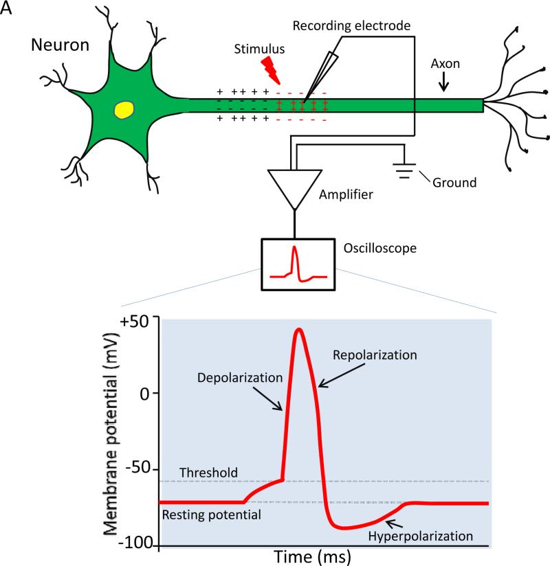
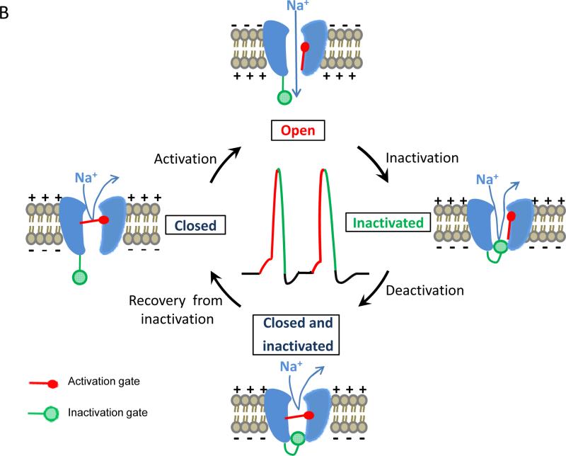
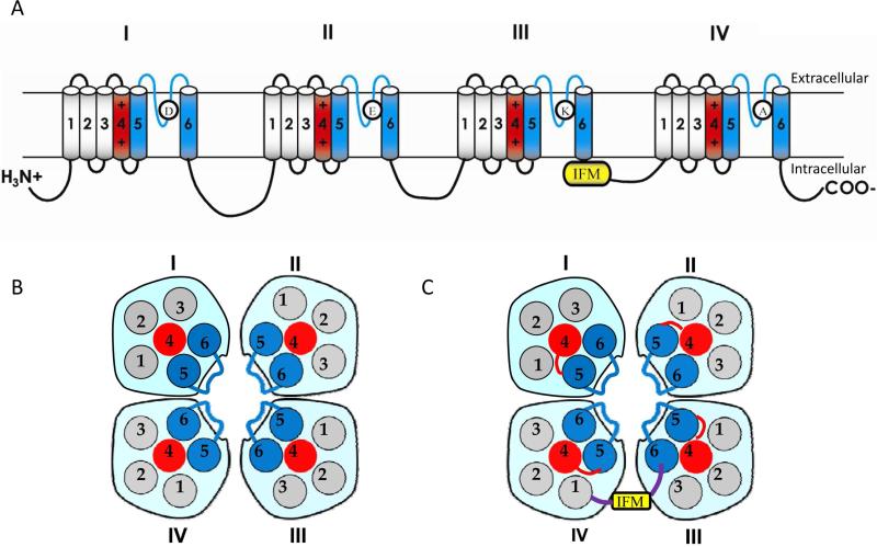
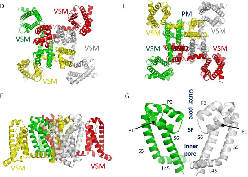
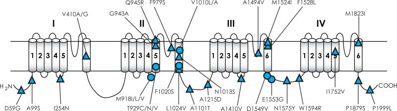
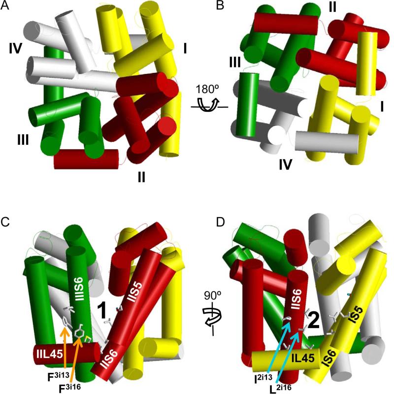
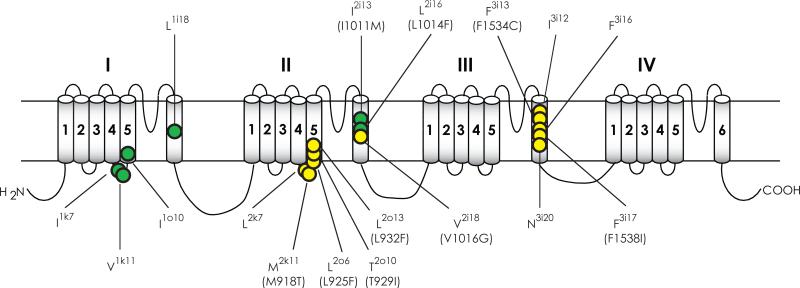
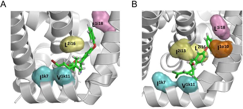
Voltage-gated sodium channels are essential for the initiation and propagation of the action potential in neurons and other excitable cells. Because of their critical roles in electrical signaling, sodium channels are targets of a variety of naturally occurring and synthetic neurotoxins, including several classes of insecticides. This review is intended to provide an update on the molecular biology of insect sodium channels and the molecular mechanism of pyrethroid resistance. Although mammalian and insect sodium channels share fundamental topological and functional properties, most insect species carry only one sodium channel gene, compared to multiple sodium channel genes found in each mammalian species. Recent studies showed that two posttranscriptional mechanisms, alternative splicing and RNA editing, are involved in generating functional diversity of sodium channels in insects. More than 50 sodium channel mutations have been identified to be responsible for or associated with knockdown resistance (kdr) to pyrethroids in various arthropod pests and disease vectors. Elucidation of molecular mechanism of kdr led to the identification of dual receptor sites of pyrethroids on insect sodium channels. Many of the kdr mutations appear to be located within or close to the two receptor sites. The accumulating knowledge of insect sodium channels and their interactions with insecticides provides a foundation for understanding the neurophysiology of sodium channels in vivo and the development of new and safer insecticides for effective control of arthropod pests and human disease vectors.

Keywords: Alternative splicing; Knockdown resistance; Pyrethroid receptor sites; Pyrethroids; RNA editing; Sodium channel.


Figures

Fig. 1
Fig. 1
Voltage-gated sodium channels and the action potential. (A) Recording of an action potential. A recording microelectrode, which inserts inside the axon (intracellular recording), is connected to an amplifier. The amplifier compares the potential difference between the tip of the recording electrode and another electrode (called ground) placed in the solution bathing the neuron. The potential difference can be displayed using an oscilloscope. In response to membrane depolarization, such as a depolarizing stimulus indicated in A, sodium channels open (i.e., activated), resulting in further depolarization of the membrane as indicated by the rising phase of the action potential. Sodium channel inactivation together with potassium channel activation helps terminate the action potential (repolarization and hyperpolarization). (B) Gating (i.e., opening and closing) of voltage-gated sodium channels. Please see the text for explanation.
Fig. 1
Fig. 1
Voltage-gated sodium channels and the action potential. (A) Recording of an action potential. A recording microelectrode, which inserts inside the axon (intracellular recording), is connected to an amplifier. The amplifier compares the potential difference between the tip of the recording electrode and another electrode (called ground) placed in the solution bathing the neuron. The potential difference can be displayed using an oscilloscope. In response to membrane depolarization, such as a depolarizing stimulus indicated in A, sodium channels open (i.e., activated), resulting in further depolarization of the membrane as indicated by the rising phase of the action potential. Sodium channel inactivation together with potassium channel activation helps terminate the action potential (repolarization and hyperpolarization). (B) Gating (i.e., opening and closing) of voltage-gated sodium channels. Please see the text for explanation.
Fig. 2
Fig. 2
Structure of the voltage-gated sodium channel. (A) The topology of the sodium channel indicating the sequence features that are critical for channel function. The sodium channel protein contains four homologous repeats (I-IV), each having six transmembrane segments (1-6). The isoluecine in the IFM motif in mammalian sodium channels is substituted with a methionine in insect sodium channels. (B and C) Schematic representations of extracellular (B) and intracellular (C) views of sodium channels. (D-G) The X-ray structure of the closed NavAb sodium channel (Payandeh et al., 2011). Four subunits of NavAb in yellow, red, green and grey, respectively, correspond to domains I, II, III and IV in eukaryotic four-domain sodium channels. (D) Extracellular view. (E) Intracellular view indicating the four voltage-sensing modules (VSM), and the pore module (PM). (F) Side view. (G) Expanded side view of the pore module showing only two subunits for clarity. The inner and outer pores are separated by the selectivity-filter region (SF).
Fig. 2
Fig. 2
Structure of the voltage-gated sodium channel. (A) The topology of the sodium channel indicating the sequence features that are critical for channel function. The sodium channel protein contains four homologous repeats (I-IV), each having six transmembrane segments (1-6). The isoluecine in the IFM motif in mammalian sodium channels is substituted with a methionine in insect sodium channels. (B and C) Schematic representations of extracellular (B) and intracellular (C) views of sodium channels. (D-G) The X-ray structure of the closed NavAb sodium channel (Payandeh et al., 2011). Four subunits of NavAb in yellow, red, green and grey, respectively, correspond to domains I, II, III and IV in eukaryotic four-domain sodium channels. (D) Extracellular view. (E) Intracellular view indicating the four voltage-sensing modules (VSM), and the pore module (PM). (F) Side view. (G) Expanded side view of the pore module showing only two subunits for clarity. The inner and outer pores are separated by the selectivity-filter region (SF).
Fig. 3
Fig. 3
Functional characterization of insect sodium channels expressed in Xenopus oocytes using the voltage-clamp technique. (A) Expression of insect sodium channels in Xenopus oocytes. Full-length cDNA clones encoding insect sodium channels are used as templates to synthesize capped-RNA (cRNA) in vitro. The cRNA is then injected into oocytes using a microinjector, and is often co-injected with cRNA of either TipE or a TipE-ortholog to enhance sodium channel expression. Insect sodium channels are expressed on the surface of the injected oocytes several days post-injection. (B) Two-electrode voltage clamp. The membrane of the oocyte is penetrated by two microelectrodes, one for measuring the voltage across the membrane while the other for injecting current into the cell to keep the voltage constant (i.e., voltage clamp). By measuring the amount of current injected, the system can determine the amplitude and time course of the ionic current flowing across the membrane at a given depolarization step, as shown in C. (C) A trace of an inward sodium current showing the rapid activation and inactivation of DmNav sodium channels. The current was elicited by a step depolarization to −10 mV from a holding potential of −120 mV. (D and E) Voltage dependence of activation (D) and inactivation (E) of three DmNav variants with different gating properties. (F and G) Activation kinetics (F) and deactivation kinetics (G) of BgNav1-1 (control) and mutants carrying V409M or V409L mutation (i.e., V410M/L in Vssc1). The kinetics of activation and deactivation (i.e., the rate of opening and closing of the activation gate were determined by the cell-attached macropatch (Oliveira et al., 2013; Warmke et al., 1997). Both mutations slow activation kinetics and accelerate deactivation kinetics (Oliveira et al., 2013).
Fig. 3
Fig. 3
Functional characterization of insect sodium channels expressed in Xenopus oocytes using the voltage-clamp technique. (A) Expression of insect sodium channels in Xenopus oocytes. Full-length cDNA clones encoding insect sodium channels are used as templates to synthesize capped-RNA (cRNA) in vitro. The cRNA is then injected into oocytes using a microinjector, and is often co-injected with cRNA of either TipE or a TipE-ortholog to enhance sodium channel expression. Insect sodium channels are expressed on the surface of the injected oocytes several days post-injection. (B) Two-electrode voltage clamp. The membrane of the oocyte is penetrated by two microelectrodes, one for measuring the voltage across the membrane while the other for injecting current into the cell to keep the voltage constant (i.e., voltage clamp). By measuring the amount of current injected, the system can determine the amplitude and time course of the ionic current flowing across the membrane at a given depolarization step, as shown in C. (C) A trace of an inward sodium current showing the rapid activation and inactivation of DmNav sodium channels. The current was elicited by a step depolarization to −10 mV from a holding potential of −120 mV. (D and E) Voltage dependence of activation (D) and inactivation (E) of three DmNav variants with different gating properties. (F and G) Activation kinetics (F) and deactivation kinetics (G) of BgNav1-1 (control) and mutants carrying V409M or V409L mutation (i.e., V410M/L in Vssc1). The kinetics of activation and deactivation (i.e., the rate of opening and closing of the activation gate were determined by the cell-attached macropatch (Oliveira et al., 2013; Warmke et al., 1997). Both mutations slow activation kinetics and accelerate deactivation kinetics (Oliveira et al., 2013).
Fig. 4
Fig. 4
Alternative splicing of DmNav transcripts. Optional exons are illustrated in blue blocks; and mutually exclusive exons (c/d and l/k) are highlighted in green. Four optional exons detected in larvae of D. melanogaster (Lin et al., 2009) are not indicated.
Fig. 5
Fig. 5
Mutations in sodium channels associated with pyrethroid resistance in arthropod species. (A) Mutations that have been examined in Xenopus oocytes. (B) Mutations that have not been examined in oocytes. Circles denote those detected in more than one species and triangles indicate those found in a single species. See Table 1 for details on these mutations. The four mutations marked with # have been examined in oocytes, but do not reduce the sensitivity of AaNav1-1 channels to permethrin or deltamethrin. The two mutations marked with * alone do not confer pyrethroid resistance, but enhanced pyrethroid resistance mediated by L1014F or V410M. References on functional characterization of mutations in Xenopus oocytes are listed below: V410L/M (Oliveira et al., 2013; (Lee et al., 1999; Lee and Soderlund, 2001; Liu et al., 2002; Oliveira et al., 2013; Zhao et al., 2000); E435K + C785R + L1014F (Tan et al., 2002b); M827I, T929I and L932F (Usherwood et al., 2007; Vais et al., 2001; Yoon et al., 2008); M918T (Lee et al., 1999; Usherwood et al., 2005; Vais et al., 2001); L925I and I936V (Usherwood et al., 2007); L1014F/S/H (Burton et al., 2011; Du et al., 2013; Smith et al., 1997; Tan et al., 2002b; Tan et al., 2005; Usherwood et al., 2005; Vais et al., 2000); V1016G/I, I1011M/V, S989P , and D1763Y (Du et al., 2013); F1534C (Du et al., 2013; Hu et al., 2011); F1538I (Tan et al., 2005).
Fig. 5
Fig. 5
Mutations in sodium channels associated with pyrethroid resistance in arthropod species. (A) Mutations that have been examined in Xenopus oocytes. (B) Mutations that have not been examined in oocytes. Circles denote those detected in more than one species and triangles indicate those found in a single species. See Table 1 for details on these mutations. The four mutations marked with # have been examined in oocytes, but do not reduce the sensitivity of AaNav1-1 channels to permethrin or deltamethrin. The two mutations marked with * alone do not confer pyrethroid resistance, but enhanced pyrethroid resistance mediated by L1014F or V410M. References on functional characterization of mutations in Xenopus oocytes are listed below: V410L/M (Oliveira et al., 2013; (Lee et al., 1999; Lee and Soderlund, 2001; Liu et al., 2002; Oliveira et al., 2013; Zhao et al., 2000); E435K + C785R + L1014F (Tan et al., 2002b); M827I, T929I and L932F (Usherwood et al., 2007; Vais et al., 2001; Yoon et al., 2008); M918T (Lee et al., 1999; Usherwood et al., 2005; Vais et al., 2001); L925I and I936V (Usherwood et al., 2007); L1014F/S/H (Burton et al., 2011; Du et al., 2013; Smith et al., 1997; Tan et al., 2002b; Tan et al., 2005; Usherwood et al., 2005; Vais et al., 2000); V1016G/I, I1011M/V, S989P , and D1763Y (Du et al., 2013); F1534C (Du et al., 2013; Hu et al., 2011); F1538I (Tan et al., 2005).
Fig. 6
Fig. 6
Modeling the pyrethroid receptor sites in the AaNav1-1 channel. (A and B) An NavAb-based model of the pore-forming module of the mosquito sodium channel AaNav1-1. Helices are shown as cylinders and repeat domains I, II, III, and IV are yellow, red, green and gray, respectively. The four repeat domains are arranged clockwise in the extracellular view (A) and counterclockwise at the cytoplasmic view (B). Side views of the sodium channel, where side chains of pyrethroid-sensing residues in Site 1 (C) and Site 2 (D) are shown by sticks. Note that Site 1 is located at the interface between domains II and III (O'Reilly et al., 2006; Usherwood et al., 2007); whereas Site 2, is located at the interface between domains I and II domain (Du et al., 2013).
Fig. 7
Fig. 7
Pyrethroid-sensing residues in Site 1 or Site 2 of an insect sodium channel. Pyrethroid-sensing residues are indicated in solid yellow circles for Site 1 residues and solid green circles for Site 2 residues. Mutations that are detected in pyrethroid-resistant field populations at the corresponding positions are indicated in brackets. See Table 2 for the amino acid sequence alignment of Kv1.2, NavAb, and AaNav1-1 channels in these regions that contain pyrethroid-sensing residues.
Fig. 8
Fig. 8
A Kv1.2-based model of the open AaNav1–1 channel with deltamethrin (A) and 1R-cis-permethrin (B) bound to Site 2. Cyan, orange, pink, and yellow surfaces are side chains of pyrethroid-sensing residues in segments IL45, IS5, IS6, and IIS6, respectively. The halogen atoms of pyrethroids bind between IL45 and IIS6. The terminal phenyl rings of deltamethrin and 1R-cis-permethrin bind between IS6 and IIS6. Reproduced with permission from PNAS.

References

    1. Bass BL. RNA editing. Oxford University Press; 2001.
    1. Bende NS, Kang E, Herzig V, Bosmans F, Nicholson GM, Mobli M, King GF. The insecticidal neurotoxin Aps III is an atypical knottin peptide that potently blocks insect voltage-gated sodium channels. Biochem Pharmacol. 2013;85:1542–1554. - PMC - PubMed
    1. Bosmans F, Tytgat J. Sea anemone venom as a source of insecticidal peptides acting on voltage-gated Na+ channels. Toxicon. 2007;49:550–560. - PMC - PubMed
    1. Bourdin CM, Moignot B, Wang L, Murillo L, Juchaux M, Quinchard S, Lapied B, Guerineau NC, Dong K, Legros C. Intron retention in mRNA encoding ancillary subunit of insect voltage-gated sodium channel modulates channel expression, gating regulation and drug sensitivity. PLoS One. 2013;8:e67290. - PMC - PubMed
    1. Brackenbury WJ, Isom LL. Na Channel β Subunits: Overachievers of the Ion Channel Family. Front Pharmacol. 2011;2:53. - PMC - PubMed

Publication types

LinkOut - more resources

Cite

AltStyle によって変換されたページ (->オリジナル) /