This site needs JavaScript to work properly. Please enable it to take advantage of the complete set of features!
Skip to main page content
U.S. flag

An official website of the United States government

Dot gov

The .gov means it’s official.
Federal government websites often end in .gov or .mil. Before sharing sensitive information, make sure you’re on a federal government site.

Https

The site is secure.
The https:// ensures that you are connecting to the official website and that any information you provide is encrypted and transmitted securely.

NIH NLM Logo
Log in
Access keys NCBI Homepage MyNCBI Homepage Main Content Main Navigation
. 2014 Mar 31:7:148.
doi: 10.1186/1756-3305年7月14日8.

Expression of microRNA-454 in TGF-β1-stimulated hepatic stellate cells and in mouse livers infected with Schistosoma japonicum

Affiliations

Expression of microRNA-454 in TGF-β1-stimulated hepatic stellate cells and in mouse livers infected with Schistosoma japonicum

Dandan Zhu et al. Parasit Vectors. .

Abstract

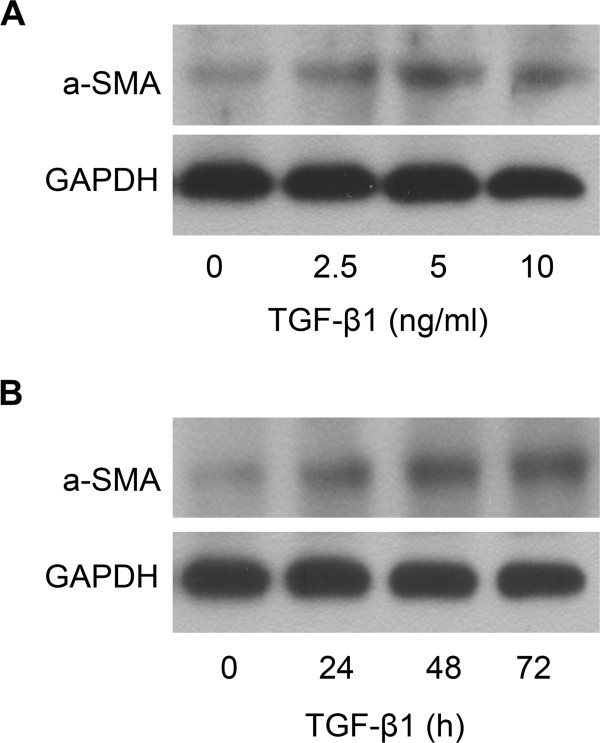
Background: In the process of hepatic fibrosis, hepatic stellate cells (HSCs) can be activated by many inflammatory cytokines. The transforming growth factor-β1 (TGF-β1) is one of the main profibrogenic mediators. Recently, some studies have also shown that microRNAs (miRNAs) play essential roles in the progress of liver fibrosis by being involved in the differentiation, fat metabolism and ECM production of HSCs.

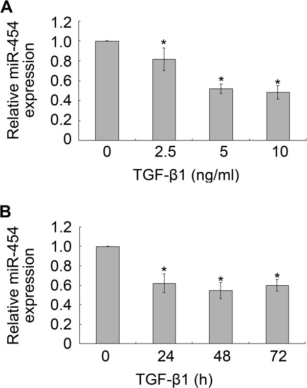
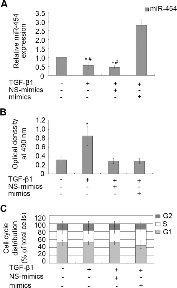
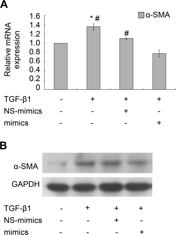
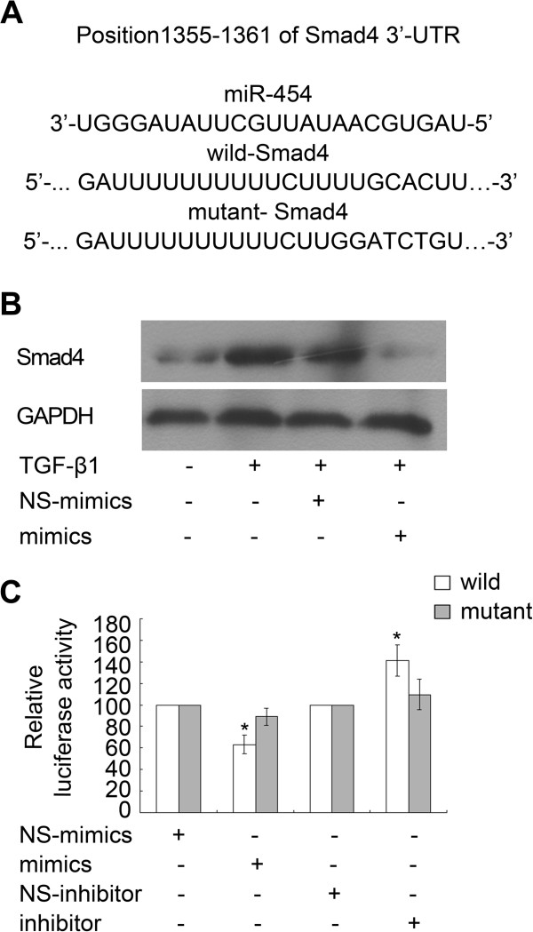
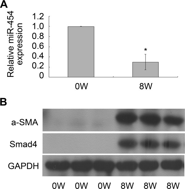
Methods: The expression of miR-454 in LX-2 cells treated with TGF-β1 and in the fibrotic livers with Schistosoma japonicum infection was detected by qRT-PCR. The role of miR-454 on LX-2 cells was then analyzed by Western blot, flow cytometry and luciferase assay.

Results: The results showed that the expression of miR-454 was down-regulated in the TGF-β1-treated LX-2 cells and miR-454 could inhibit the activation of HSCs by directly targeting Smad4. However, we found that miR-454 had no effect on cell cycle and cell proliferation in TGF-β1-treated LX-2. Besides these, miR-454 was found to be regulated in the process of Schistosoma japonicum infection.

Conclusions: All the results suggested that miR-454 could provide a novel therapeutic approach for treating liver fibrosis, especially the liver fibrosis induced by Schistosoma japonicum.


Figures

Figure 1
Figure 1
TGF-β1 can up-regulate the expression of α-SMA in LX-2. (A) Dose-dependent effect of TGF-β1 on α-SMA protein expression was analyzed by Western blot. (B) Time-dependent effect of TGF-β1 on α-SMA protein expression was analyzed by Western blot.
Figure 2
Figure 2
The expression of miR-454 was down-regulated in TGF-β1-treated LX-2 cells. (A, B) The expression of miR-454 in LX-2 cells which were treated with TGF-β1 at the indicated concentration or for the indicated time were detected by qRT-PCR. * P <0.05 vs control (cells with no stimulus).
Figure 3
Figure 3
The effect of miR-454 on proliferation and cell cycle distribution of TGF-β1-treated LX-2. (A) The expression of miR-454 in LX-2 cells was analyzed by qRT-PCR. The expression of miR-454 could be down-regulated in the cells treated with TGF-β1 and in the cells co-treated with TGF-β1 and NS-miRNA. * P <0.05 vs control (cells with no stimulus). However, the down-regulation of miR-454 expression induced by TGF-β1 could be reversed in the cells transfected with miR-454 mimics (# P <0.05, compared with the group of the cells co-treated with TGF-β1 and miR-454 mimics). (B) The proliferation capacity of LX-2 cells was increased when the cells were treated with TGF-β1. * P <0.05 vs control (cells with no stimulus). However, miR-454 mimics could not regulate the proliferation of LX-2 cells by MTT assay (P>0.05, compared with the group of the cells co-treated with TGF-β1 and NS-miRNA). (C) The effect of miR-454 on cell cycle of LX-2 cells was analyzed by flow cytometry. P>0.05 vs control (cells with no stimulus).
Figure 4
Figure 4
miR-454 mimics could inhibit TGF-β1–induced α-SMA expression in LX-2 cells. (A) miR-454 mimics reduced the levels of α-SMA mRNA expression in TGF-β1-treated LX-2 cells as detected by qRT-PCR. * P <0.05 vs control (cells with no stimulus). # P <0.05 vs the group of TGF-β1 + mimics+. (B) Western blot analysis confirmed that α-SMA protein expression was inhibited by miR-454 mimics in TGF-β1-treated LX-2 cells.
Figure 5
Figure 5
miR-454 directly targeted Smad4 in LX-2 cells. (A) Sequence alignment of putative miR-454-binding site in the 3′-UTR of Smad4 and the sequence of the mutant 3′-UTR of Smad4 were shown. (B) The expression of Smad4 protein was reduced by miR-454 in TGF-β1-treated LX-2 cells,as determined by Western blot. (C) Over-expression of miR-454 could induce the decrease of the luciferase activity of the wild-type Luc-Smad4 reporter, but not that of the mutant-type Luc-Smad4 reporter. Meanwhile, miR-454 inhibitor could increase luciferase activity of the wild-type 3′-UTR of Smad4. * P <0.05 vs one’s own control group (cells transfected with NS-miRNA and wild or mutant plasmids).
Figure 6
Figure 6
miR-454 was down-regulated in S. japonicum-infected fibrotic livers. (A) The expression of the miR-454 was down-regulated in the fibrotic tissues infected with S. japonicum for 8 weeks as detected by qRT-PCR. * P <0.05 vs control (normal mice). (B) The levels of α-SMA and Smad4 expression were all up-regulated in the fibrotic livers infected with S. japonicum for 8 weeks as determined by Western blot.

References

    1. Lee UE, Friedman SL. Mechanisms of hepatic fibrogenesis. Best Pract Res Clin Gastroenterol. 2011;25(2):195–206. doi: 10.1016/j.bpg.201102005. - DOI - PMC - PubMed
    1. Wynn TA, Barron L. Macrophages: master regulators of inflammation and fibrosis. Semin Liver Dis. 2010;30(3):245–257. doi: 10.1055/s-0030-1255354. - DOI - PMC - PubMed
    1. Popov Y, Schuppan D. Targeting liver fibrosis: strategies for development and validation of antifibrotic therapies. Hepatology. 2009;50(4):1294–1306. doi: 10.1002/hep.23123. - DOI - PubMed
    1. Bataller R, Brenner DA. Liver fibrosis. J Clin Invest. 2005;115(2):209–218. doi: 10.1172/JCI24282. - DOI - PMC - PubMed
    1. El-Lakkany NM, Hammam OA, El-Maadawy WH, Badawy AA, Ain-Shoka AA, Ebeid FA. Anti-inflammatory/anti-fibrotic effects of the hepatoprotective silymarin and the schistosomicide praziquantel against Schistosoma mansoni-induced liver fibrosis. Parasit Vectors. 2012;5:9–22. doi: 10.1186/1756-3305年5月9日. - DOI - PMC - PubMed

Publication types

LinkOut - more resources

Cite

AltStyle によって変換されたページ (->オリジナル) /