This site needs JavaScript to work properly. Please enable it to take advantage of the complete set of features!
Skip to main page content
U.S. flag

An official website of the United States government

Dot gov

The .gov means it’s official.
Federal government websites often end in .gov or .mil. Before sharing sensitive information, make sure you’re on a federal government site.

Https

The site is secure.
The https:// ensures that you are connecting to the official website and that any information you provide is encrypted and transmitted securely.

NIH NLM Logo
Log in
Access keys NCBI Homepage MyNCBI Homepage Main Content Main Navigation
. 2014 Jun;85(6):858-65.
doi: 10.1124/mol.114.091884. Epub 2014 Mar 19.

Direct activation of β-cell KATP channels with a novel xanthine derivative

Affiliations

Direct activation of β-cell KATP channels with a novel xanthine derivative

Rene Raphemot et al. Mol Pharmacol. 2014 Jun.

Abstract

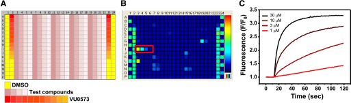
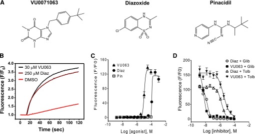
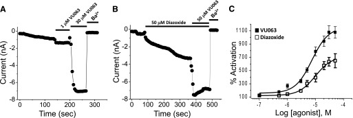
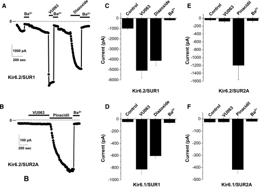
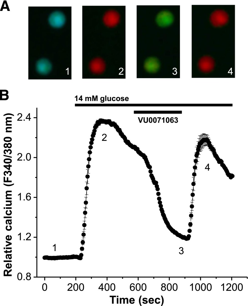
ATP-regulated potassium (KATP) channel complexes of inward rectifier potassium channel (Kir) 6.2 and sulfonylurea receptor (SUR) 1 critically regulate pancreatic islet β-cell membrane potential, calcium influx, and insulin secretion, and consequently, represent important drug targets for metabolic disorders of glucose homeostasis. The KATP channel opener diazoxide is used clinically to treat intractable hypoglycemia caused by excessive insulin secretion, but its use is limited by off-target effects due to lack of potency and selectivity. Some progress has been made in developing improved Kir6.2/SUR1 agonists from existing chemical scaffolds and compound screening, but there are surprisingly few distinct chemotypes that are specific for SUR1-containing KATP channels. Here we report the serendipitous discovery in a high-throughput screen of a novel activator of Kir6.2/SUR1: VU0071063 [7-(4-(tert-butyl)benzyl)-1,3-dimethyl-1H-purine-2,6(3H,7H)-dione]. The xanthine derivative rapidly and dose-dependently activates Kir6.2/SUR1 with a half-effective concentration (EC50) of approximately 7 μM, is more efficacious than diazoxide at low micromolar concentrations, directly activates the channel in excised membrane patches, and is selective for SUR1- over SUR2A-containing Kir6.1 or Kir6.2 channels, as well as Kir2.1, Kir2.2, Kir2.3, Kir3.1/3.2, and voltage-gated potassium channel 2.1. Finally, we show that VU0071063 activates native Kir6.2/SUR1 channels, thereby inhibiting glucose-stimulated calcium entry in isolated mouse pancreatic β cells. VU0071063 represents a novel tool/compound for investigating β-cell physiology, KATP channel gating, and a new chemical scaffold for developing improved activators with medicinal chemistry.


Figures

Fig. 1.
Fig. 1.
Discovery of VU0071063 in a Tl+ flux assay of Kir6.2/SUR1. (A) Plate map used in CRC analyses. DMSO (solvent) and broad-spectrum Kir channel inhibitor VU0573 (Raphemot et al., 2011) were used as controls. The 4-point test compound CRCs were distributed horizontally, whereas 11-point VU0573 CRCs were distributed vertically. (B) Fluorescence heat map recorded from the assay plate containing four doses of VU0071063 (red box). Fluorescence intensity is indicated by the pseudocolored scale (right), with cooler (blue) to hotter (red) colors corresponding to low high Tl+ flux, respectively. (C) Representative time versus normalized (F/F0) fluorescence intensity in wells containing the indicated concentrations of VU0071063.
Fig. 2.
Fig. 2.
Characterization of VU0071063 activity against Kir6.2/SUR1 in Tl+ flux assays. (A) Chemical structures of VU0071063 (VU063), diazoxide (Diaz), and pinacidil (Pin). (B) Representative Tl+ flux experiment demonstrating activation of Kir6.2/SUR1 by 30 μM VU0071063 or 250 μM diazoxide, but not the vehicle control DMSO (0.3%). Fluorescence data have been normalized (F/F0) to baseline values recorded before Tl+ addition. (C) Dose-dependent activation of Kir6.2/SUR1 by VU0071063 and diazoxide, but not pinacidil (n = 4–6 independent experiments, each performed in triplicate). (D) Dose-dependent inhibition of VU0071063- and diazoxide-dependent Tl+ flux by glibenclamide (Glib) and tolbutamide (Tolb) (n = 3 independent experiments, each performed in triplicate).
Fig. 3.
Fig. 3.
Characterization of VU0071063 activity against Kir6.2/SUR1 with patch-clamp electrophysiology. (A) Transfected cells expressing Kir6.2/SUR1 were voltage-clamped at −75 mV and stepped every 5 seconds to −120 mV to elicit inward current. Minor current run-up was observed following establishment of the whole-cell configuration and dialysis with the pipette solution. Addition of 1 or 30 μM VU0071063 led to rapid activation of inward current. (B) In contrast, addition of 50 mM diazoxide activated Kir6.2/SUR1 slowly. After achieving steady-state activation, addition of 50 μM VU0071063 led to further activation of Kir6.2/SUR1. Inward currents were blocked with 2 mM Ba2+. (C) Mean ± S.E.M. dose-response data fitted with 4-parameter logistic functions to derive EC50 values of 7 and 11 for VU0071063 (▪) and diazoxide (しろいしかく), respectively (n = 5–10 per concentration). Data are normalized and expressed as percent (%) activation from baseline current in the absence of agonist.
Fig. 4.
Fig. 4.
VU0071063 is selective for SUR1-containing KATP channels. (A) Representative whole-cell patch-clamp experiment showing the effects of 50 μM VU0071063, 50 μM diazoxide, and 2 mM Ba2+ on Kir6.2/SUR1 current at −120 mV. Note the differences in the kinetics of activation of VU0071063 and diazoxide. (B) Representative recording showing effects of 50 μM VU0071063, 50 μM pinacidil, and 2 mM Ba2+ on Kir6.2/SUR2A currents. (C–F) Mean ± S.E.M./ current at −120 mV recorded from cells transfected with Kir6.2/SUR1, Kir6.1/SUR1, Kir6.2/SUR2A, or Kir6.1, SUR2A, respectively (n = 4–7).
Fig. 5.
Fig. 5.
VU0071063 inhibits glucose-stimulated β-cell calcium entry. (A) Two representative β cells loaded with FURA-2, displayed as a fluorescent ratio (340/380 nM) in response to 2 mM glucose (1), 14 mM glucose (2), 14 mM glucose + 10 μM VU0071063 (3), and 14 mM glucose (4). (B) Relative calcium responses of mouse islet cells following treatment with 2 mM glucose and as indicated by the conditions labeled above (black lines, n = 239 islet cells over 3 days and 13 plates of cells).

References

    1. Adebiyi A, McNally EM, Jaggar JH. (2008) Sulfonylurea receptor-dependent and -independent pathways mediate vasodilation induced by ATP-sensitive K+ channel openers. Mol Pharmacol 74:736–743 - PMC - PubMed
    1. Alemzadeh R, Fledelius C, Bodvarsdottir T, Sturis J. (2004) Attenuation of hyperinsulinemia by NN414, a SUR1/Kir6.2 selective K-adenosine triphosphate channel opener, improves glucose tolerance and lipid profile in obese Zucker rats. Metabolism 53:441–447 - PubMed
    1. Ashcroft FM. (1988) Adenosine 5′-triphosphate-sensitive potassium channels. Annu Rev Neurosci 11:97–118 - PubMed
    1. Ashcroft FM. (2007) The Walter B. Cannon Physiology in Perspective Lecture, 2007. ATP-sensitive K+ channels and disease: from molecule to malady. Am J Physiol Endocrinol Metab 293:E880–E889 - PubMed
    1. Carr RD, Brand CL, Bodvarsdottir TB, Hansen JB, Sturis J. (2003) NN414, a SUR1/Kir6.2-selective potassium channel opener, reduces blood glucose and improves glucose tolerance in the VDF Zucker rat. Diabetes 52:2513–2518 - PubMed

Publication types

Cite

AltStyle によって変換されたページ (->オリジナル) /