Nucleic acid test to diagnose cryptosporidiosis: lab assessment in animal and patient specimens
- PMID: 24479858
- PMCID: PMC3958140
- DOI: 10.1021/ac403750z
Nucleic acid test to diagnose cryptosporidiosis: lab assessment in animal and patient specimens
Abstract
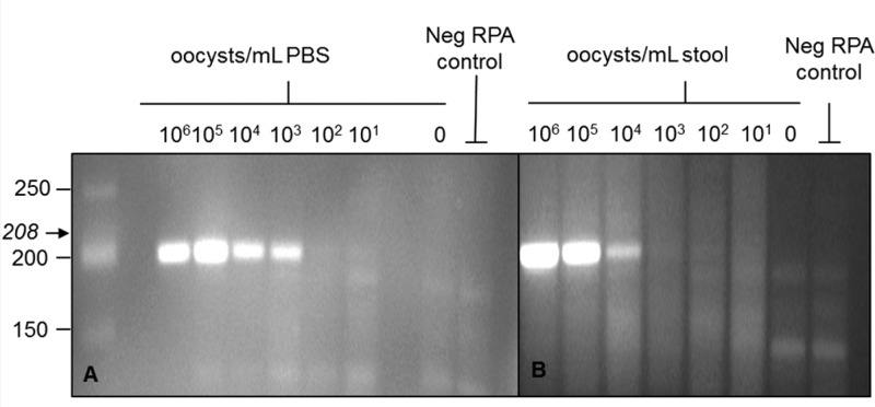
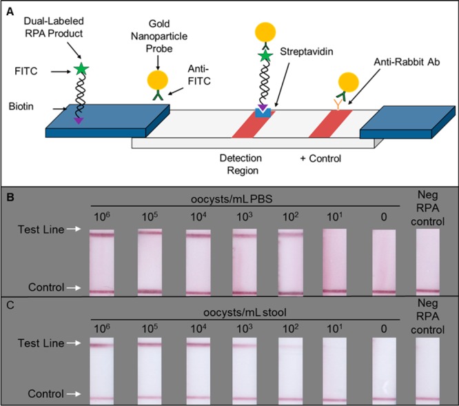
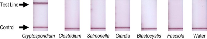
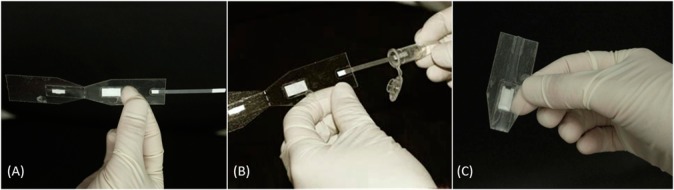
Diarrheal diseases cause more morbidity and mortality around the world than human immunodeficiency virus (HIV), malaria, or tuberculosis. Given that effective treatment of persistent diarrheal illness requires knowledge of the causative organism, diagnostic tests are of paramount importance. The protozoan parasites of the genus Cryptosporidium are increasingly recognized to be responsible for a significant portion of diarrhea morbidity. We present a novel nucleic acid test to detect the presence of Cryptosporidium species in DNA extracted from stool samples. The assay uses the isothermal amplification technique recombinase polymerase amplification (RPA) to amplify trace amounts of pathogen DNA extracted from stool to detectable levels in 30 min; products are then detected visually on simple lateral flow strips. The RPA-based Cryptosporidium assay (RPAC assay) was developed and optimized using DNA from human stool samples spiked with pathogen. It was then tested using DNA extracted from the stool of infected mice where it correctly identified the presence or absence of 27 out of 28 stool samples. It was finally tested using DNA extracted from the stool of infected patients where it correctly identified the presence or absence of 21 out of 21 stool samples. The assay was integrated into a foldable, paper and plastic device that enables DNA amplification with only the use of pipets, pipet tips, and a heater. The performance of the integrated assay is comparable to or better than polymerase chain reaction (PCR), without requiring the use of thermal cycling equipment. This platform can easily be adapted to detect DNA from multiple pathogens.
Figures
References
-
- Lozano R.; Naghavi M.; Foreman K.; Lim S.; Shibuya K.; Aboyans V.; Abraham J.; Adair T.; Aggarwal R.; Ahn S. Y.; Alvarado M.; Anderson H. R.; Anderson L. M.; Andrews K. G.; Atkinson C.; Baddour L. M.; Barker-Collo S.; Bartels D. H.; Bell M. L.; Benjamin E. J.; Bennett D.; Bhalla K.; Bikbov B.; Bin Abdulhak A.; Birbeck G.; Blyth F.; Bolliger I.; Boufous S.; Bucello C.; Burch M.; Burney P.; Carapetis J.; Chen H.; Chou D.; Chugh S. S.; Coffeng L. E.; Colan S. D.; Colquhoun S.; Colson K. E.; Condon J.; Connor M. D.; Cooper L. T.; Corriere M.; Cortinovis M.; de Vaccaro K. C.; Couser W.; Cowie B. C.; Criqui M. H.; Cross M.; Dabhadkar K. C.; Dahodwala N.; De Leo D.; Degenhardt L.; Delossantos A.; Denenberg J.; Des Jarlais D. C.; Dharmaratne S. D.; Dorsey E. R.; Driscoll T.; Duber H.; Ebel B.; Erwin P. J.; Espindola P.; Ezzati M.; Feigin V.; Flaxman A. D.; Forouzanfar M. H.; Fowkes F. G. R.; Franklin R.; Fransen M.; Freeman M. K.; Gabriel S. E.; Gakidou E.; Gaspari F.; Gillum R. F.; Gonzalez-Medina D.; Halasa Y. A.; Haring D.; Harrison J. E.; Havmoeller R.; Hay R. J.; Hoen B.; Hotez P. J.; Hoy D.; Jacobsen K. H.; James S. L.; Jasrasaria R.; Jayaraman S.; Johns N.; Karthikeyan G.; Kassebaum N.; Keren A.; Khoo J.-P.; Knowlton L. M.; Kobusingye O.; Koranteng A.; Krishnamurthi R.; Lipnick M.; Lipshultz S. E.; Ohno S. L.; Mabweijano J.; MacIntyre M. F.; Mallinger L.; March L.; Marks G. B.; Marks R.; Matsumori A.; Matzopoulos R.; Mayosi B. M.; McAnulty J. H.; McDermott M. M.; McGrath J.; Mensah G. A.; Merriman T. R.; Michaud C.; Miller M.; Miller T. R.; Mock C.; Mocumbi A. O.; Mokdad A. A.; Moran A.; Mulholland K.; Nair M. N.; Naldi L.; Narayan K. M. V.; Nasseri K.; Norman P.; O’Donnell M.; Omer S. B.; Ortblad K.; Osborne R.; Ozgediz D.; Pahari B.; Pandian J. D.; Rivero A. P.; Padilla R. P.; Perez-Ruiz F.; Perico N.; Phillips D.; Pierce K.; Pope C. A. 3rd; Porrini E.; Pourmalek F.; Raju M.; Ranganathan D.; Rehm J. T.; Rein D. B.; Remuzzi G.; Rivara F. P.; Roberts T.; De Leon F. R.; Rosenfeld L. C.; Rushton L.; Sacco R. L.; Salomon J. A.; Sampson U.; Sanman E.; Schwebel D. C.; Segui-Gomez M.; Shepard D. S.; Singh D.; Singleton J.; Sliwa K.; Smith E.; Steer A.; Taylor J. A.; Thomas B.; Tleyjeh I. M.; Towbin J. A.; Truelsen T.; Undurraga E. A.; Venketasubramanian N.; Vijayakumar L.; Vos T.; Wagner G. R.; Wang M.; Wang W.; Watt K.; Weinstock M. A.; Weintraub R.; Wilkinson J. D.; Woolf A. D.; Wulf S.; Yeh P.-H.; Yip P.; Zabetian A.; Zheng Z.-J.; Lopez A. D.; Murray C. J. L. Lancet 2013, 380(9859), 2095–2128. - PMC - PubMed
-
- White A. C. Cryptosporidium Species. In Mandell, Douglas, and Bennett’s Principles and Practice of Infectious Diseases, 7th ed.; Mandell G. L., Bennett J. E., Eds.; Elsevier: Philadelphia, PA, 2009; pp 3547–3560.
-
- Guerrant D. I.; Moore S. R.; Lima A. A. M.; Patrick P. D.; Schorling J. B.; Guerrant R. L. Am. J. Trop. Med. Hyg. 1999, 61(5), 707–713. - PubMed
- Putignani L.; Menichella D. Interdiscip. Perspect. Infect. Dis. 2010, 2010, 753512. - PMC - PubMed
- Mondal D.; Haque R.; Sack R. B.; Kirkpatrick B. D.; Petri W. A. Jr. Am. J. Trop. Med. Hyg. 2009, 80(5), 824–826. - PMC - PubMed
-
- Chalmers R. M.; Campbell B. M.; Crouch N.; Charlett A.; Davies A. P. J. Med. Microbiol. 2011, 60(11), 1598–1604. - PubMed
Publication types
MeSH terms
Substances
Grants and funding
LinkOut - more resources
Full Text Sources
Other Literature Sources
Medical
Miscellaneous